Smoking, Tobacco & Nicotine Impact on Gum Recession
Tobacco smoking and nicotine exposure represent one of the most destructive modifiable risk factors for severe periodontal destruction and accelerated receding gums overview. Nicotine exerts profound vasoconstrictive effects on the microvasculature of the gingiva, suppressing classical warning signs such as bleeding and redness while accelerating deep periodontal attachment loss. Understanding this biological disconnect is crucial for patients who use combustible tobacco, cigars, or electronic nicotine delivery systems.
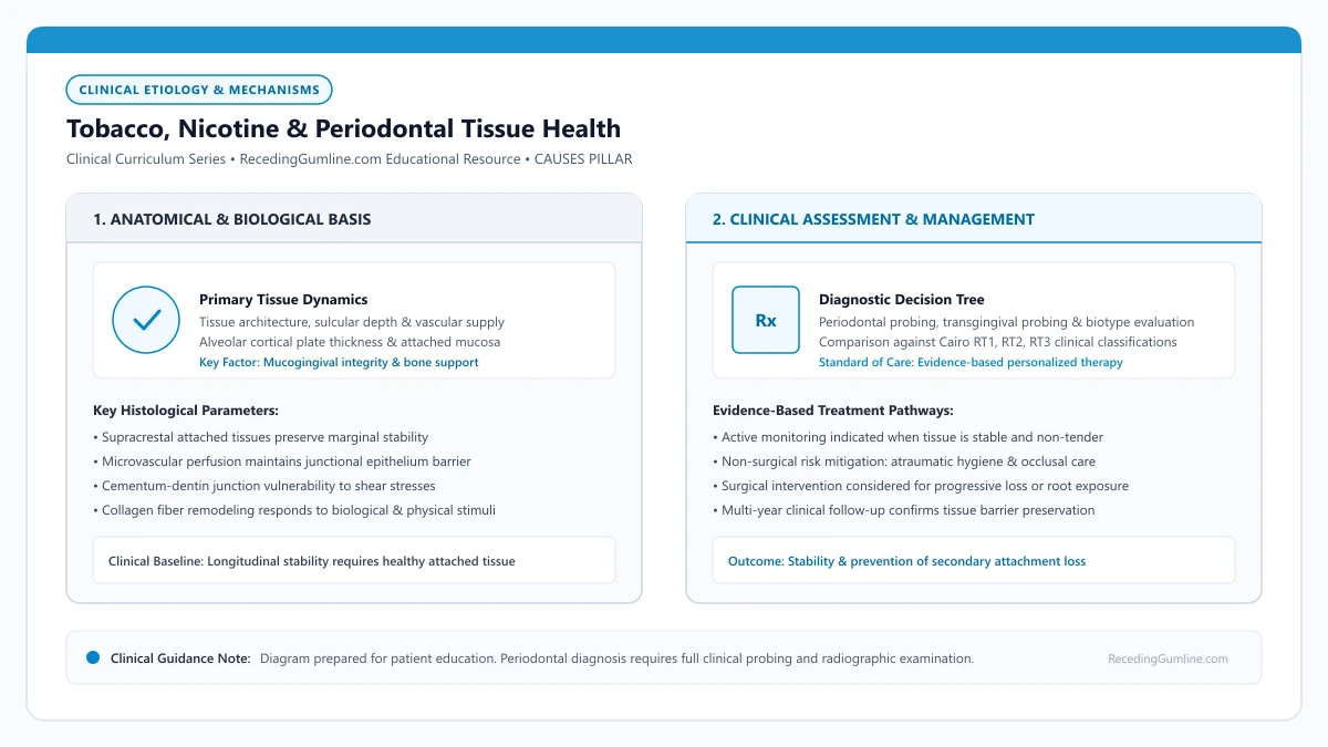
Educational illustration: Tobacco, Nicotine & Periodontal Tissue Health. Clinical management requires comprehensive periodontal evaluation rather than isolated self-assessment.
Source: RecedingGumline.com Clinical Editorial Team (Proprietary educational diagram for RecedingGumline.com)
Microvascular Vasoconstriction & Gingival Ischemia
Nicotine is a potent sympathomimetic alkaloid that binds to nicotinic acetylcholine receptors, stimulating local and systemic release of catecholamines (epinephrine and norepinephrine). In the oral cavity, this induces rapid vasoconstriction of the terminal arterioles and capillary loops supplying the free and attached gingiva, drastically curtailing microvascular perfusion.
Laser Doppler flowmetry studies demonstrate that acute smoking causes a prolonged reduction in gingival blood flow. This persistent state of localized ischemia deprives periodontal fibroblasts and epithelial cells of vital oxygen and nutrients, impairing normal cellular turnover, collagen synthesis, and physiological barrier maintenance.
Because blood flow is constricted, smokers characteristically exhibit pale, dense, thickened gingival margins with an absence of inflammatory bleeding. This creates a dangerous clinical illusion: patients mistakenly assume their gums are healthy because they do not bleed during brushing, while destructive attachment loss silently advances beneath the surface.
Key Etiological Insights
- Nicotine triggers localized catecholamine release, constricting terminal capillary loops in marginal gingiva.
- Reduced microvascular perfusion creates chronic tissue hypoxia, impairing cellular repair mechanisms.
- The absence of gingival bleeding in smokers masks underlying progressive attachment and bone loss.
Host Immune Suppression & Neutrophil Dysfunction
In addition to vascular constriction, tobacco smoke fundamentally disrupts the innate and adaptive immune responses within the periodontal sulcus. Polymorphonuclear leukocytes (neutrophils) serve as the body's frontline defense against invading periodontal bacteria. In smokers, nicotine and volatile combustion products paralyze neutrophil migration, suppress chemotaxis, and diminish phagocytic engulfment.
While neutrophil defense capabilities are crippled, their production of destructive collagenolytic enzymes—including matrix metalloproteinase-8 (MMP-8) and neutrophil elastase—is paradoxically elevated. This uncoupling results in massive bystander destruction of connective tissue attachment fibers and alveolar bone without effectively clearing bacterial pathogens.
Furthermore, tobacco smoke promotes subgingival colonization by virulent anaerobic pathogens, including Porphyromonas gingivalis, Treponema denticola, and Tannerella forsythia (the classical "Red Complex"). The synergistic combination of an aggressive microbial biofilm and a paralyzed host immune response accelerates tissue destruction by up to three- to five-fold compared to non-smokers.
Key Etiological Insights
- Neutrophil chemotaxis and phagocytosis are blunted, compromising first-line antimicrobial defenses.
- Elevated release of matrix metalloproteinases (MMP-8) accelerates the breakdown of collagen attachment fibers.
- Smokers exhibit higher prevalence and colonization density of high-virulence red-complex anaerobic pathogens.
Fibrotic Tissue Remodeling & Clinical Presentation
Chronic exposure to smoke chemicals induces phenotypic alterations in gingival fibroblasts, driving them toward excessive production of dense, collagenous extracellular matrix. As a result, the marginal gingiva of chronic smokers often appears thickened, heavily stippled, and fibrotic, lacking the delicate scalloped architecture of healthy tissue.
Despite this apparent structural bulk, the tissue's biological attachment to the root surface is severely weakened. The junctional epithelium migrates apically along the cementum, forming deep, silent periodontal pockets or translating directly into extensive buccal recession, particularly in the anterior maxilla where smoke directly impinges on the tissue.
When recession manifests in smokers, root coverage grafting procedures face significantly compromised prognoses. Because connective tissue grafts rely entirely on collateral blood supply from the recipient bed during the first 14 days of healing, nicotine-induced vasoconstriction dramatically increases the incidence of partial or complete graft necrosis.
Key Etiological Insights
- Chronic smoking promotes fibrotic tissue thickening that conceals ongoing deep periodontal attachment loss.
- Maxillary anterior facial surfaces sustain direct thermal and chemical insult, accelerating localized recession.
- Surgical root coverage grafting exhibits significantly lower complete coverage rates in active smokers due to compromised perfusion.
Impact of Cessation on Periodontal Regeneration & Stability
Smoking cessation produces immediate and profound improvements in periodontal microvascular dynamics and clinical outcomes. Within days of quitting, peripheral vasoconstriction subsides, restoring normal blood flow and nutrient delivery to the gingiva. Patients frequently notice a temporary increase in gingival bleeding during the first several weeks of cessation—a positive clinical sign reflecting re-establishment of normal microvascular responsiveness and immune surveillance.
Longitudinal epidemiological studies confirm that former smokers who quit experience rates of attachment loss that decelerate toward those of non-smokers. While lost alveolar bone and receded gum tissue do not spontaneously regenerate, cessation stabilizes the remaining periodontal foundation and halts further tissue retreat.
For patients contemplating surgical root coverage (such as connective tissue grafting or coronally advanced flaps), smoking cessation for a minimum of 4 weeks pre-operatively and 8 weeks post-operatively is essential to maximize recipient-bed revascularization and prevent graft failure.
Key Etiological Insights
- Cessation restores normal gingival microcirculation, which may initially manifest as transient brushing bleeding.
- Longitudinal disease progression rates in former smokers decelerate to match those of non-smokers over time.
- Pre- and post-surgical cessation is essential for soft-tissue graft revascularization and clinical success.
Vaping & E-Cigarettes Impact on Periodontal Gum Recession
As electronic cigarettes and vaping devices have surged in popularity, clinical periodontists are witnessing distinct oral health consequences. In any objective receding gumline assessment, vaping delivers concentrated nicotine, propylene glycol, vegetable glycerin, and chemical flavorants directly to oral tissues. Emerging scientific research demonstrates that vaping impairs gingival microcirculation, masks early bleeding signs, and accelerates attachment loss.
• Nicotine delivered via e-cigarette aerosols acts as a potent systemic and local vasoconstrictor.
• Impaired microvascular blood flow deprives gingival tissues of oxygen and vital immune cells.
• Vaping masks gingival inflammation by reducing bleeding on probing, giving a false appearance of tissue health.
• Aerosolized humectants and flavoring chemicals induce oxidative stress and suppress fibroblast attachment.
Microvascular Impairment: The Vasoconstrictive Choke Point
Nicotine is a sympathomimetic alkaloid that stimulates alpha-1 adrenergic receptors in peripheral blood vessels. When inhaled via e-cigarette vapor, nicotine is absorbed almost instantly through the oral mucosa and pulmonary capillaries, triggering immediate constriction of microvascular capillary beds in the gingiva.
This vasoconstriction reduces gingival blood flow by 30% to 50% for several hours after vaping. Starved of adequate arterial blood, marginal gum tissues experience cellular hypoxia (oxygen deprivation) and reduced delivery of defensive polymorphonuclear leukocytes (white blood cells).
Aerosolized nicotine from electronic vapor devices induces acute peripheral vasoconstriction, drastically restricting blood flow through delicate gingival microcapillaries. This severe reduction in tissue perfusion deprives the marginal gums of oxygen and essential immunological defense cells.
Key Etiological Insights
- Nicotine triggers alpha-adrenergic receptors causing peripheral capillary constriction
- Reduces gingival microvascular blood flow by up to 50%
- Tissue hypoxia impairs cellular turnover and suppresses immune defenses against bacteria
The Masked Bleeding Phenomenon: The Illusion of Health
One of the most dangerous clinical aspects of vaping is the "masked bleeding" phenomenon. Under normal circumstances, accumulating bacterial plaque causes gingivitis, marked by bleeding when brushing or flossing—an early warning signal that alerts patients to seek dental care.
Because nicotine restricts blood flow to surface capillaries, vapers with extensive plaque and deep pockets often experience zero bleeding. The gums may look pale and pink despite active destruction occurring deep beneath the surface, delaying diagnosis until severe recession or tooth mobility develops.
Propylene glycol and vegetable glycerin vapor bases act as hygroscopic desiccants, drying out oral mucosal membranes and lowering protective salivary flow. Reduced salivary lubrication amplifies friction during brushing and accelerates mechanical tearing of thin gingival tissues.
Key Etiological Insights
- Nicotine prevents normal capillary engorgement, eliminating early warning bleeding
- Vapers may have active periodontal attachment loss despite non-bleeding gums
- Delays diagnosis because patients mistakenly assume lack of bleeding equals health
Chemical Cytotoxicity: Propylene Glycol and Flavoring Aldehydes
Beyond nicotine, the carrier base of all vape liquids consists of propylene glycol (PG) and vegetable glycerin (VG). Propylene glycol is a powerful humectant that absorbs water molecules, causing severe and chronic dry mouth (xerostomia). Saliva is the mouth's primary defense; without it, virulent periodontal bacteria proliferate rapidly.
Furthermore, popular chemical flavorings—especially cinnamon, menthol, vanilla, and sweet aldehydes—have been shown in vitro to be cytotoxic to human periodontal ligament fibroblasts. These chemicals induce DNA oxidative stress and inhibit the cells responsible for repairing the collagen fibers that anchor teeth.
Patients who vape frequently present with masked gingival symptoms, exhibiting minimal bleeding on probing despite underlying periodontal degradation. Periodontal specialists advise complete smoking and vaping cessation at least four weeks prior to any soft-tissue grafting procedure to ensure graft microvascularization.
Key Etiological Insights
- Propylene glycol strips moisture from oral tissues, causing chronic xerostomia
- Lack of saliva allows pathogenic anaerobic bacteria to flourish along the gumline
- Flavoring aldehydes cause oxidative damage and inhibit collagen-producing fibroblasts
Aerosol Chemistry: Propylene Glycol, Glycerin & Vascular Constriction
Electronic nicotine delivery systems (e-cigarettes) aerosolize liquids containing propylene glycol, vegetable glycerin, nicotine, and flavor chemicals. Nicotine absorbed through the oral mucosa stimulates sympathetic adrenergic receptors, inducing profound microvascular vasoconstriction in gingival capillaries.
This localized ischemia deprives periodontal tissues of vital oxygen and nutrients while blunting the early inflammatory warning signs—such as bleeding—that typically alert patients to underlying tissue breakdown. Consequently, periodontal attachment loss and marginal recession progress silently beneath a pale, deceptively healthy-looking tissue exterior.
Furthermore, propylene glycol and glycerin humectants cause severe cellular dehydration in oral epithelial cells, compromising cell-to-cell tight junctions and accelerating the penetration of toxic bacterial lipopolysaccharides into deeper connective tissue layers.
Key Etiological Insights
- Aerosolized nicotine causes severe gingival vasoconstriction, masking tissue bleeding.
- Ischemia starves periodontal tissues of oxygen while permitting silent attachment loss.
- Humectants disrupt epithelial barrier junctions, facilitating cytotoxic bacterial penetration.
Impact on Periodontal Surgery & Connective Tissue Graft Healing
Vaping introduces severe clinical complications for patients requiring periodontal surgery. Successful soft-tissue grafting depends on rapid revascularization—the in-growth of micro-capillaries from the recipient bed into the avascular donor tissue within the first 48 to 72 hours postoperatively.
Nicotine and toxic carbonyl byproducts (such as acrolein and formaldehyde) present in vape vapor inhibit fibroblast proliferation, decrease collagen production, and paralyze neutrophil phagocytic function. This hostile biological environment dramatically increases the risk of partial graft necrosis, flap dehiscence, and surgical failure.
Periodontists strongly recommend a minimum cessation window of four weeks prior to and four weeks following periodontal plastic surgery to re-establish physiological tissue perfusion and optimize graft survival.
Key Etiological Insights
- Graft revascularization requires uncompromised microcapillary perfusion in the first 72 hours.
- Acrolein and formaldehyde in vapor suppress fibroblast migration and collagen synthesis.
- A strict 4-week pre- and post-surgical cessation protocol is necessary for predictable graft healing.
Clinical Reality Check
Because smoking suppresses classical inflammatory symptoms like bleeding and edema, a lack of symptoms in a smoker must never be interpreted as clinical stability. Full 6-point periodontal charting and full-mouth radiographs are mandatory to assess true bone and attachment levels in tobacco users.
Questions to Ask Your Dentist or Periodontist
- Has smoking masked underlying periodontal bone loss around my receded gumlines?
- How does my current smoking status affect my eligibility for surgical root coverage or gum grafting?
- What specific changes in my gums should I expect if I begin a smoking cessation program?
- Are electronic cigarettes or nicotine pouches less harmful to my periodontal attachment than combustible cigarettes?
- What is my current periodontal probing depth and clinical attachment level status?
- Do you see evidence of reduced blood flow or tissue changes related to my vaping habits?
- What were my actual periodontal pocket measurements, regardless of whether my gums bled during probing?
- Are my exposed tooth roots showing increased vulnerability to decay due to vaping-induced dry mouth?
- How long after quitting nicotine does gingival microcirculation return to normal baseline levels?
Unsure What Your Gum Changes Mean?
Take our free, evidence-based Gum Recession Assessment — approximately 3 minutes. Identify potential risk factors, evaluate symptoms, and receive personalized discussion questions for your dentist or periodontist.
Non-diagnostic educational triage. Private, secure, completed in your browser.
Related Educational Topics
Scientific Literature & Clinical Guidelines
4sources · Hide ▲
- Pihlstrom BL, Michalowicz BS, Johnson NW (2005).
"Periodontal diseases." The Lancet.
Clinical relevance: Peer-reviewed Lancet seminar review synthesizing global periodontal epidemiology, microbial etiology, host immunopathology, and systemic interactions, emphasizing prevention, biofilm disruption, and early risk factor modification.
- Tonetti MS, Greenwell H, Kornman KS (2018).
"Staging and grading of periodontitis: Framework and proposal of a new classification and case definition." Journal of Clinical Periodontology.
Clinical relevance: Consensus framework establishing the multidimensional staging (severity and extent of periodontal tissue breakdown) and grading (biological rate of disease progression, incorporating smoking and diabetes as grade modifiers) for periodontitis. It addresses periodontitis diagnosis and staging, not the classification of localized gingival recession defects.
- Caton JG, Armitage G, Berglundh T, Chapple ILC, Jepsen S, Kornman KS, et al. (2018).
"A new classification scheme for periodontal and peri-implant diseases and conditions - Introduction and key changes from the 1999 classification." Journal of Clinical Periodontology.
Clinical relevance: Introductory consensus overview of the 2018 international classification scheme, introducing periodontal phenotype (incorporating gingival thickness and keratinized tissue width), defining gingival recession independently of inflammatory periodontitis, and standardizing mucogingival diagnostic terminology.
- Chapple ILC, Mealey BL, Van Dyke TE, Bartold PM, Dommisch H, Eickholz P, et al. (2018).
"Periodontal health and gingival diseases and conditions on an intact and a reduced periodontium: Consensus report of workgroup 1 of the 2017 World Workshop on the Classification of Periodontal and Peri-Implant Diseases and Conditions." Journal of Clinical Periodontology.
Clinical relevance: Consensus report establishing diagnostic criteria for periodontal health and gingivitis across intact and reduced periodontia, defining clinical gingival health as <10% bleeding on probing without attachment loss and strictly differentiating gingivitis from periodontitis.
Important Medical Notice
The contents of RecedingGumline.com, including text, graphics, self-assessment calculators, and other materials, are intended solely for educational and informational purposes. This content is not intended to replace professional dental examination, diagnosis, or treatment. Always seek the advice of a qualified dentist, periodontist, or other licensed oral healthcare provider with any questions you may have regarding a medical or dental condition. Never disregard professional medical advice or delay seeking it because of something you read on this website.