Periodontal Attachment Apparatus & Architecture in Gum Recession
The periodontium is a complex, highly specialized anatomical unit engineered to support teeth within the maxilla and mandible, resist heavy mechanical masticatory forces, and provide an impervious biological seal against microbial invasion (see our receding gumline options). Composed of four distinct tissues—gingiva, periodontal ligament (PDL), root cementum, and alveolar bone—the periodontal attachment apparatus functions as an integrated biological system. Understanding this structural architecture is essential for comprehending how a receding gumline develops and why lost attachment cannot spontaneously regrow.
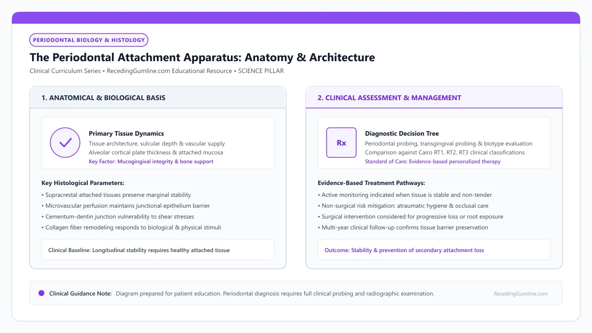
Educational illustration: The Periodontal Attachment Apparatus: Anatomy & Architecture. Clinical management requires comprehensive periodontal evaluation rather than isolated self-assessment.
Source: RecedingGumline.com Clinical Editorial Team (Proprietary educational diagram for RecedingGumline.com)
The Four Functional Tissues of the Periodontal Complex
The periodontium is architecturally divided into two primary functional components: the investing soft-tissue envelope (the gingiva) and the supporting attachment apparatus (periodontal ligament, cementum, and alveolar bone proper). Each tissue possesses a unique histological composition and embryological origin, deriving from the ectomesenchyme of the dental follicle, with the exception of the oral epithelium which arises from surface ectoderm.
The gingiva provides the visible external barrier, sealing underlying bone and connective tissues from oral fluids and microbial contaminants. The root cementum is an avascular mineralized tissue covering the anatomical root, providing the molecular landing surface for inserting collagen fibers.
The periodontal ligament (PDL) is an intensely cellular, vascular connective tissue space that suspends the root within the alveolar socket. Finally, the alveolar bone forms the structural bony crypt (lamina dura) that absorbs and dissipates mechanical stresses generated during mastication and parafunction.
Key Scientific Insights
- The periodontium originates embryologically from dental follicle ectomesenchyme and surface ectoderm.
- The gingiva provides a protective soft-tissue seal; bone, cementum, and PDL provide structural support.
- All four tissues operate as a synchronized biological unit; injury to one impacts the stability of all.
Supracrestal Attached Tissues & Biological Width Dimensions
A cornerstone concept in periodontal biology is the dimensional stability of the supracrestal attached tissues—classically termed the "biological width." In a landmark 1961 histological study of human cadaver dentitions, Anthony Gargiulo and colleagues established the mean physiological dimensions of this dentogingival junction.
Gargiulo demonstrated that the junctional epithelium averages approximately 0.97 mm in vertical height, while the supracrestal connective tissue attachment averages 1.07 mm, establishing a combined biological width of approximately 2.04 mm coronal to the alveolar bone crest. The physiological gingival sulcus accounts for an additional 0.69 mm.
The body fiercely defends this 2.04 mm biological dimension. If restorative margins, calculus deposits, or mechanical trauma impinge within this zone, the host immune system triggers localized osteoclast activation, resorbing alveolar bone crests apically to re-establish the mandatory 2 mm clearance. This protective bone resorption inevitably results in permanent apical migration of the gingival margin.
Key Scientific Insights
- Gargiulo (1961) defined the mean biological width: 0.97 mm junctional epithelium + 1.07 mm connective tissue.
- The combined supracrestal attached tissue dimension averages 2.04 mm above the alveolar bone crest.
- Iatrogenic or mechanical violation of biological width triggers immediate bone resorption and recession.
Periodontal Ligament Fiber Architecture & Sharpey's Fibers
The periodontal ligament space measures merely 0.15 to 0.38 mm in width, yet it represents one of the most mechanically sophisticated tissues in the human body. The PDL is populated by dense bundles of type I and type III collagen fibers organized into principal fiber groups: alveolar crest, horizontal, oblique, periapical, and interradicular fibers.
The terminal ends of these principal collagen bundles mineralize and embed directly into the root cementum on one side and the cribriform plate of the alveolar bone on the other. These embedded anchors are termed Sharpey's fibers. The oblique fiber group comprises the vast majority of the PDL, angling coronally from root to bone to suspend the tooth in a hammock-like sling that transforms axial compressive chewing forces into tensile strain on alveolar bone.
In addition to mechanical support, the PDL is richly innervated with Ruffini-like mechanoreceptors and nociceptive nerve endings that provide exquisite proprioceptive tactile feedback, modulating chewing force and protecting teeth from traumatic impact.
Key Scientific Insights
- Principal collagen fiber bundles are categorized into alveolar crest, horizontal, oblique, and apical groups.
- Sharpey's fibers mineralize directly into cementum and alveolar bone, anchoring the tooth suspended.
- Rich mechanoreceptive innervation provides tactile proprioception, regulating biting force vectors.
Root Cementum & Alveolar Cortical Plate Interactions
Root cementum is unique among mineralized tissues because it is completely avascular and lacks innervation. Acellular extrinsic fiber cementum (AEFC) covers the coronal and middle thirds of the root, forming slowly throughout life. Because cementum lacks internal remodeling blood supply, once it is worn away by toothbrush abrasion or aggressive root planing, it cannot regenerate spontaneously.
The alveolar bone consists of the alveolar bone proper (bundle bone) lining the socket and the outer cortical plates. The facial cortical plate in the anterior maxilla and mandible is remarkably thin—frequently measuring under 0.5 mm in thickness.
In patients with prominent root positions, the cortical plate may naturally feature congenital dehiscences (marginal V-shaped clefts) or fenestrations (isolated windows in bone). When soft-tissue margins over these thin bony plates are challenged by inflammation or physical friction, the absence of underlying bone guarantees rapid, severe gingival recession.
Key Scientific Insights
- Root cementum is avascular and cannot regenerate spontaneously once lost to abrasion or instrumentation.
- The facial cortical bone plate in anterior teeth is often razor-thin (< 0.5 mm).
- Congenital bone dehiscences eliminate skeletal support, predisposing overlying margins to rapid recession.
Pathophysiology of Attachment Breakdown
Gingival recession represents the physical manifestation of breakdown within the periodontal attachment apparatus. Whether initiated by mechanical abrasion, occlusal abfraction, or bacterial periodontitis, the common biological pathway involves disruption of the supracrestal connective tissue fiber attachment.
Once inserting collagen fibers detach from root cementum, the junctional epithelium proliferates and migrates apically along the root to establish a new hemidesmosomal seal. However, if the overlying attached gingiva is thin and unbacked by alveolar bone, the epithelial collar cannot maintain its coronal position.
The soft-tissue margin collapses and contracts apically toward the level of the remaining alveolar bone, exposing root cementum and creating the clinical defect recognized as gum recession. Arresting this process requires eliminating the underlying mechanical or microbial cause before the attachment apparatus is permanently destroyed.
Key Scientific Insights
- Recession initiates when supracrestal connective tissue fibers detach from root cementum.
- The junctional epithelium proliferates apically to create a new seal, pulling the margin downward.
- Halting recession requires identifying and eliminating the specific mechanical or microbial insult.
Gingival Margin Microanatomy in Gum Recession: Free Gingiva, Sulcus & Junctional Epithelium
In any comprehensive receding gums overview, the gingival margin represents the biological interface between the external microbial environment of the mouth and the sterile internal connective tissues of the human body. Understanding its microscopic architecture—consisting of the keratinized free gingival margin, the shallow gingival sulcus, and the specialized junctional epithelium—explains how health is maintained and how early recession initiates.
• The free gingiva is the unattached coronal border of tissue that collars the tooth like a turtleneck sweater.
• The gingival sulcus is the microscopic 1 to 3 mm crevice between the tooth and the inner lining of the free gingiva.
• The junctional epithelium forms a biological adhesive seal directly onto the tooth surface via hemidesmosomes.
• Gingival crevicular fluid (GCF) flows outward through the junctional epithelium, flushing bacteria and delivering antibodies.
Macroscopic and Microscopic Architecture: Free Gingiva and Sulcus
The gingival margin is divided into unattached and attached compartments. The free gingiva (marginal gingiva) is the most coronal ribbon of tissue, approximately 1.0 to 1.5 millimeters wide, that wraps around the neck of the tooth. It forms the soft-tissue wall of the gingival sulcus.
The gingival sulcus is the microscopic space bounded by the tooth surface on one side and the sulcular epithelium on the other. In a state of pristine periodontal health, the sulcular depth measures between 1 and 3 millimeters. It is continually bathed by gingival crevicular fluid (GCF), a serum exudate rich in antibodies and antimicrobial proteins.
The terminal edge of the free gingiva forms a delicate knife-edged scalloped border that contours around the cervical enamel of each tooth. This unattached marginal cuff extends coronally from the free gingival groove to form the external soft-tissue boundary of the gingival sulcus.
Key Scientific Insights
- Free gingiva forms the terminal 1.0 to 1.5 mm unattached border around the tooth
- Sulcular depth of 1 to 3 mm represents normal, non-pathological periodontal anatomy
- Gingival crevicular fluid flows continuously outward, delivering immunoglobulins and neutrophils
The Junctional Epithelium: The Body's Specialized Adhesive Gasket
At the base of the sulcus lies the junctional epithelium (JE)—a specialized non-keratinized epithelial band that is 15 to 30 cells thick coronally, tapering to only 1 to 3 cells thick at its apical termination. The JE attaches directly to the enamel or cementum via an internal basal lamina and cellular anchoring units called hemidesmosomes.
Unlike the protective outer skin, the junctional epithelium is highly porous, possessing wide intercellular spaces. This deliberate biological design allows host immune defenders (neutrophils) to migrate freely from deep blood vessels into the sulcus to patrol for invading bacteria.
Internally, the sulcular epithelium transitions seamlessly into the specialized non-keratinized junctional epithelium. The junctional epithelium forms an organic biological seal along the tooth surface via hemidesmosomes and an internal basal lamina, shielding alveolar structures from microbial invasion.
Key Scientific Insights
- Attaches directly to tooth mineral via hemidesmosomes and internal basal lamina
- Wide intercellular spaces facilitate rapid transit of defensive white blood cells
- Highly permeable nature makes it the primary pathway where bacterial toxins penetrate
The Onset of Recession: Apical Migration of the Epithelial Seal
When bacterial biofilms linger at the margin, persistent leukocytic infiltration and inflammatory cytokines dissolve the coronal hemidesmosomal attachments. The junctional epithelium detaches from the enamel surface, transforming into a pocket epithelium.
To protect the underlying connective tissue, the basal epithelial cells proliferate and migrate apically down the root surface onto the cementum. Once this epithelial seal migrates below the cementoenamel junction, the anatomical tooth crown is permanently lengthened, and clinical gingival recession becomes visible.
Variations in marginal gingival contour reflect the underlying architectural profile of the alveolar bone crest and root prominence. Maintaining continuous microvascular perfusion through the supraperiosteal capillary plexus is essential to preserve marginal tissue height against ischemic atrophy.
Key Scientific Insights
- Enzyme activation dissolves hemidesmosomes, detaching the coronal epithelial seal
- Junctional epithelium proliferates apically down the root to seal off deeper tissues
- Apical migration below the CEJ represents the irreversible onset of attachment loss
Gingival Crevicular Fluid (GCF) Dynamics & Local Host Defense
The gingival crevice is not a static anatomical gutter; it is a dynamically perfused micro-environment flushed by Gingival Crevicular Fluid (GCF). GCF is a serum transudate in health that transforms into an inflammatory exudate during microbial challenge.
Emerging from post-capillary venules in the dentogingival plexus, GCF flows continuously through the junctional epithelium into the oral cavity. It delivers circulating immunoglobulins (predominantly IgG), complement cascade factors, and migrating polymorphonuclear neutrophils (PMNs) to neutralize subgingival bacteria.
In active periodontitis or localized recession, GCF flow rates increase dramatically, transporting host-derived collagenases (MMP-8) and pro-inflammatory cytokines that can be sampled as biochemical biomarkers of active tissue destruction.
Key Scientific Insights
- GCF transitions from a physiological serum transudate to an inflammatory exudate under microbial challenge.
- Delivers host immunoglobulins (IgG), complement proteins, and neutrophils to defend the sulcular margin.
- Elevated GCF flow transports destructive host collagenases (MMP-8) during active attachment loss.
The Free Gingival Groove & Marginal Epithelial Histology
The boundary separating the free marginal gingiva from the attached gingiva is marked clinically in about 50% of individuals by the free gingival groove—a shallow, scalloped depression running parallel to the gingival margin.
Histologically, this groove corresponds to the coronal termination of dense collagen fibers inserting into the periosteum. The marginal tissue is surfaced by parakeratinized stratified squamous epithelium characterized by prominent rete pegs that interlock with connective tissue papillae, providing mechanical resistance against masticatory shear.
Coronal to the attached tissue, the marginal collar tapers to a knife-edge tip. Loss of this parakeratinized barrier through toothbrush abrasion or chronic inflammation leaves the underlying vascular plexus unprotected, facilitating rapid marginal retraction.
Key Scientific Insights
- The free gingival groove demarcates free marginal tissue from firmly anchored attached gingiva.
- Parakeratinized epithelium with dense rete pegs resists mechanical shear forces from mastication.
- Loss of the keratinized epithelial barrier leaves the underlying capillary bed vulnerable to mechanical recession.
Attached Gingiva in Gum Recession: Structural Barrier, Mucogingival Junction & Functional Role
As noted in our receding gumline guide, not all oral gum tissue is created equal. While the inner lips and cheeks are lined by loose, elastic, movable mucosa, the tissue immediately surrounding the teeth is a specialized, rigid, keratinized band known as attached gingiva. Firmly bound to the underlying alveolar bone and cementum, attached gingiva serves as an indispensable physical shock absorber against mechanical wear and bacterial invasion.
• Attached gingiva is firmly bound to the underlying periosteum and cementum by dense collagen fibers.
• The Mucogingival Junction (MGJ) represents the sharp anatomical boundary separating attached gingiva from movable alveolar mucosa.
• Attached gingiva features a heavily keratinized, stratified squamous epithelium that resists mechanical chew trauma.
• Absence of attached gingiva allows lip and cheek muscle movements to pull directly on the marginal seal, accelerating recession.
Histological Architecture: Masticatory Mucosa vs. Alveolar Mucosa
The oral cavity features two fundamentally different mucosal types meeting at the mucogingival junction (MGJ). Attached gingiva is categorized as masticatory mucosa: its surface consists of a thick, orthokeratinized or parakeratinized stratified squamous epithelium supported by dense, collagenous lamina propria.
Crucially, attached gingiva possesses zero submucosal layer; its deep collagen fibers insert directly into the periosteum of the alveolar bone and the supra-alveolar root cementum. This dense anchoring makes it completely immobile. In contrast, alveolar mucosa is non-keratinized, thin, red, and rich in elastic fibers, allowing free facial movement.
Attached gingiva is firmly bound to the underlying alveolar periosteum and root cementum by dense networks of coarse type-I collagen fiber bundles. This immovable fibrous anchor prevents dynamic muscle forces from the lips, cheeks, and tongue from pulling directly on the free marginal cuff.
Key Scientific Insights
- Attached gingiva lacks a submucosa, inserting directly into bone periosteum
- Heavily keratinized surface epithelium protects against abrasive food and brushes
- Alveolar mucosa contains abundant elastin fibers that make it loose and stretchable
The Mucogingival Junction: The Critical Anatomical Landmark
The demarcation line between immobile pink attached gingiva and dark red, movable alveolar mucosa is the Mucogingival Junction (MGJ). Clinicians identify this boundary by gently rolling the patient's lip; the mucosa moves freely, while the attached gingiva remains stationary.
The MGJ is genetically determined and remains relatively constant in position throughout adult life. When gingival recession progresses past the MGJ—leaving zero millimeters of attached tissue—the movable mucosa pulls directly on the marginal sulcus, causing rapid pocket deepening and tooth instability.
A stippled orange-peel surface texture frequently characterizes healthy attached gingiva, reflecting the microscopic anchoring of collagen rete pegs within the underlying connective tissue papillae. The presence of dense keratinization provides formidable resistance to frictional shear during mastication.
Key Scientific Insights
- MGJ represents the sharp physiological boundary between fixed and movable tissues
- Remains in a constant anatomical position relative to the basal bone
- Recession extending past the MGJ allows facial muscles to pull open the gum pocket
Functional Purpose: Shock Absorber and Hygiene Buffer
Attached gingiva performs two critical protective functions. First, it serves as a mechanical shock absorber. When you chew tough foods or brush your teeth, the forces are absorbed by this tough, keratinized collar without pulling the tissue away from the root.
Second, attached gingiva facilitates plaque removal. Because the tissue is immobile and firm, toothbrush bristles can sweep across it effectively without causing pain or bruising. If attached tissue is missing, brushing movable mucosa is intensely uncomfortable, causing patients to avoid cleaning the area.
When the zone of attached gingiva is lost or reduced to zero millimeters, the movable alveolar mucosa directly borders the tooth. Without an attached collar, routine functional movements of the vestibule pull open the sulcular seal, predisposing the site to progressive recession and inflammation.
Key Scientific Insights
- Absorbs muscular traction from lips, cheeks, and tongue during chewing and speaking
- Provides a painless, firm foundation that allows comfortable daily toothbrushing
- Prevents bacterial plaque from migrating apically into deep connective tissue spaces
The Mucogingival Junction: Histochemical & Elastic Properties
The mucogingival junction (MGJ) marks the definitive biological boundary between attached gingiva and movable alveolar mucosa. This demarcation is readily visualized clinically using Schiller's iodine solution; the non-keratinized alveolar mucosa stains dark brown due to abundant intracellular glycogen, whereas glycogen-deficient keratinized gingiva remains unstained.
Histologically, attached gingiva is dense, fibrous, and tightly bound to the underlying alveolar periosteum and cementum via collagenous Sharpey's fibers, containing zero elastic fibers. In stark contrast, the alveolar mucosa is loose, highly vascular, and rich in elastin fibers, permitting wide mobility during speech and chewing.
The attached gingiva acts as a rigid buffer that absorbs the dynamic pull of facial muscles, preventing mechanical forces from being transmitted to the delicate junctional epithelial attachment.
Key Scientific Insights
- Schiller's iodine staining differentiates glycogen-rich alveolar mucosa from attached gingiva.
- Attached gingiva is dense and inelastic, anchored directly to periosteum via collagen bundles.
- The attached tissue band acts as a static shock absorber against facial and lip muscular pull.
Historical Thresholds vs. Contemporary Phenotype Perspectives
In 1972, Lang and Löe published a landmark investigation concluding that at least 2.0 mm of keratinized gingiva (corresponding to ≥ 1.0 mm of attached gingiva) is biologically necessary to maintain clinical gingival health and prevent inflammation under standard oral hygiene.
While this 2 mm rule served as a universal surgical guideline for decades, contemporary periodontal consensus—formalized in the 2017 World Workshop—takes a more nuanced, individualized view. In individuals with exemplary plaque control and non-abrasive brushing habits, sites with less than 1 mm of attached gingiva can remain stable without progressive recession for years.
However, when thin attached tissue coincides with restorative margin placement, subgingival preparation, or planned orthodontic expansion, surgical augmentation remains strongly indicated to prevent rapid marginal breakdown.
Key Scientific Insights
- Lang & Löe (1972) historically proposed 2 mm of keratinized tissue (≥ 1 mm attached) as a health threshold.
- Modern consensus recognizes that plaque-free sites with narrow attached gingiva can remain stable.
- Augmentation is indicated when narrow tissue faces restorative margins or orthodontic expansion forces.
Keratinized Tissue Width: The Historical 2mm Threshold and Modern Clinical Views
Few clinical debates in periodontics have generated more research and discussion than the question: "How much keratinized tissue does a tooth need to stay healthy?" In the long-term management of receding gums, a landmark 1972 study proposed that at least 2 mm of keratinized tissue was mandatory. Decades of subsequent clinical trials have refined this concept, establishing a nuanced, patient-specific understanding of keratinized tissue width (KTW).
• Keratinized tissue width (KTW) is measured from the gingival margin to the mucogingival junction.
• Lang & Löe (1972) established the classic historical reference point: ≥ 2 mm of keratinized tissue (including 1 mm attached) for gingival health.
• Wennström (1987) demonstrated that sites with < 1 mm of KTW can remain stable indefinitely if plaque control is pristine.
• Modern periodontics does not treat width alone, but considers phenotype thickness, patient symptoms, and restorative plans.
The Historical Benchmark: Lang & Löe's 1972 Landmark Trial
In 1972, Dr. Niklaus Lang and Dr. Harald Löe conducted a seminal study at the Royal Dental College in Aarhus, Denmark. They examined 148 tooth surfaces in dental students who performed meticulous plaque control, measuring the relationship between keratinized tissue width and gingival inflammation.
Their data showed that tooth surfaces possessing 2.0 mm or more of keratinized gingiva (which typically provided at least 1.0 mm of firmly attached tissue) remained completely healthy and free of bleeding. Surfaces with less than 2.0 mm of keratinized tissue frequently displayed persistent marginal redness and exudate, establishing the famous "2 mm rule."
The total width of keratinized tissue extends from the coronal gingival margin apically to the mucogingival junction, encompassing both free and attached gingiva. Clinical periodontics historically regards a minimum dimension of two millimeters of keratinized tissue (including one millimeter of attached tissue) as the threshold for mechanical stability.
Key Scientific Insights
- Lang & Löe (1972) established the classic 2 mm keratinized tissue reference point
- Calculated as 1.0 mm of free marginal gingiva plus 1.0 mm of attached gingiva
- Led to decades of routine prophylactic gum grafting to achieve this 2 mm number
The Paradigm Shift: Wennström's Longitudinal Evidence
During the 1980s, Dr. Jan Wennström and colleagues at the University of Gothenburg challenged this dogma through extensive animal and human longitudinal trials. They surgically excised all attached gingiva around teeth, leaving movable alveolar mucosa extending directly to the margin.
They monitored these patients for years. Wennström discovered that as long as the patients maintained excellent, plaque-free oral hygiene without traumatic scrubbing, the teeth with zero attached gingiva experienced zero attachment loss and zero recession. The lack of attached tissue did not automatically cause breakdown.
While plaque-free sites can occasionally maintain attachment with minimal keratinized mucosa, real-world patients often struggle with plaque control when tissue is thin and mobile. Frictional toothbrushing against non-keratinized alveolar mucosa generates stinging discomfort, causing patients to avoid cleaning the cervical margin.
Key Scientific Insights
- Wennström demonstrated that teeth with zero attached gingiva can remain stable indefinitely
- Plaque control and absence of mechanical trauma are more decisive than width alone
- Disproved the notion that narrow keratinized tissue is an automatic surgical emergency
Modern Consensus: When Does Narrow Width Actually Matter?
The 2017 AAP/EFP World Workshop established the contemporary consensus: keratinized tissue width must be evaluated in conjunction with gingival thickness (phenotype) and environmental stressors. Narrow width (< 2 mm) does matter under specific clinical conditions.
Surgical augmentation is recommended when narrow keratinized tissue coincides with: (1) documented progressive apical recession across checkups, (2) severe discomfort when brushing, or (3) planned orthodontic movement or subgingival crown restorations that will subject the margin to mechanical and bacterial stress.
Periodontal plastic surgical procedures such as free gingival grafts or apically repositioned flaps are indicated when inadequate keratinized tissue width compromises restorative margins or orthodontic stability. Augmenting the keratinized collar creates an impenetrable mechanical defense against further apical migration.
Key Scientific Insights
- Narrow width is evaluated alongside tissue thickness and patient-specific risk factors
- Surgical grafting is indicated if active, progressive recession is documented across visits
- Indicated when subgingival crown margins or orthodontic expansion are planned
Anatomical Delineation: Keratinized Tissue Width vs. Attached Gingiva
In clinical periodontics, "keratinized tissue" and "attached gingiva" are distinct anatomical entities that are frequently confused. Keratinized Tissue Width (KTW) represents the total vertical measurement from the coronal margin of the gingiva to the mucogingival junction.
Attached gingiva, however, represents only that portion of keratinized tissue that is firmly bound to the underlying tooth cementum and periosteum. It is calculated by subtracting the sulcular probing depth from the total keratinized tissue width (Attached Gingiva = KTW − Probing Depth).
If a tooth displays 3.0 mm of total keratinized tissue but exhibits a 3.0 mm periodontal pocket, the effective attached gingiva is zero. In this scenario, the tissue is unattached and vulnerable to dynamic muscle pull despite the presence of surface keratin.
Key Scientific Insights
- Total Keratinized Tissue Width (KTW) extends from the soft-tissue margin to the mucogingival junction.
- Attached gingiva equals total KTW minus sulcular probing depth.
- Deep periodontal pockets can completely eliminate attached gingiva even when keratinized tissue is visible.
Clinical Significance Around Natural Teeth vs. Dental Implants
The width of keratinized tissue plays an essential protective role around both natural teeth and dental implants, but with distinct biological dynamics. Around natural teeth, the presence of a connective tissue attachment perpendicular to the root provides robust resistance against mechanical disruption.
Around dental implants, however, supracrestal connective tissue fibers run exclusively parallel to the titanium abutment without inserting into the metal surface, creating a weaker mucosal seal. Multiple systematic reviews confirm that implant sites lacking at least 2.0 mm of keratinized mucosa exhibit significantly higher plaque accumulation, bleeding on probing, mucosal recession, and patient brushing discomfort.
Augmenting keratinized tissue width via free gingival or connective tissue grafting prior to or during restorative therapy ensures long-term tissue stability and cleansability around both teeth and implants.
Key Scientific Insights
- Natural teeth have perpendicular inserting fibers; implants have only parallel non-inserting collagen fibers.
- Peri-implant sites with < 2 mm keratinized tissue exhibit higher inflammation and recession rates.
- Soft-tissue grafting around teeth and implants prevents plaque accumulation and protects restorations.
Gingival Phenotype Classification: Biotypes in Gum Recession
For evaluating diverse receding gumline conditions, the 2018 AAP/EFP global classification updated diagnostic criteria across the periodontium. Among the most impactful changes was replacing the subjective term "biotype" with the standardized, multidimensional concept of periodontal phenotype. This classification provides objective criteria to stratify patients into thin or thick categories, directly predicting recession risk and surgical outcomes.
• The 2018 classification replaced "biotype" with "periodontal phenotype," recognizing that phenotype can be modified by clinical intervention.
• Periodontal phenotype encompasses three distinct parameters: gingival thickness, keratinized tissue width, and bone morphotype.
• Thin-scalloped phenotypes exhibit delicate margins, narrow keratinized tissue (< 1 mm), and thin or dehiscenced cortical bone.
• Thick-flat phenotypes exhibit dense fibrous margins, broad keratinized tissue (≥ 2 mm), and thick cortical bone plates.
Terminology Evolution: Why "Phenotype" Replaced "Biotype"
For decades, dentists used the term "gingival biotype," coined in the 1970s. However, in genetics and biology, a biotype represents a genetically fixed, immutable profile. This was scientifically inaccurate because dental surgeons regularly alter soft-tissue dimensions through grafting.
At the 2017 World Workshop in Chicago, the AAP and EFP officially adopted periodontal phenotype. Phenotype accurately describes the observable physical characteristics resulting from the interaction between genetic predisposition and environmental factors, acknowledging that clinicians can modify tissue thickness surgically.
The 2017 AAP/EFP World Workshop standardized the terminology of periodontal phenotype, recognizing it as a composite of gingival thickness, keratinized tissue width, and alveolar bone morphotype. Clinicians assess phenotype objectively using standard periodontal probe transparency through the sulcular margin.
Key Scientific Insights
- Replaced "biotype" with "periodontal phenotype" in the 2018 global classification
- Acknowledges that tissue thickness can be modified through periodontal plastic surgery
- Integrates soft-tissue dimensions with underlying alveolar bone architecture
The Three Phenotypic Categories: Thin, Thick, and Scalloped
The 2018 classification stratifies periodontal architecture into three primary categories: Thin Scalloped, Thick Flat, and Thick Scalloped.
The Thin Scalloped phenotype features delicate, translucent soft tissue (thickness < 1.0 mm), a narrow zone of keratinized tissue, pronounced architectural scalloping, triangular tooth crowns, and a paper-thin underlying cortical bone plate. The Thick Flat phenotype features dense, fibrous tissue (thickness ≥ 1.0 mm), a broad keratinized band, flatter gingival contours, square tooth crowns, and a thick cortical plate.
Patients exhibiting a thin scalloped phenotype possess delicate, highly translucent gingiva, narrow zones of keratinized tissue, and thin, scalloped cortical bone plates. In contrast, thick flat phenotypes feature dense, fibrotic tissue, broad keratinized collars, and thick, square alveolar bone architecture.
Key Scientific Insights
- Thin Scalloped: Delicate tissue (< 1 mm), triangular crowns, thin cortical bone, high recession risk
- Thick Flat: Dense fibrous tissue (≥ 1 mm), square crowns, thick bone, low recession risk
- Thick Scalloped: Thick tissue with pronounced architectural scalloping and intermediate bone thickness
Clinical Diagnosis and Phenotype Modification Therapy
To determine phenotype chairside, clinicians rely on the probe transparency method: inserting a periodontal probe into the sulcus. If the probe outline shines through the tissue, it is thin (< 1 mm); if invisible, it is thick (≥ 1 mm). High-frequency ultrasound and CBCT can also measure thickness to the tenth of a millimeter.
Identifying a thin phenotype allows periodontists to implement Phenotype Modification Therapy. By performing a connective tissue graft or placing a collagen matrix, the clinician thickens the delicate margin to > 1.5 mm, permanently converting a high-risk thin phenotype into a durable, recession-resistant thick phenotype.
Recognizing phenotypic characteristics is paramount before initiating implant placement, orthodontic movement, or cosmetic crown preparations. Thin phenotypes carry a significantly heightened risk of rapid recession and bone fenestration when subjected to surgical or mechanical trauma.
Key Scientific Insights
- Probe transparency test provides instantaneous chairside diagnosis of thickness
- Thin phenotypes require ultra-gentle hygiene and caution during orthodontics
- Phenotype modification surgery thickens tissue to permanently resist recession
The 2017 AAP/EFP Paradigm: GT, KTW & Bone Morphotype Integration
The 2017 World Workshop on Periodontal and Peri-Implant Diseases formally retired the historical term "biotype" in favor of "periodontal phenotype." This update reflects that while genetic factors establish baseline anatomy, the phenotype is dynamic and modifiable by environmental factors and clinical interventions.
The standardized classification assesses three core components: (1) Gingival Thickness (GT), clinically categorized as thin (≤ 1.0 mm) or thick (> 1.0 mm) using the probe transparency test or ultrasonic pulse-echo measurement; (2) Keratinized Tissue Width (KTW); and (3) Bone Morphotype, evaluated via CBCT.
Patients with a thin scalloped phenotype combine thin translucent gingiva, narrow KTW, and delicate underlying cortical bone with frequent dehiscences, representing the highest risk category for progressive recession under physical stress.
Key Scientific Insights
- The 2017 World Workshop replaced "biotype" with the dynamic, modifiable "periodontal phenotype."
- Diagnostic criteria integrate gingival thickness (≤ 1 mm thin; > 1 mm thick), KTW, and bone morphotype.
- Thin scalloped phenotypes combine delicate soft tissue with thin cortical bone, maximizing recession risk.
Phenotype Conversion: Biology of Connective Tissue Integration
One of the most powerful capabilities of periodontal plastic surgery is phenotype conversion—the surgical transformation of a thin, vulnerable phenotype into a thick, protective tissue band. Placing a subepithelial connective tissue graft (SCTG) under the thin margin achieves this objective.
Histologic studies reveal that the donor connective tissue harvested from the palate retains its genetically programmed phenotypic memory. When grafted into the recipient bed, donor fibroblasts dictate epithelial differentiation, instructing the overlying mucosal cells to express cytokeratins characteristic of thick, parakeratinized gingiva.
This biological conversion permanently increases mucosal thickness beyond 1.5 to 2.0 mm, creating a dense collagenous barrier that resists mechanical toothbrush trauma and shields the underlying alveolar bone from inflammatory resorption.
Key Scientific Insights
- Surgical grafting transforms a fragile thin phenotype into a thick, durable protective tissue band.
- Grafted palatal fibroblasts retain phenotypic memory, instructing overlying cells to keratinize.
- Thickened tissue (> 1.5 mm) provides lifelong resistance against mechanical abrasion and inflammation.
The Periodontal Attachment Apparatus: Cementum, PDL & Alveolar Bone Complex
A natural tooth does not sit rigidly fused to the jawbone like a post in concrete. Instead, it is suspended within a sophisticated, living biological shock absorber known as the periodontal attachment apparatus (periodontium). Composed of three intricately integrated tissues—root cementum, the periodontal ligament (PDL), and the alveolar bone proper—this complex anchors the tooth and dissipates heavy chewing forces.
• The attachment apparatus comprises three interdependent structures: cementum, the periodontal ligament, and alveolar bone.
• Sharpey's fibers are terminal collagen bundles of the PDL that embed deeply into both cementum and alveolar bone.
• The periodontal ligament contains mechanoreceptors that provide proprioceptive feedback, protecting teeth from fracture.
• Gingival recession is not merely a loss of surface gum tissue; it represents the destruction of this underlying attachment complex.
The Tripartite Anchoring Complex: Cementum, PDL & Bundle Bone
The connection between tooth and skeleton is achieved through a tripartite biological unit. On the tooth side sits root cementum—a specialized calcified avascular tissue covering the dentin. On the jawbone side sits the alveolar bone proper (also called bundle bone or cribriform plate).
Bridging the 0.2-millimeter gap between these two hard surfaces is the Periodontal Ligament (PDL)—a dense, vascular fibrous connective tissue containing collagen bundles, cellular repair units, blood vessels, and neural sensory receptors. This tripartite complex functions as a single harmonious organ.
The periodontal attachment apparatus comprises four distinct biological tissues functioning as a synchronized anatomical unit: root cementum, periodontal ligament (PDL), alveolar bone, and the junctional epithelium. Together, these tissues suspend the tooth in its socket and dissipate masticatory shock.
Key Scientific Insights
- Cementum covers the root, alveolar bone forms the socket, and PDL bridges the gap
- PDL space averages 0.2 mm wide and is heavily vascularized and innervated
- All three tissues must be present and integrated for a tooth to possess normal attachment
Sharpey's Fibers: The Molecular Rivets of Tooth Suspension
The primary functional elements of the PDL are Type I collagen fiber bundles known as principal fibers. When these collagen bundles reach the root, their terminal ends become mineralized and embedded deep within the cementum matrix.
On the opposite side, the other terminal ends embed directly into the bundle bone of the socket wall. These mineralized embedded fiber ends are called Sharpey's fibers. Acting like microscopic suspension bridge cables, Sharpey's fibers convert compressive biting forces into tensile pull on the bone, stimulating healthy bone maintenance.
Sharpey’s fibers embedded within the root cementum traverse the periodontal ligament space to anchor securely into the bundle bone of the alveolar wall. This dense fibrous network converts destructive compressive chewing forces into physiological tensile stimuli that promote homeostatic bone remodeling.
Key Scientific Insights
- Sharpey's fibers are terminal collagen bundles mineralized into cementum and bone
- Function like suspension cables, allowing the tooth microscopic physiological mobility
- Convert compressive chewing forces into tensile bone stimulation that prevents atrophy
The Destruction of the Apparatus in Gum Recession
When gum recession occurs, it is not merely a superficial aesthetic shift; it marks the permanent unraveling of the attachment apparatus. Bacterial enzymes or mechanical friction sever the supracrestral connective tissue fibers, exposing root cementum to oral fluids.
Once exposed, the fragile cementum undergoes cytotoxic degradation, losing its embedded Sharpey's fibers. Osteoclasts resorb the alveolar bone crest, and the PDL space is permanently obliterated in the receded zone. Healing following standard treatments produces an epithelial scar (long junctional epithelium) rather than reforming this complex.
When bacterial toxins or mechanical trauma disrupt the coronal junctional epithelial seal, periodontal destruction initiates. The subsequent degradation of Sharpey’s fibers and crestal bone resorption leads directly to clinical attachment loss and visible root exposure.
Key Scientific Insights
- Recession severs Sharpey's fibers and detaches the periodontal ligament from cementum
- Exposed cementum loses its vitality and becomes contaminated with oral bacteria
- Standard healing creates an epithelial seal rather than regenerating new PDL fibers and bone
The Tripartite Attachment Apparatus: Cementum, PDL & Alveolar Bone
The tooth is anchored within the jaw through the periodontal attachment apparatus—a specialized tripartite organ comprising root cementum, the periodontal ligament (PDL), and alveolar bone proper (cribriform plate). These three tissues function as a unified biological stress-distribution complex.
The periodontal ligament is a specialized fibrous connective tissue spanning the 0.2 to 0.4 mm space between the root and bone. Highly organized Type I collagen fibers are embedded deeply into both the mineralized matrix of the root cementum and the lamellar alveolar bone as Sharpey's fibers.
This continuous physical linkage transforms compressive masticatory loads into tensile traction on the bone, stimulating continuous physiological bone remodeling and preventing bone resorption.
Key Scientific Insights
- The attachment apparatus functions as an integrated unit: root cementum, PDL, and alveolar bone.
- Sharpey's collagen fibers insert deeply into both cementum and alveolar bone proper.
- Transforms masticatory forces into tensile stress, maintaining healthy bone and tooth stability.
Mechanisms of Pathological Detachment & Margin Migration
The breakdown of the attachment apparatus during gum recession proceeds through an uncoupling of tissue turnover. When bacterial plaque toxins or mechanical friction induce localized chronic inflammation, host neutrophils release collagenolytic enzymes, primarily MMP-8 and MMP-13.
These matrix metalloproteinases cleave the coronal Sharpey's fibers anchoring the junctional epithelium and principal gingival fibers to the cementum. Deprived of connective tissue anchorage, the junctional epithelial cells migrate apically along the root surface to find an intact collagen attachment.
Simultaneously, pro-inflammatory cytokines stimulate osteoclast differentiation via the RANKL pathway, resorbing the crestal alveolar bone. As both bone and fiber support are lost apically, the soft-tissue margin collapses, producing clinical recession.
Key Scientific Insights
- Host collagenase enzymes (MMPs) cleave Sharpey's fibers anchoring the tissue to cementum.
- Junctional epithelial cells proliferate apically along the root as connective tissue fibers detach.
- RANKL-mediated osteoclast activation resorbs alveolar crest bone, causing marginal soft-tissue collapse.
Clinical Attachment Level (CAL) vs. Probing Depth: True Measurement of Periodontal Stability
In dental examinations, patients frequently hear their hygienist call out numbers like "2, 2, 3" or "4, 5, 4." During a formal receding gumline assessment, while numbers represent probing pocket depths, probing depth alone can be dangerously deceptive. A tooth can have an apparently "healthy" 2 mm pocket while suffering from severe, advanced recession. Understanding Clinical Attachment Level (CAL)—the established benchmark clinical metric in periodontics—reveals the true measurement of tooth stability.
• Probing pocket depth measures from the gingival margin to the base of the sulcus; CAL measures from a fixed anatomical landmark: the cementoenamel junction (CEJ).
• Clinical Attachment Loss is calculated as: Probing Depth + Recession Distance.
• A tooth with 2 mm probing depth and 4 mm of recession has 6 mm of clinical attachment loss.
• The 2017/2018 AAP/EFP World Workshop establishes CAL as the primary parameter for staging periodontitis severity.
The Diagnostic Trap: Why Probing Depth Alone Is Deceptive
When a clinician slides a probe into the gingival sulcus, the measurement recorded is probing pocket depth (PPD). This is the distance from the crest of the gingival margin down to the bottom of the pocket.
However, the gingival margin is a moving, variable boundary. If a patient has severe gingival recession—say, 4 millimeters of exposed root—the tissue margin has moved far down the tooth. If the probe measures a shallow 2 mm pocket from this receded margin, an uneducated patient might assume their gums are in pristine health, completely missing the fact that 6 millimeters of biological support has been destroyed.
Clinical Attachment Level (CAL) represents the true biological distance from a fixed anatomical landmark—the cementoenamel junction (CEJ)—to the bottom of the periodontal pocket. Unlike probing pocket depth, which fluctuates with gingival swelling or recession, CAL provides an immutable baseline for longitudinal tracking.
Key Scientific Insights
- Probing depth uses the moving gingival margin as its reference point
- A shallow pocket (2 mm) does not mean a tooth is healthy if the gum has receded 4 mm
- Relies on a variable boundary that changes with inflammation, swelling, or recession
The CAL Formula: Anchoring to the Cementoenamel Junction
To overcome this flaw, periodontists calculate Clinical Attachment Level (CAL). CAL uses an unchanging, permanent anatomical landmark: the Cementoenamel Junction (CEJ)—the exact microscopic line where the white enamel crown meets the root cementum.
CAL measures the distance from the CEJ down to the bottom of the periodontal pocket. The clinical formula is straightforward: CAL = Probing Depth + Recession. If a tooth has a 3 mm probing depth and 3 mm of marginal recession, the Clinical Attachment Loss is 6 millimeters.
In a patient with gingival overgrowth, a 5 mm probing depth might exist with zero attachment loss because the tissue margin has moved coronally above the CEJ. Conversely, a tooth with severe 4 mm recession and a shallow 2 mm pocket has suffered 6 mm of irreversible clinical attachment loss.
Key Scientific Insights
- CEJ serves as the permanent, unshifting anatomical landmark on the tooth
- Formula: Clinical Attachment Loss (CAL) = Probing Pocket Depth + Recession Distance
- Reflects the true total millimeter loss of periodontal ligament fibers and bone support
Staging Disease: How CAL Determines Treatment Urgency
Under the 2018 AAP/EFP global staging framework, CAL is the definitive metric that stages periodontal disease. Stage I (mild) involves 1 to 2 mm of CAL; Stage II (moderate) involves 3 to 4 mm of CAL; and Stage III or IV (severe) involves 5 mm or more of CAL.
Tracking CAL across successive recall appointments allows your dental team to calculate your disease progression rate. If your CAL increases by 1 to 2 mm over 12 months, your disease is classified as rapidly progressing (Grade C), signaling an immediate need for aggressive periodontal therapy.
The AAP/EFP staging framework relies strictly on interdental clinical attachment loss to classify periodontitis severity from Stage I through Stage IV. Monitoring CAL over sequential six-month recall appointments allows dental clinicians to definitively verify disease arrest or active progression.
Key Scientific Insights
- Stage I: 1–2 mm CAL; Stage II: 3–4 mm CAL; Stage III/IV: ≥ 5 mm CAL
- Sequential increases in CAL ≥ 2 mm prove active, ongoing periodontal disease progression
- Dictates whether conservative monitoring, deep cleaning, or surgical grafting is required
The Diagnostic established clinical benchmark: CAL vs. Pocket Probing Depth
In periodontics, pocket probing depth (PPD) alone is an unreliable measure of true cumulative tissue destruction. Probing depth measures only the distance from the free gingival margin to the base of the sulcus; because the gingival margin can move coronally (due to inflammatory swelling) or apically (due to recession), PPD fluctuates independently of true bone support.
Clinical Attachment Level (CAL) is the definitive biological standard because it measures the distance from a fixed, permanent anatomical landmark—the cementoenamel junction (CEJ)—to the base of the sulcular pocket. CAL is mathematically calculated as: CAL = Probing Depth + Recession Depth.
For example, a tooth with a shallow 2 mm probing depth but 4 mm of exposed root recession has 6 mm of clinical attachment loss. Recognizing this true attachment loss ensures that severe recession is never misdiagnosed as healthy based on shallow pocket depths alone.
Key Scientific Insights
- Pocket probing depth fluctuates with tissue swelling and recession, making it an incomplete metric.
- Clinical Attachment Level (CAL) measures true biological loss from the fixed cementoenamel junction.
- CAL = Probing Depth + Recession Depth; shallow pockets can mask severe attachment loss.
Identifying the CEJ in Worn, Restored & Abfraction Sites
Accurately measuring CAL requires unambiguous identification of the cementoenamel junction. In virgin, unworn teeth, the CEJ is easily detected as a subtle horizontal step or textural transition where smooth enamel meets softer root cementum.
However, in patients with severe toothbrush abrasion, abfraction notches, or cervical restorations, the anatomical CEJ is frequently obliterated. In these complex scenarios, periodontists locate the reference baseline using adjacent anatomical landmarks.
Clinicians extrapolate the CEJ position by measuring the clinical crown length of contralateral homologous teeth, projecting the curvature of adjacent interproximal enamel margins, or referencing the restoration margin if pre-operative records document its biological placement.
Key Scientific Insights
- The CEJ provides the fixed reference point separating anatomical crown enamel from root cementum.
- Abfraction notches and cervical fillings often obliterate or mask the true anatomical CEJ.
- Clinicians use contralateral crown lengths and interdental curvature projections to identify baselines.
Root Cementum Biology & Exposure in Gum Recession
Essential to understanding gum recession, covering the entire anatomical root surface of the human tooth is a micro-thin, specialized calcified tissue known as root cementum. Functioning as the biological glue of the periodontium, cementum anchors the principal periodontal ligament fibers to the tooth. When gum recession exposes cementum to the harsh oral cavity, this fragile layer undergoes rapid physical wear and bacterial contamination.
• Root cementum is a mineralized, avascular connective tissue covering root dentin.
• Acellular afibrillar cementum and acellular extrinsic fiber cementum (AEFC) cover the coronal third, measuring only 20 to 50 micrometers thick.
• Cementum lacks blood vessels, lymphatics, and nerves, making it incapable of self-repair once worn away.
• Exposure to oral biofilms saturates cementum with bacterial lipopolysaccharides (endotoxins), requiring mechanical root planing.
Histological Architecture: Acellular vs. Cellular Cementum
Root cementum is classified into two major histological varieties based on the presence of embedded cells (cementocytes). Acellular extrinsic fiber cementum (AEFC) covers the coronal and middle third of the root. It contains zero cells and forms slowly during tooth eruption, providing the primary anchor for inserting Sharpey's fibers.
Crucially, AEFC at the cervical cementoenamel junction is razor-thin, measuring only 20 to 50 micrometers in thickness (a human hair is approximately 70 micrometers). Cellular intrinsic fiber cementum (CIFC) covers the apical third of the root, containing cementocytes that continue depositing layers throughout life to compensate for occlusal wear.
Root cementum is a specialized, avascular, mineralized connective tissue covering the anatomical roots of teeth, categorized into acellular extrinsic fiber cementum and cellular intrinsic fiber cementum. Acellular cementum covers the coronal root third and serves as the critical anchor for inserting principal periodontal fibers.
Key Scientific Insights
- Acellular extrinsic fiber cementum (AEFC) covers the vulnerable cervical third of the root
- Cervical cementum thickness is only 20 to 50 µm—thinner than a human hair
- Cellular cementum is restricted to the root tip and contains living cementocytes
Degradation Upon Exposure: Abrasion and Endotoxin Saturation
Under healthy conditions, cementum is shielded beneath attached gingiva. When gum recession exposes cervical cementum to the oral environment, two destructive processes occur almost simultaneously: mechanical abrasion and biochemical contamination.
Because cementum is only 45% to 50% inorganic hydroxyapatite, it is significantly softer than enamel. Daily toothbrushing with standard toothpaste scrubs away this 30-micrometer layer within weeks. Concurrently, bacterial biofilms saturate the porous cementum with lipopolysaccharides (endotoxins), creating a toxic surface that prevents soft tissue from reattaching.
Because cementum contains only about 50% hydroxyapatite mineral content by weight, it is significantly softer and more porous than dental enamel. When gingival recession exposes the cementum to the oral environment, the thin layer is easily worn away by toothbrush friction or dissolved by dietary acids within weeks.
Key Scientific Insights
- Low mineral content (50%) makes cementum soft and easily abraded by toothbrushing
- Ordinary brushing can completely wear through cervical cementum in under six months
- Bacterial endotoxins permeate porous cementum, creating a biologically toxic root surface
Clinical Management: Root Planing and Chemical Decontamination
The rapid loss of cementum explains why receded roots develop acute cold sensitivity: once cementum is abraded, underlying dentinal tubules are exposed. Furthermore, when periodontists perform root coverage surgery, they must thoroughly debride the root surface.
Surgeons gently plane the root to remove cytotoxic endotoxins and often condition the surface with EDTA (ethylenediaminetetraacetic acid) or citric acid. This chemical etching dissolves smear plugs, exposes native collagen fibers, and creates a biocompatible surface that allows newly transplanted gum grafts to attach securely.
Once cementum is lost, the underlying radicular dentin is exposed, opening thousands of microscopic dentinal tubules to the oral cavity. Exposed dentin is highly vulnerable to rapid cervical root caries, erosion, and excruciating hydrodynamic thermal hypersensitivity.
Key Scientific Insights
- Loss of cementum denudes underlying dentin, triggering hydrodynamic cold sensitivity
- Root planing removes necrotic, endotoxin-impregnated cementum prior to surgery
- EDTA chemical conditioning decontaminates the root to promote new soft-tissue adhesion
Histological Diversity: Acellular vs. Cellular Intrinsic Fiber Cementum
Root cementum is a specialized, avascular mineralized tissue covering the anatomical root. Histologically, it exists in two primary functional varieties: Acellular Extrinsic Fiber Cementum (AEFC) and Cellular Intrinsic Fiber Cementum (CIFC).
AEFC covers the coronal two-thirds of the root, forming a thin, delicate layer (20 to 50 micrometers thick) that contains densely packed Sharpey's fibers. AEFC is the primary tissue responsible for anchoring the tooth to alveolar bone in the cervical region where recession occurs.
Because AEFC is exceptionally thin and completely devoid of living cementocytes, once it is destroyed by toothbrush abrasion or aggressive root planing, it cannot regenerate spontaneously from within. Regeneration requires recruitment of new cementoblast precursors from the periodontal ligament space.
Key Scientific Insights
- Acellular Extrinsic Fiber Cementum (AEFC) covers the cervical two-thirds of roots where recession occurs.
- AEFC is paper-thin (20-50 micrometers) and contains dense inserting Sharpey's fibers.
- Acellular cementum possesses no living cells; once stripped away, it cannot spontaneously regenerate.
The Denuded Root: Toxic Endotoxin Adsorption & Smear Layers
When gingival recession exposes acellular cementum to the oral environment, the tissue undergoes rapid physical and chemical degradation. Exposure to oral fluids and saliva leads to alternating cycles of surface demineralization and hypermineralization.
Simultaneously, subgingival gram-negative bacteria release lipopolysaccharides (endotoxins) that adsorb directly into the porous organic matrix of the superficial cementum. This endotoxin-impregnated cementum is cytotoxic, actively preventing periodontal ligament fibroblasts from attaching to the root.
During root coverage surgery, periodontists must detoxify this contaminated root layer using ultrasonic scaling, sharp curettes, and chemical conditioning agents (such as citric acid or EDTA) to expose clean collagen fibrils receptive to new soft-tissue attachment.
Key Scientific Insights
- Exposed cementum adsorbs cytotoxic bacterial lipopolysaccharides (endotoxins).
- Endotoxin contamination prevents fibroblast adhesion and prohibits soft-tissue reattachment.
- Chemical and mechanical root conditioning detoxifies the root to enable surgical graft integration.
Periodontal Ligament Principal Fibers & Architecture in Gum Recession
Critical to the stability of teeth susceptible to receding gums, suspended within the microscopic 0.2 mm space between the tooth root and the alveolar bone is one of the most mechanically sophisticated tissues in the human body: the Periodontal Ligament (PDL). Composed of specialized collagen fiber bundles arranged in precise mathematical orientations, the PDL absorbs masticatory shocks, cushions biting forces, and provides sensory proprioception.
• The PDL consists of five distinct principal fiber groups: alveolar crest, horizontal, oblique, apical, and interradicular fibers.
• Oblique fibers constitute the largest group, suspended at an angle to resist vertical chewing forces.
• Sharpey's fibers are terminal collagen ends embedded deeply into root cementum and alveolar bundle bone.
• Periodontal breakdown in gum disease and recession progressively severs these fiber bundles from coronal to apical.
The Functional Geometry: Five Principal Fiber Groups
The collagen fibers of the PDL are not arranged randomly; they are organized into five distinct anatomical groups designed to counter forces from every three-dimensional vector. The Alveolar Crest fibers extend from the cervical cementum downward to the alveolar crest, resisting lateral tilting forces.
Horizontal fibers run perpendicularly from cementum to bone to resist horizontal loads. Oblique fibers represent the largest and most powerful group: running diagonally upward from bone to cementum, they suspend the tooth like a hammock, converting heavy downward chewing shocks into tensile bone stimulation. The Apical fibers stabilize the root tip, while Interradicular fibers stabilize the furcation of multi-rooted molars.
The periodontal ligament is a complex, vascularized cellular connective tissue that surrounds the tooth root, connecting root cementum to the alveolar bone socket. Its principal collagen fibers are arranged in distinct anatomical groups: alveolar crest, horizontal, oblique, periapical, and interradicular bundles.
Key Scientific Insights
- Alveolar crest and horizontal fibers resist lateral tilting and rotational forces
- Oblique fibers form a diagonal hammock that converts vertical chewing into bone tension
- Apical and interradicular fibers stabilize root tips and multi-rooted molar furcations
Sharpey's Fibers and Neurosensory Proprioception
At either end of each principal fiber bundle, the collagen fibers insert deeply into mineralized tissue: cementum on the tooth, and bundle bone on the socket wall. These mineralized insertions are called Sharpey's fibers. This structural arrangement ensures that chewing loads never tear the ligament at the bone-cementum interface.
Furthermore, the PDL is richly supplied with sensory nerves containing Ruffini-like mechanoreceptors. These receptors detect microscopic pressure variations as subtle as a grain of sand between your teeth. When biting forces become excessively heavy, these receptors instantly trigger a reflex that relaxes jaw-closing muscles to prevent tooth fracture.
The oblique fiber group forms the largest contingent, running obliquely from bone down to cementum to absorb and dissipate vertical occlusal forces. Embedded within this collagen matrix are mechanoreceptive nerve endings and undifferentiated mesenchymal stem cells capable of osteogenic and fibroblastic differentiation.
Key Scientific Insights
- Sharpey's fibers mineralize directly into bone and cementum matrices
- Ruffini-like mechanoreceptors provide precise tactile feedback on chewing pressure
- Triggers protective muscular reflexes to prevent tooth cracking under heavy loads
Destruction in Periodontal Disease: The Coronal-to-Apical Unzipping
In periodontal disease and severe recession, tissue destruction follows an "unzipping" pattern from coronal to apical. Plaque-induced matrix metalloproteinases first degrade the alveolar crest and horizontal fibers.
As bone resorbs, the oblique fiber bundles are progressively severed. With fewer fibers remaining to suspend the tooth, occlusal loads place excessive mechanical stress on the remaining apical attachment, leading to progressive tooth hypermobility (looseness) and eventual tooth loss if clinical treatment is not rendered.
In active periodontal breakdown, matrix metalloproteinases enzymatically sever these principal fibers near the alveolar crest. The loss of ligamentous attachment eliminates the tensile suspension mechanism, accelerating alveolar bone resorption and progressive tooth hypermobility.
Key Scientific Insights
- Enzymes degrade fibers in a progressive coronal-to-apical direction
- Loss of oblique fibers destroys the tooth's shock-absorbing suspension hammock
- Excessive strain on remaining fibers produces tooth looseness and mobility
Principal Fiber Groups: From Alveolar Crest to Apical Bundles
The periodontal ligament (PDL) is dominated by dense, wavy bundles of Type I collagen organized into five distinct principal fiber groups: alveolar crest, horizontal, oblique, periapical, and interradicular fibers.
The alveolar crest and horizontal fibers reside in the most coronal portion of the PDL, directly beneath the junctional epithelium. These fibers are the first biological line of defense against lateral rotational forces and marginal bacterial invasion.
The oblique fibers constitute the largest group, running obliquely downward from alveolar bone to root cementum. This hammock-like configuration suspends the tooth within its osseous socket, converting vertical axial masticatory impact into lateral tensile strain on the alveolar bone.
Key Scientific Insights
- PDL collagen fibers are organized into five anatomically distinct principal bundle groups.
- Alveolar crest and horizontal fibers form the coronal defense barrier against mechanical displacement.
- Oblique fibers suspend the tooth like a biological hammock, absorbing vertical biting forces.
Sensory Proprioception & Neuromuscular Bite Regulation
In addition to its structural role, the periodontal ligament is a highly specialized sensory receptor organ. Mechanoreceptors (Ruffini-like endings) distributed throughout the PDL fibers detect minute physical displacements and directional loads as small as 1 to 3 grams.
These mechanoreceptors transmit rapid afferent impulses via the trigeminal nerve to the central nervous system, coordinating the jaw-opening reflex and regulating the intensity of masseter and temporalis muscle contraction.
When severe gum recession and bone loss destroy coronal PDL fibers, periodontal proprioception is diminished. The loss of sensory feedback can lead to uncoordinated bite forces and traumatic occlusal overload, accelerating further attachment loss.
Key Scientific Insights
- Ruffini-like mechanoreceptors in PDL fibers detect microscopic tooth movements as small as 1 gram.
- Sensory signals regulate masticatory muscle force and trigger protective jaw-opening reflexes.
- Loss of coronal PDL fibers impairs sensory proprioception, increasing vulnerability to bite trauma.
Alveolar Process Bone Morphology: Cortical Plate, Trabecular Bone & Dehiscence
The alveolar process is the specialized ridge of bone that forms the sockets (alveoli) supporting the teeth in the maxilla and mandible. Highly dynamic and tooth-dependent, alveolar bone exists solely to support the dentition; when teeth erupt, it forms, and when teeth are extracted, it atrophies. Understanding the distinct layers of alveolar bone clarifies why bone loss occurs and how it triggers gum recession.
• Alveolar bone consists of three distinct layers: outer cortical plates, central trabecular spongy bone, and the inner alveolar bone proper (bundle bone).
• The facial cortical plate covering anterior teeth is exceptionally thin (often < 0.5 mm), offering minimal resistance against resorption.
• Alveolar bone crest normally resides 1.5 to 2.0 mm apical to the cementoenamel junction in health.
• Alveolar bone resorption inevitably causes the overlying soft tissue to collapse, manifesting as gum recession.
Anatomical Triad: Cortical Plates, Trabecular Spongiosa & Bundle Bone
The alveolar process is composed of three distinct bone compartments. The outer boundaries consist of facial and lingual cortical bone plates, made of dense, compact lamellar bone covered by vascular periosteum. Between these plates lies the trabecular (cancellous) bone—a spongy network of marrow spaces.
Lining the tooth socket itself is the alveolar bone proper (also called bundle bone or cribriform plate). This thin layer of bone is perforated by hundreds of microscopic Volkmann's canals that carry blood vessels into the PDL. On dental x-rays, the bundle bone appears as a continuous, radio-opaque white line known as the lamina dura.
The alveolar process consists of the alveolar bone proper (bundle bone lining the socket) and the supporting alveolar bone (facial and lingual cortical plates surrounding central trabecular cancellous bone). The thickness of the facial cortical bone varies dramatically depending on tooth position and individual skeletal anatomy.
Key Scientific Insights
- Outer cortical plates provide dense physical protection for the jaw ridge
- Central trabecular bone contains rich marrow and blood vessels that nourish the periodontium
- Alveolar bone proper (bundle bone) forms the socket wall where Sharpey's fibers anchor
The Paper-Thin Facial Plate: Why Front Teeth Are Vulnerable
In human anatomy, the facial cortical plate covering anterior teeth (incisors, canines, and premolars) is remarkably thin. In over 70% of healthy individuals, this bone plate measures under 0.8 millimeters, and frequently less than 0.3 millimeters near the crest.
Because bone requires its own microvascular network to stay alive, a bone plate thinner than 0.5 mm contains zero internal Haversian blood vessels. It survives entirely on blood diffusing from the outer periosteum and inner PDL. Any minor insult—such as aggressive brushing, orthodontic tipping, or slight plaque inflammation—cuts off this blood supply, causing the bone plate to vanish.
In anterior teeth and prominent canine regions, the facial bone plate is frequently paper-thin (often less than 0.5 mm) or completely absent in areas of anatomical dehiscence. This absence of cancellous marrow limits the intrinsic microvascular blood supply available to support overlying soft tissues.
Key Scientific Insights
- Facial bone plates over front teeth frequently measure under 0.5 mm in thickness
- Ultra-thin bone lacks internal blood vessels and relies entirely on diffusion to survive
- Minor mechanical friction or plaque easily triggers complete cortical resorption
The Bone-Gum Relationship: Why Gums Follow Bone
A fundamental biological principle in periodontics is that soft tissue follows bone. Under healthy physiological conditions, the alveolar bone crest sits 1.5 to 2.0 mm apical to the cementoenamel junction, and the gingival margin sits 1.5 to 2.0 mm coronal to the bone crest.
When host osteoclasts dissolve the alveolar bone crest—whether from periodontitis, mechanical trauma, or tooth flexure—the overlying gingival tissue loses its structural scaffolding. Within weeks to months, the gingival margin collapses downward to re-establish its biological relationship with the new bone level, creating visible gum recession.
When localized bone resorption occurs, the height and morphology of the interproximal bone crest dictate the regenerative potential of the defect. Preserving interdental bone architecture is the absolute prerequisite for maintaining the interdental soft-tissue papillae and aesthetic gingival contours.
Key Scientific Insights
- Soft-tissue contours faithfully mirror the underlying skeletal bone architecture
- Healthy bone crest sits 1.5 to 2.0 mm below the cementoenamel junction
- When bone resorbs, the overlying gum tissue inevitably recedes to follow the bone
Osseous Microarchitecture: Bundle Bone vs. Lamellar Cortical Plates
The alveolar bone housing comprises two distinct histological types of bone: the inner alveolar bone proper (bundle bone or cribriform plate) and the outer cortical plates (lamellar bone).
Bundle bone lines the inner tooth socket and is named for the dense bundles of Sharpey's fibers embedded within it. Crucially, bundle bone is a tooth-dependent structure; its embryological development and vascular nutrition depend entirely on the presence of the periodontal ligament.
When a tooth is extracted or when severe recession destroys the coronal PDL, the bundle bone undergoes rapid osteoclastic resorption. Because the facial cortical plate in anterior teeth is composed almost entirely of thin bundle bone, any loss of attachment results in permanent loss of facial bone height.
Key Scientific Insights
- Bundle bone lines the tooth socket and embeds the inserting Sharpey's fibers of the PDL.
- Bundle bone is tooth-dependent; its survival requires viable periodontal ligament blood flow.
- Loss of attachment causes rapid bundle bone resorption, permanently reducing facial bone height.
Developmental Defects: Fenestrations vs. Dehiscences
Anatomical variations in alveolar bone architecture strongly influence recession susceptibility. The two most significant osseous defects are fenestrations and dehiscences.
A fenestration is an isolated "window" defect in the cortical bone plate where the root surface is exposed, but the marginal bone crest remains fully intact coronally. In contrast, an alveolar dehiscence is a continuous V-shaped or U-shaped defect where the marginal bone crest is missing, leaving the root covered only by periosteum and soft tissue.
Clinical studies demonstrate that dehiscences are present in up to 20% of teeth in patients with thin phenotypes. A pre-existing dehiscence leaves the overlying gingiva unsupported, predisposing the site to sudden, extensive recession if subjected to minor trauma.
Key Scientific Insights
- A fenestration is an isolated bone window defect with an intact coronal bone crest.
- A dehiscence is a continuous absence of marginal cortical bone extending apically from the crest.
- Pre-existing dehiscences deprive soft tissue of bone support, leading to rapid recession under trauma.
Interdental Bone Height as the Determinant of Root Coverage: The Cairo 2011 Paradigm
In 2011, Dr. Francesco Cairo and colleagues revolutionized periodontal plastic surgery by establishing interdental clinical attachment level as the definitive prognostic factor for surgical root coverage. Before Cairo's work, clinicians struggled with subjective classification systems. The Cairo paradigm established an objective biological rule: the height of the bone between adjacent teeth sets the absolute ceiling for how high gums can be surgically restored.
• Interdental bone and papilla height establish the biological ceiling for predictable surgical root coverage.
• Recession Type 1 (RT1) exhibits intact interdental bone, allowing predictable 100% complete root coverage.
• Recession Type 2 (RT2) exhibits interdental attachment loss less than or equal to buccal loss, limiting coverage to interdental bone height.
• Recession Type 3 (RT3) exhibits interdental attachment loss greater than buccal loss, where root coverage is biologically impossible.
The Vascular "Tent-Pole" Biology: Why Lateral Bone Matters
To understand why interdental bone governs surgical success, one must examine surgical microvascular biology. When an autogenous connective tissue graft is sutured over an exposed root, the root surface itself possesses zero blood vessels (it is avascular mineralized dentin).
The transplanted tissue can survive only if it receives collateral blood supply diffusing from adjacent tissues. The primary vascular donors are the two lateral interdental papillae flanking the receded tooth. These papillae are supported directly by the underlying interdental alveolar bone crest.
The Cairo classification of gingival recession defects categorizes tissue loss based on the relationship between facial attachment loss and interdental clinical attachment levels. Recession Type 1 (RT1) exhibits facial recession with zero loss of interproximal clinical attachment, representing an ideal candidate for 100% root coverage.
Key Scientific Insights
- Exposed root surfaces are completely avascular and cannot provide blood flow to a graft
- The graft relies entirely on collateral circulation diffusing from adjacent interdental papillae
- Interdental papilla height is anchored by the underlying interdental alveolar bone crest
The Cairo 2011 Categories: Biological Predictability Rules
Dr. Cairo categorized recession defects into three distinct groups based on the relationship between buccal and interdental Clinical Attachment Level (CAL):
Recession Type 1 (RT1): Gingival recession with zero interdental clinical attachment loss. The interdental bone is at its full physiological height. Complete 100% root coverage is biologically predictable. Recession Type 2 (RT2): Gingival recession accompanied by interdental attachment loss, but the interdental loss is less than or equal to the buccal recession. Complete coverage is unattainable, but partial coverage up to the interdental bone height is predictable. Recession Type 3 (RT3): Interdental attachment loss is greater than buccal loss; root coverage is biologically impossible.
Recession Type 2 (RT2) defects feature facial recession accompanied by interdental attachment loss that is less than or equal to the facial loss. In RT2 defects, partial root coverage can be achieved, but complete 100% soft-tissue restoration is limited by the reduced interdental bone height.
Key Scientific Insights
- RT1: Intact interdental bone = Predictable 100% complete root coverage
- RT2: Interdental bone loss ≤ buccal loss = Partial root coverage up to interdental level
- RT3: Interdental bone loss > buccal loss = Zero predictable root coverage
Clinical Decision-Making: Aligning Goals with Biological Reality
The beauty of the Cairo classification is that it removes guesswork and protects patients from unrealistic promises. If a patient presents with an RT1 defect, the surgeon can confidently aim for complete cosmetic root coverage and restoration of the original gumline.
If the defect is RT2, the periodontist reframes the surgical goals before picking up a scalpel. The patient is informed that 60% to 70% coverage will be achieved, and that the primary purpose of surgery is augmenting attached tissue thickness to stop progressive tooth loss and eliminate sensitivity. In RT3, surgery focuses strictly on non-surgical stabilization.
Recession Type 3 (RT3) defects present severe interdental attachment loss that exceeds the extent of facial recession, typically caused by advanced periodontitis. Complete surgical root coverage in Cairo RT3 defects is biologically impossible because there is no interproximal vascular bone scaffold to nourish an advanced tissue flap.
Key Scientific Insights
- RT1 defects justify cosmetic plastic surgery with 100% root coverage expectations
- RT2 defects focus on tissue thickening, stability, and partial coverage (60–80%)
- Protects patients from misleading marketing claims promising 100% coverage in bone loss cases
The Cairo Classification Paradigm Shift: Objective Interdental CAL
For over three decades, the 1985 Miller classification was the standard system used to categorize gingival recession. However, Miller's system relied on the mucogingival junction—a landmark that can be ambiguous or absent—and suffered from poor inter-examiner reproducibility.
In 2011, Cairo and colleagues introduced a paradigm shift based entirely on objective clinical measurements of interproximal clinical attachment loss. The Cairo system recognizes that the interdental bone height and papilla integrity represent the true biological foundation for root coverage.
Cairo RT1 defects feature zero interproximal attachment loss; RT2 defects feature interproximal loss less than or equal to buccal loss; and RT3 defects feature interproximal loss greater than buccal loss. This objective framework provides unparalleled predictive accuracy for surgical outcomes.
Key Scientific Insights
- Cairo classification replaced Miller by utilizing objective interdental clinical attachment loss.
- Interdental bone and papilla stability govern the biological predictability of root coverage.
- Cairo RT1 offers 100% predictable coverage; RT2 permits partial coverage; RT3 is non-predictable.
Interproximal Bone as the Absolute Biological Ceiling for Coverage
The central biological principle established by the Cairo classification is that soft tissue cannot predictably survive coronal to the underlying interproximal bone crest. When a surgical flap is coronally advanced over an avascular root, its collateral blood supply is derived from the adjacent interdental vascular plexuses.
In RT1 defects, the intact interproximal bone and papilla provide a rich, multi-directional blood supply that nourishes the advanced flap, enabling complete root coverage. In RT2 defects, where interdental bone has resorbed, the vascular bed is reduced, and the newly formed gingival margin will heal only up to the level of the interdental attachment.
Understanding this biological ceiling prevents clinicians from promising unachievable complete coverage in patients with underlying interdental bone loss.
Key Scientific Insights
- Soft tissue cannot predictably survive coronal to the adjacent interproximal bone height.
- Interdental bone provides the primary collateral vascular supply nourishing coronally advanced flaps.
- The interdental attachment level establishes the biological maximum for surgical root coverage.
Brännström's Hydrodynamic Theory of Dentin Sensitivity: Fluid Movement & Nerve Activation
In the clinical evaluation of root exposure from gum recession, dentists debated for over a century how exposed tooth roots could feel sharp, intense pain despite having no living nerve fibers on their outer surface. In 1966, Swedish researcher Martin Brännström formulated the Hydrodynamic Theory of Dentin Sensitivity. Validated by decades of subsequent physiological research, this theory explains how physical fluid movement within microscopic dentinal tubules triggers rapid nerve impulses.
• Dentinal tubules are microscopic channels spanning from the pulp-dentin junction to the outer root surface.
• Brännström's theory proves that thermal, tactile, and osmotic stimuli cause rapid movement of dentinal fluid inside tubules.
• Outward fluid displacement at velocities of 2 to 3 millimeters per second deforms mechanosensitive A-delta nerve endings.
• Physical or chemical occlusion of tubule apertures stops fluid movement and completely abolishes sensitivity.
Microscopic Architecture: The Dentinal Tubule Highway
Dentin is not a solid mineral mass; it is a porous, living tissue permeated by millions of microscopic channels called dentinal tubules. In the cervical root region exposed by gum recession, tubule density averages between 20,000 and 30,000 tubules per square millimeter.
Each tubule has an average diameter of 0.8 to 2.0 micrometers and spans the full distance from the outer cementum boundary to the inner dental pulp. Inside each tubule resides dentinal fluid (an extracellular fluid communicating with the pulpal interstitial space) and the cytoplasmic process of an odontoblast cell.
Brännström’s hydrodynamic theory provides the scientifically accepted mechanism for dentin hypersensitivity following gingival recession and cementum loss. The theory posits that external thermal, tactile, or osmotic stimuli cause rapid micro-displacement of fluid within open dentinal tubules.
Key Scientific Insights
- Dentin contains 20,000 to 30,000 microscopic tubules per square millimeter
- Tubules span the entire distance from the outer root surface to the vascular pulp
- Filled with dentinal fluid acting as a continuous hydraulic column
The Hydrodynamic Mechanics: Fluid Velocity and Shear Stress
Brännström's genius was recognizing that dentinal fluid obeys basic fluid mechanics (Poiseuille's law). When a cold liquid or burst of cold air touches an open tubule, the fluid contracts rapidly. Following capillary laws, this volumetric contraction pulls fluid outward toward the tooth surface at velocities of 2 to 3 millimeters per second.
This rapid fluid displacement creates mechanical shear stress at the base of the tubule. Wrapped around the odontoblastic cell bodies are unmyelinated endings of A-delta sensory nerve fibers (and some A-beta fibers). The hydrodynamic shear physically deforms these mechanosensitive nerve membranes, opening stretch-activated ion channels that fire an electrical action potential to the brain.
This inward or outward fluid movement deforms nerve terminals wrapped around odontoblast cell bodies within the subodontoblastic plexus of Raschkow. The resulting mechanical deformation activates mechanoreceptive A-beta and A-delta sensory nerve fibers, triggering sharp, rapid pulpal pain sensations.
Key Scientific Insights
- Cold stimuli cause rapid fluid contraction and outward displacement at 2 to 3 mm/s
- Rapid fluid movement generates mechanical shear stress at the pulp-dentin border
- Stretch-activated ion channels on A-delta nerve endings depolarize, firing sharp pain signals
Clinical Application: Occlusion vs. Nerve Depolarization
Understanding hydrodynamic theory directly guides modern sensitivity treatments. Because fluid flow is proportional to the fourth power of the tubule radius, even partial occlusion of the tubule opening dramatically abolishes fluid movement and stops pain.
This explains why treatments that deposit calcium fluoride crystals, glutaraldehyde protein plugs, or resin adhesives provide immediate relief—they seal the hydraulic pipeline. In contrast, potassium nitrate toothpastes do not stop fluid flow; they diffuse potassium ions down the pipeline to raise the electrical firing threshold of the A-delta nerves.
Cold stimuli trigger outward fluid contraction, which elicits the fastest fluid velocity and consequently the most excruciating pain response in patients. Effective desensitizing agents work fundamentally by either occluding the open tubule lumens or chemically depolarizing nerve membrane potentials.
Key Scientific Insights
- Halving tubule radius decreases fluid flow by 94% according to Poiseuille's law
- Tubule-occluding agents (fluoride, Gluma, bonding resins) physically seal the hydraulic pipe
- Potassium ions alter nerve membrane polarity to prevent action potential firing
Brännström's Hydrodynamic Theory: Fluid Velocities & Shear Forces
Dentin hypersensitivity is explained scientifically by the hydrodynamic theory formulated by Martin Brännström in the 1960s. Dentin is traversed by 20,000 to 45,000 microscopic tubules per square millimeter, each filled with dentinal fluid (an ultrafiltrate of plasma).
When physical, thermal, or osmotic stimuli are applied to an exposed root surface, they induce rapid shifts in dentinal fluid volume. Cold stimuli cause fluid contraction, generating an outward flow velocity of 2 to 3 millimeters per second; heat causes expansion, producing an inward flow.
This rapid fluid movement generates mechanical shear stresses against the odontoblast process and nearby nerve endings within the inner tubule and pulpal interface, triggering mechanosensitive ion channels.
Key Scientific Insights
- Exposed dentin contains 20,000 to 45,000 microscopic fluid-filled tubules per square millimeter.
- Thermal and osmotic stimuli create rapid outward or inward fluid displacement (2-3 mm/sec).
- Fluid shear stresses activate mechanosensitive ion channels on pulpal nerve endings.
The Subodontoblastic Plexus of Raschkow & Neural Signaling
The pain response in dentin hypersensitivity is mediated primarily by myelinated A-delta and unmyelinated A-beta sensory nerve fibers originating in the subodontoblastic plexus of Raschkow located in the dental pulp.
Terminal neurofilaments extend 100 to 200 micrometers into the pulpal end of dentinal tubules, intimately associated with the odontoblast cell membrane. When hydrodynamic fluid displacement deforms the odontoblast and its nerve terminal, mechanosensitive piezo channels and transient receptor potential (TRP) channels open, initiating rapid sodium influx.
This depolarizes the nerve membrane, transmitting a sharp, stabbing, short-duration pain signal along the trigeminal pathway to the primary somatosensory cortex.
Key Scientific Insights
- Myelinated A-delta sensory fibers from the plexus of Raschkow mediate sharp dentinal pain.
- Nerve terminals extend into inner tubule lumens, monitoring fluid mechanical movement.
- Hydrodynamic deformation triggers piezo and TRP ion channels, initiating immediate neural depolarization.
Dentinal Tubules & Smear Layer in Gum Recession Sensitivity
When roots become exposed by gum recession, the primary biological barrier preventing excruciating pain is a micro-thin layer of mineralized debris known as the smear layer. Composed of microscopic hydroxyapatite particles, denatured collagen, and salivary glycoproteins, this natural coating forms a protective plug inside open dentinal tubules. Understanding smear layer dynamics explains why sensitivity flares up, calms down, and vanishes.
• The smear layer is an amorphous, 1- to 2-micrometer thick mineralized coating that covers cut or abraded dentin.
• Smear plugs extend 1 to 5 micrometers deep into tubule orifices, reducing hydraulic fluid permeability by 85% to 90%.
• Dietary acids and abrasive toothpastes dissolve and scour away this smear layer, reopening tubules.
• Salivary calcium, phosphate, and specialized toothpastes continuously regenerate natural mineral plugs.
Microscopic Composition: The Smear Layer and Smear Plugs
Whenever tooth structure is cut, scraped, or abraded by dental instruments or daily toothbrushing, microscopic mineral fragments do not simply wash away. Instead, they form an amorphous, adherent coating approximately 1 to 2 micrometers thick known as the smear layer.
Crucially, this mineral debris is packed deep into the openings of the dentinal tubules, forming dense cylindrical stoppers called smear plugs that extend 1 to 5 micrometers into the channels. These plugs act like corks in wine bottles, physically blocking fluid movement and rendering an exposed root completely asymptomatic.
Human root dentin contains between 20,000 and 45,000 microscopic dentinal tubules per square millimeter, each radiating outward from the dental pulp chamber toward the cementoenamel junction. In health, these tubule apertures are hermetically sealed by root cementum and a protective amorphous smear layer.
Key Scientific Insights
- Amorphous 1–2 µm coating formed from abraded mineral crystals and collagen fragments
- Smear plugs penetrate 1–5 µm into tubule mouths, corking the microscopic channels
- Reduces fluid movement by 85% to 90%, preventing thermal and tactile nerve firing
The Dissolution Cycle: How Acids Strip the Smear Layer
The smear layer is in a dynamic, continuous state of turnover. The greatest threat to smear layer stability is dietary acidity. Whenever you drink wine, citrus juices, sports drinks, or carbonated sodas, the acidic pH quickly dissolves the sub-micron calcium phosphate particles that compose the smear plugs.
Within seconds, the tubule openings are un-corked and open to the oral cavity. If you immediately brush your teeth with a stiff brush, the softened surface dentin is scoured away. Conversely, if left undisturbed, natural saliva containing supersaturated calcium and phosphate ions slowly remineralizes and re-plugs the tubules over 24 to 48 hours.
The natural smear layer consists of microcrystalline mineral debris and salivary glycoproteins that occlude tubule orifices, reducing hydraulic fluid conductance by over 80%. When patients consume acidic foods or scrub vigorously with abrasive toothpastes, this fragile smear layer is rapidly stripped away.
Key Scientific Insights
- Dietary acids dissolve smear plugs in seconds, reopening tubules to oral fluids
- Brushing immediately after acidic exposure scrubs away the softened dentin permanently
- Salivary calcium and phosphate naturally precipitate new mineral plugs if protected from acid
Therapeutic Remineralization: Rebuilding Smear Plugs with Modern Pastes
Modern desensitizing toothpastes are engineered to artificially replicate and reinforce the natural smear layer. Formulations containing stannous fluoride, calcium sodium phosphosilicate (NovaMin), or arginine-calcium carbonate precipitate insoluble, acid-resistant mineral crystals inside tubule orifices.
These artificial smear plugs are far more durable than natural salivary plugs, resisting subsequent acid challenges and permanently calming sensitive receded roots. Applying these pastes directly with a fingertip before bed allows uninterrupted overnight mineral precipitation.
Removing the smear layer increases tubular fluid flow exponentially according to Poiseuille’s law, resulting in severe clinical dentin hypersensitivity. Therapeutic interventions focus on re-establishing an artificial smear layer or precipitating mineral complexes to permanently seal patent tubules.
Key Scientific Insights
- NovaMin and arginine-calcium toothpastes deposit synthetic mineral plugs deep into tubules
- Stannous fluoride forms an acid-resistant tin-fluoride barrier that resists dietary acids
- Bedtime topical application without rinsing maximizes tubule occlusion efficiency
The Smear Layer: Composition, Thickness & Natural Hydrodynamic Plugs
The smear layer is a 1.0 to 2.0 micrometer thick microcrystalline debris layer that coats dentin surfaces following mechanical instrumentation, cutting, or abrasive toothbrushing. It consists of pulverized hydroxyapatite crystals, denatured collagen fragments, and saliva proteins.
Importantly, portions of this debris are burnished into the orifices of dentinal tubules, forming dense "smear plugs" that extend 2 to 5 micrometers into the tubule lumens. These smear plugs reduce the hydraulic conductance (permeability) of dentin by up to 85% to 90%.
In health, this natural barrier prevents oral fluids from contacting pulpal nerve endings. When recession occurs, preserving or chemically reinforcing the smear layer is the front-line biological defense against hypersensitivity.
Key Scientific Insights
- The smear layer is a 1-2 micrometer thick microcrystalline coating of hydroxyapatite and collagen.
- Smear plugs extend into tubule lumens, reducing dentin permeability by up to 90%.
- Preserving smear plugs prevents fluid movement and protects against root sensitivity.
Acid Dissolution Kinetics & In-Office Biomimetic Remineralization
While the smear layer provides effective natural insulation, it is acid-labile. Exposure to dietary acids (citrus, soda, vinegar) or bacterial lactic acid with a pH below 6.0 rapidly dissolves the smear layer and dissolves the protective intratubular smear plugs within minutes.
Once dissolved, the functional diameter of tubule lumens triples, dramatically increasing hydrodynamic fluid flow and provoking acute hypersensitivity. Restoring this lost barrier requires biomimetic remineralization.
In-office treatments with calcium phosphate technologies, bioactive glass (NovaMin), and potassium oxalate precipitate insoluble mineral complexes inside the dissolved tubule lumens, recreating artificial mineral plugs that withstand subsequent acid challenges.
Key Scientific Insights
- Dietary and bacterial acids (pH < 6.0) dissolve smear plugs and triple tubule lumen diameter.
- Loss of smear plugs exposes dentin tubules to rapid hydrodynamic fluid shifts and pain.
- Biomimetic agents (bioactive glass, oxalates) precipitate mineral complexes to recreate durable plugs.
Biology of Periodontal Wound Healing in Gum Recession
Following periodontal plastic surgery, scaling, or soft-tissue grafting, the body initiates a complex, highly coordinated biological repair sequence. Unlike wound healing in skin, oral tissues must heal in an open, warm, saliva-filled environment populated by hundreds of bacterial species while constantly exposed to mechanical movement from speech and chewing. Understanding this wound healing cascade is vital for safeguarding recovery.
• Periodontal wound healing progresses through four distinct phases: hemostasis, inflammation, proliferation, and maturation.
• The initial fibrin clot is the critical biological scaffold that anchors surgical flaps and guides capillary ingrowth.
• Epithelial cells migrate at a rapid rate of 0.5 mm per day, racing to seal the surface wound.
• Mechanical stability of the wound during the first 14 days is the decisive factor determining surgical success.
Phase 1 & 2: Hemostasis, Fibrin Clot Scaffolding & Inflammation
Within seconds of a surgical incision, platelets aggregate at severed capillaries, releasing clotting factors that convert soluble fibrinogen into an insoluble mesh of fibrin strands. This creates a stable blood clot between the tooth root and the overlying gum flap.
This fibrin clot is not just a temporary plug; it is a vital biological highway. Platelets trapped in the clot degranulate, releasing growth factors such as Platelet-Derived Growth Factor (PDGF) and Transforming Growth Factor-beta (TGF-β). Neutrophils and macrophages infiltrate the clot within 24 to 48 hours to clean necrotic tissue and kill bacteria.
Periodontal wound healing following surgical intervention is a tightly orchestrated biological cascade progressing through four distinct phases: hemostasis, inflammation, proliferation, and tissue remodeling. Within seconds of surgical incision, a fibrin clot forms, providing a temporary provisional matrix for migrating cells.
Key Scientific Insights
- Platelet aggregation creates an insoluble fibrin mesh that anchors the surgical flap
- Release of PDGF and TGF-β growth factors recruits healing repair cells
- Neutrophils and macrophages debride the wound space within 24 to 48 hours
Phase 3: Angiogenesis, Granulation Tissue & Rapid Epithelial Migration
Between days 3 and 7, capillary endothelial cells sprout from adjacent vascular beds, forming new capillary loops (angiogenesis) that invade the fibrin mesh. Concurrently, fibroblasts proliferate and secrete a provisional extracellular matrix rich in Type III collagen and hyaluronic acid, transforming the clot into vascular granulation tissue.
Simultaneously, basal epithelial cells at the wound margins begin migrating across the surface at a rapid rate of 0.5 to 1.0 millimeter per day. This rapid epithelial migration seals the external barrier within 7 to 10 days, shielding the delicate granulation tissue beneath.
Epithelial cells exhibit the fastest migration velocity of all periodontal tissues, migrating across wound margins at approximately 0.5 mm per day to re-establish a surface barrier. If the epithelial front reaches the root surface first, it forms a long junctional epithelium rather than new fibrous connective tissue attachment.
Key Scientific Insights
- Capillary sprouting (angiogenesis) establishes active blood flow by days 4 to 7
- Fibroblasts synthesize provisional Type III collagen, forming granulation tissue
- Epithelial cells migrate rapidly across the surface, closing the external wound within 10 days
Phase 4: Collagen Maturation and Tissue Remodeling
From week 2 through month 6, the wound enters the prolonged remodeling phase. Fibroblasts replace weak provisional Type III collagen with dense, highly organized bundles of mature Type I collagen. Blood vessel density gradually diminishes, transitioning red granulation tissue into firm, pale pink, healthy gingiva.
Over weeks 8 to 12, the new tissue achieves functional tensile strength, and the junctional epithelium forms a permanent hemidesmosomal seal against the root surface. Creeping attachment—the gradual coronal migration of the margin—may continue for up to a year.
Periodontal regenerative therapies utilize barrier membranes to physically exclude rapidly migrating epithelial cells from the root surface. This exclusion creates protected space that allows slower-migrating pluripotential cells from the periodontal ligament and alveolar bone to regenerate authentic attachment tissues.
Key Scientific Insights
- Weak Type III collagen is gradually replaced by dense, mature Type I collagen bundles
- Vascular density normalizes, transitioning pink tissue from red granulation tissue
- Complete collagen reorganization and functional tissue maturation requires 6 to 12 months
Melcher's Four-Compartment Theory: The Cellular Race to the Root
In 1976, A.H. Melcher published a foundational biological concept governing periodontal wound healing: the four-compartment theory. Melcher demonstrated that the nature of periodontal repair depends entirely on which of four distinct cellular compartments repopulates the root surface first following surgery.
The four cellular compartments are: (1) oral epithelium, (2) gingival connective tissue, (3) alveolar bone, and (4) periodontal ligament (PDL). Epithelial cells possess the fastest migration velocity, advancing across a denuded root at approximately 0.5 to 1.0 mm per day.
If epithelial cells win the race, they form a "long junctional epithelium"—a weak, adhesion-based seal without true fibrous reattachment. True periodontal regeneration requires excluding epithelial cells to allow slower-moving PDL and bone cells to repopulate the root.
Key Scientific Insights
- Melcher's theory identifies four cellular compartments: epithelium, gingival CT, bone, and PDL.
- Epithelial cells migrate fastest (0.5-1.0 mm/day), forming a non-regenerative long junctional epithelium.
- True regeneration requires blocking epithelial downgrowth to allow PDL and bone cells to colonize the root.
Barrier Mechanics: Guiding Cellular Repopulation and Stability
The clinical application of Melcher's biological theory led directly to the development of Guided Tissue Regeneration (GTR) and contemporary periodontal plastic surgical flaps. By placing a physical biocompatible barrier membrane over the alveolar bone and PDL, epithelial down-growth is mechanically blocked.
This barrier isolation creates a protected, secluded space over the root, allowing undifferentiated mesenchymal stem cells from the remaining periodontal ligament space and adjacent bone marrow to migrate, proliferate, and differentiate.
These cells differentiate into functional cementoblasts, osteoblasts, and fibroblasts, synthesizing new acellular extrinsic fiber cementum, inserting new Sharpey's fibers, and regenerating genuine alveolar bone.
Key Scientific Insights
- Barrier membranes physically exclude rapidly migrating epithelial cells from the root surface.
- Protected secluded spaces allow slower-migrating PDL and bone mesenchymal cells to colonize.
- Differentiating progenitor cells regenerate true acellular cementum, Sharpey's fibers, and alveolar bone.
Long Junctional Epithelium vs. True Gum Regeneration in Periodontics
In our comprehensive guide to receding gumline conditions, a vital scientific distinction separates periodontal repair from true periodontal regeneration. When gums heal after deep cleaning or traditional surgery, the body almost universally heals by repair—forming an elongated, scar-like junctional epithelium along the root. Understanding Dr. Antony Melcher's landmark 1976 hypothesis reveals the cellular competition that governs whether tissues merely heal or truly regenerate.
• Periodontal repair heals defects through tissue adaptation (long junctional epithelium) without reconstructing lost bone, cementum, or PDL fibers.
• True periodontal regeneration is the de novo biological reconstruction of all lost tissues: new root cementum, new inserting PDL fibers, and new alveolar bone.
• The Melcher Hypothesis (1976) proved that the specific cell type populating the healing root determines whether repair or regeneration occurs.
• Epithelial cells migrate ten times faster than bone and PDL cells, explaining why repair is the default biological outcome.
The Melcher Hypothesis: Four Competing Cell Compartments
In 1976, Dr. Antony Melcher published a revolutionary paper in the Journal of Periodontology. He proposed that the periodontium contains four distinct cellular compartments participating in wound healing: (1) oral epithelium, (2) gingival connective tissue, (3) alveolar bone, and (4) periodontal ligament (PDL).
Melcher hypothesized that the ultimate nature of periodontal healing is determined exclusively by which of these four cell types migrates first onto the instrumented root surface. If epithelial cells populate the root, an epithelial seal forms. If gingival connective tissue cells populate the root, root resorption occurs. Only if cells originating from the PDL and perivascular bone populate the root can true regeneration occur.
In periodontal biology, clinical healing occurs via two fundamentally distinct pathways: tissue repair or true biological regeneration. Periodontal repair describes the healing of a wound by tissue that does not fully restore the original architecture or function, typically manifesting as a long junctional epithelium.
Key Scientific Insights
- Four competing cellular compartments: epithelium, gingival connective tissue, bone, and PDL
- The phenotype of the cell that first colonizes the root surface dictates the healing outcome
- Only periodontal ligament-derived cells possess the genetic capacity to form new cementum and PDL
The Cellular Race: Why Long Junctional Epithelium Is the Default
In natural healing, there is an unfair biological race. Epithelial cells are rapid sprinters, migrating across wound surfaces at speeds of 0.5 to 1.0 millimeter per day. In contrast, osteoblasts and periodontal ligament progenitor cells are slow crawlers.
Consequently, after conventional scaling or flap surgery, epithelial cells easily win the race. They migrate down the instrumented root surface, interposing themselves between the tooth and the healing bone. This creates a Long Junctional Epithelium (LJE)—a thin, elongated epithelial sheet attached via hemidesmosomes. This is periodontal repair: it seals the pocket, but regenerates zero bone or inserting fibers.
True periodontal regeneration represents the complete reconstitution of lost structural tissues, including new alveolar bone, new cementum, and functionally oriented periodontal ligament fibers inserting into both. While soft-tissue grafting repairs the mucosal margin, it rarely achieves histological regeneration on denuded root surfaces.
Key Scientific Insights
- Fast-moving epithelial cells (0.5–1.0 mm/day) consistently out-race slow bone and PDL cells
- Epithelium sheets down the root surface, forming a Long Junctional Epithelium (LJE)
- LJE provides a healthy, functional biological seal, but is a repair mechanism, not regeneration
Achieving True Regeneration: Guided Tissue Regeneration (GTR)
To overcome this epithelial takeover, periodontal researchers developed Guided Tissue Regeneration (GTR). The surgeon places a biocompatible barrier membrane (collagen or PTFE) over the bone defect beneath the gum flap.
The membrane acts as a physical shield, mechanically blocking fast-moving epithelial cells and gingival connective tissue from touching the root. This creates a protected, secluded space where slow-moving PDL cells and osteoprogenitor cells can colonize the root, successfully regenerating new cementum, new Sharpey's fibers, and true alveolar bone.
Achieving authentic regeneration requires combining osteoconductive bone scaffolds, barrier membranes for cell exclusion, and biological bioactive growth factors like enamel matrix derivatives. Meticulous surgical immobility and tension-free wound closure are mandatory to protect fragile regenerating microvascular networks.
Key Scientific Insights
- GTR barrier membranes physically exclude fast-migrating epithelial cells from the root
- Creates a secluded biological chamber where slow-moving PDL cells can thrive
- Enables true de novo regeneration of cementum, inserting Sharpey's fibers, and bone
Histologic Endpoints: Long Junctional Epithelium vs. True Regeneration
In periodontal literature, a fundamental biological distinction exists between "periodontal repair" and "true periodontal regeneration." Periodontal repair describes the healing of a defect by tissue that does not fully restore the original architecture or function.
Following conventional scaling and root planing or flap repositioning, healing almost universally occurs by repair via a Long Junctional Epithelium (LJE). Hemidesmosomes attach the epithelial cells to root cementum or dentin without forming collagen fiber insertions into bone.
True periodontal regeneration, by contrast, is defined histologically as the complete de novo reformation of all three lost attachment tissues: new cementum with inserting Sharpey's fibers, a functionally oriented periodontal ligament, and new alveolar bone crest height.
Key Scientific Insights
- Periodontal repair restores tissue continuity but fails to recreate original functional architecture.
- Long Junctional Epithelium (LJE) attaches via hemidesmosomes without true fibrous bone anchorage.
- True regeneration histologically demands new cementum, inserting Sharpey's fibers, and alveolar bone.
Biologic Mediators: Enamel Matrix Derivatives & Recombinant Growth Factors
Achieving true periodontal regeneration on exposed root surfaces requires biologic mediators that stimulate embryological developmental cascades. Enamel Matrix Derivative (EMD, derived from porcine tooth buds) contains amelogenins that mimic the biochemical signals of Hertwig's Epithelial Root Sheath.
When applied to an acid-etched, detoxified root, EMD precipitates into an insoluble protein matrix that promotes cementoblast differentiation and suppresses epithelial cell down-growth, stimulating true cementogenesis and periodontal ligament regeneration.
Recombinant human Platelet-Derived Growth Factor-BB (rhPDGF-BB) combined with an osteoconductive beta-tricalcium phosphate (β-TCP) scaffold acts as a potent mitogen and chemotactic agent, accelerating angiogenesis and recruiting osteoprogenitor cells to rebuild osseous defects.
Key Scientific Insights
- Enamel Matrix Derivatives (EMD) mimic root embryogenesis to stimulate new cementum formation.
- EMD suppresses epithelial proliferation while promoting cementoblast and fibroblast migration.
- Recombinant PDGF-BB stimulates powerful angiogenesis and recruits bone-forming osteoprogenitor cells.
Supracrestal Attached Tissues & Biologic Width in Gum Recession
In restorative and surgical dentistry, no anatomical rule is more revered than the dimension of the supracrestal attached tissues, historically and widely known as the biologic width. Measured at a microscopic average of 2.04 millimeters, this physiological zone represents the minimum vertical soft-tissue space that nature requires coronal to the alveolar bone crest to protect internal tissues from bacterial invasion.
• Gargiulo et al. (1961) established the classic mean dimensions: 0.97 mm junctional epithelium and 1.07 mm connective tissue attachment (total 2.04 mm).
• The 2017 AAP/EFP World Workshop officially updated the terminology from "biologic width" to "supracrestal attached tissues."
• Encroaching upon this 2 mm zone with crown margins or fillings triggers chronic inflammation and bone resorption.
• Surgical crown lengthening removes a precise collar of bone to re-establish this mandatory biological buffer.
The Blueprint: Gargiulo's 1961 Histological Measurements
In 1961, Dr. Anthony Gargiulo, Dr. Frank Wentz, and Dr. Balint Orban examined 287 teeth from 30 human autopsy specimens ranging from 19 to 50 years of age. They conducted meticulous microscopic measurements of the tissue layers coronal to the alveolar bone crest.
Their research established that the soft-tissue attachment consists of three specific vertical zones: an average sulcus depth of 0.69 mm, an epithelial attachment (junctional epithelium) averaging 0.97 mm, and a supracrestal connective tissue attachment averaging 1.07 mm. Combining the epithelial and connective tissue attachments yielded the famous 2.04 mm biologic width.
The biologic width, officially redesignated as the supracrestal tissue attachment by the AAP/EFP, consists of the junctional epithelium and the supracrestal connective tissue attachment. In healthy human periodontium, this biological complex measures approximately two millimeters in vertical height coronal to the alveolar bone crest.
Key Scientific Insights
- Junctional epithelium averages 0.97 mm in vertical height
- Connective tissue attachment averages 1.07 mm and is the most dimensionally stable layer
- Total supracrestal attached tissue dimension equals an average of 2.04 mm
The Connective Tissue Seal: Sharpey's Fiber Groups
The 1.07 mm connective tissue attachment is the biological anchor of the complex. Unlike the junctional epithelium (which merely sticks via cellular glue), this zone consists of dense Type I collagen fiber bundles that physically insert into root cementum.
These fibers are organized into distinct architectural groups: dentogingival fibers (radiating upward into the free gingiva), dentoperiosteal fibers (running over the alveolar crest), circular fibers (encircling the tooth like a belt), and transseptal fibers (connecting adjacent teeth over the bone crest). This dense collagen seal physically blocks bacteria from entering the bone marrow.
When dental restorations, crowns, or orthodontics encroach within this two-millimeter zone, the body perceives the foreign restorative material as a biological threat. In response, chronic inflammation develops, triggering osteoclastic bone resorption as the body attempts to recreate its physiological biological width.
Key Scientific Insights
- Dense collagen fiber bundles insert directly into root cementum above the bone crest
- Dentogingival, circular, and transseptal fiber groups form an impermeable biological gasket
- Physically separates the microbial oral environment from the sterile alveolar bone marrow
Consequences of Violation: Unpredictable Bone Resorption and Recession
When a restorative dental crown margin or veneer is placed within 2.0 mm of the alveolar crest, the body perceives the artificial restorative material as a foreign body penetrating its sterile internal connective tissue.
Because soft tissue cannot form hemidesmosomes or anchor collagen into artificial porcelain or metal, chronic inflammation ensues. Osteoclasts resorb the alveolar bone crest until the mandatory 2.04 mm distance is re-established. In thin phenotypes, this bone resorption causes the overlying gum tissue to recede apically, exposing the dark edge of the restoration.
Surgical crown lengthening or orthodontic tooth extrusion is routinely performed to re-establish adequate supracrestal space before final restorative margins are placed. Respecting this fundamental biological dimension is the most critical principle in preventing iatrogenic gingival recession.
Key Scientific Insights
- Encroaching within 2 mm of bone crest triggers foreign body inflammatory reactions
- Osteoclasts resorb the alveolar crest to re-establish nature's required 2.04 mm buffer
- Causes progressive gum recession in thin phenotypes and chronic bleeding in thick phenotypes
Gargiulo's Landmark Dimensions: The 2.04 mm Biological Constant
In 1961, Gargiulo, Wentz, and Orban published the landmark histological study establishing the dimensions of the dentogingival junction in human cadavers. They established that healthy supra-alveolar soft tissue comprises three distinct vertical components.
On average, the histological sulcular depth measures 0.69 mm; the junctional epithelial attachment measures 0.97 mm; and the supra-alveolar connective tissue attachment measures 1.07 mm. The sum of the junctional epithelium and connective tissue attachment (averaging 2.04 mm) was coined "biologic width."
In the 2017 World Workshop, this anatomical complex was formally renamed "supracrestal attached tissues" to reflect that these dimensions are biological variables that differ between individuals and tooth types.
Key Scientific Insights
- Gargiulo et al. (1961) established the classic 2.04 mm biologic width dimension in humans.
- Components comprise ~0.97 mm junctional epithelium and ~1.07 mm supra-alveolar connective tissue.
- The 2017 World Workshop renamed this biological constant "supracrestal attached tissues."
Restorative Infringement & Surgical Crown Lengthening Principles
Violating the supracrestal attached tissues by placing dental crown, veneer, or filling margins closer than 2.0 mm to the alveolar bone crest provokes severe biological consequences. Host tissues cannot tolerate foreign materials within the biological seal, triggering chronic inflammation, persistent bleeding, and unpredictable bone resorption.
In thin phenotypes, the bone resorbs rapidly to re-establish biological space for the soft-tissue attachment, leading to immediate marginal gum recession. In thick phenotypes, deep chronic periodontal pockets develop.
When restorative margins must be placed subgingivally, clinicians perform surgical crown lengthening. Removing crestal bone via ostectomy ensures that at least 3.0 mm of sound tooth structure exists between the future crown margin and the alveolar bone crest.
Key Scientific Insights
- Placing restoration margins within 2 mm of bone crest induces chronic inflammation and bone loss.
- Thin phenotypes respond to margin infringement with bone resorption and soft-tissue recession.
- Surgical crown lengthening ensures a mandatory 3 mm biological clearance from margin to bone crest.
Clinical Reality Check
Gum tissue does not recede into empty space; it recedes because the underlying alveolar bone plate and inserting connective tissue fibers have already been resorbed or were congenitally absent. Periodontal evaluation must assess bone support to determine clinical prognosis.
Questions to Ask Your Dentist or Periodontist
- What are the bone crest levels around my receded teeth on my current radiographs?
- Do I have an underlying alveolar bone dehiscence that makes my gums more vulnerable to recession?
- What is my measured biological width, and are any existing fillings or crowns impinging on it?
- Can periodontal regenerative therapies rebuild any of my lost periodontal ligament or bone?
- How does my anatomical tissue thickness affect my long-term prognosis without surgery?
- Are my sulcular probing depths within the healthy 1 to 3 millimeter physiological range?
- Has my junctional epithelial attachment migrated below the cementoenamel junction on my receded teeth?
- Is my gingival crevicular fluid showing signs of active inflammatory cell bleeding?
- How does gentle home hygiene help maintain the delicate hemidesmosomal seal?
- How many millimeters of attached keratinized gingiva do I have around my receded teeth?
- Has my recession extended past my mucogingival junction into the movable mucosa?
- Is my remaining band of attached tissue wide enough to keep my tooth stable without surgery?
- Would a soft-tissue graft be necessary to recreate attached gingiva in this area?
- What is my measured keratinized tissue width on my receded teeth?
- Is my narrow band of tissue currently stable across my checkups or is it showing progressive recession?
- Do I have enough tissue thickness to protect my roots without needing a surgical graft?
- If we choose to monitor this site instead of grafting, how frequently will you record measurements?
- Which official 2018 periodontal phenotype (thin scalloped, thick flat, or thick scalloped) describes my mouth?
- Did the probe transparency test confirm that my gingival thickness is under 1.0 millimeter?
- Does my phenotype put me at elevated risk for recession during future dental work?
- Would phenotype modification therapy (tissue grafting) benefit my long-term tooth preservation?
- How much of my periodontal attachment apparatus has been lost around my receded teeth?
- Do my dental x-rays show that the bundle bone (lamina dura) is intact around my roots?
- Are my receded teeth showing signs of periodontal ligament widening or tooth looseness?
- Can advanced regenerative therapies regenerate new periodontal ligament fibers on these roots?
- What is my maximum Clinical Attachment Loss (CAL) number when you factor in my gum recession?
- What periodontal disease stage (Stage I, II, III, or IV) does my CAL place me in?
- Has my CAL increased on any specific teeth compared to my previous checkup charts?
- Is my recession associated with deep pockets between my teeth or shallow, clean sulci?
- Has the protective cementum layer worn away on my receded teeth, exposing root dentin?
- Are my exposed roots showing signs of mechanical toothbrush abrasion into the cementum?
- Will chemical conditioning (such as EDTA) be used during my gum graft to clean the root surface?
- What gentle, non-abrasive toothpaste will help preserve my remaining root cementum?
- Are my periodontal ligament fibers showing signs of widening or damage on my x-rays?
- Has my gum recession severed the horizontal and oblique fiber bundles around my receded teeth?
- Are my teeth showing any measurable mobility resulting from lost ligament attachment?
- Can periodontal regenerative therapies (such as Emdogain or bone grafting) restore these fibers?
- Do my dental x-rays show that the lamina dura (white bone outline) is intact around my teeth?
- Is my bone loss horizontal (flat across all teeth) or vertical (deep angular craters around individual roots)?
- How thick is my facial cortical bone plate on my front teeth?
- What steps can we take to stabilize my alveolar bone and prevent further loss?
- Which Cairo category (RT1, RT2, or RT3) do my receded teeth fall into?
- Do my interproximal x-rays show pristine bone height between the teeth or horizontal bone loss?
- Given my Cairo classification, what exact percentage of root coverage is biologically realistic?
- If complete coverage is not possible, how will grafting benefit my tooth longevity?
- Does my tooth sensitivity behave according to the classic hydrodynamic model?
- Are my dentinal tubules wide open due to loss of the smear layer or acidic drinks?
- Would a physical tubule-occluding agent (varnish or resin) provide faster relief than desensitizing toothpaste?
- Could a gum graft permanently cover these open tubules and restore natural protection?
- Has my natural smear layer been stripped away by acidic foods or abrasive brushing?
- Do my receded roots show open dentinal tubules under high-magnification examination?
- Which remineralizing toothpaste technology (stannous fluoride, NovaMin, or nano-hydroxyapatite) is best for rebuilding my smear layer?
- How long does it take for saliva to naturally rebuild smear plugs after an acid attack?
- What healing phase is my surgical site currently in at my one-week checkup?
- Has the surface epithelium completely sealed over the underlying graft tissue?
- When is the fibrin clot secure enough for me to safely resume brushing with an ultra-soft brush?
- How long will full collagen maturation take before my gums reach their permanent final position?
- Will my proposed treatment result in periodontal repair (long junctional epithelium) or true regeneration?
- Am I a candidate for Guided Tissue Regeneration (GTR) using a barrier membrane?
- Does my bone loss have vertical defect walls that can support bone grafting materials?
- How does an epithelial seal protect my tooth compared to a regenerated ligament?
- Is my crown margin invading my supracrestal attached tissue (biologic width)?
- What is the measured distance between my crown margin and my alveolar bone crest on x-rays?
- Would crown lengthening surgery be needed to move the bone crest down before remaking the crown?
- Can my replacement crown be designed with a margin at or slightly above the gumline to protect the tissue?
Unsure What Your Gum Changes Mean?
Take our free, evidence-based Gum Recession Assessment — approximately 3 minutes. Identify potential risk factors, evaluate symptoms, and receive personalized discussion questions for your dentist or periodontist.
Non-diagnostic educational triage. Private, secure, completed in your browser.
Related Educational Topics
Scientific Literature & Clinical Guidelines
16sources · Hide ▲
- Gargiulo AW, Wentz FM, Orban B (1961).
"Dimensions and relations of the dentogingival junction in humans." Journal of Periodontology.Peer-Reviewed Study doi:10.1902/jop.1961.32.3.261
Clinical relevance: Foundational morphometric human autopsy investigation defining average histological dimensions of the dentogingival junction: sulcus depth of 0.69 mm, junctional epithelium of 0.97 mm, and supracrestal connective tissue attachment of 1.07 mm, establishing the biological concept of supracrestal tissue attachment.
- Tonetti MS, Greenwell H, Kornman KS (2018).
"Staging and grading of periodontitis: Framework and proposal of a new classification and case definition." Journal of Clinical Periodontology.
Clinical relevance: Consensus framework establishing the multidimensional staging (severity and extent of periodontal tissue breakdown) and grading (biological rate of disease progression, incorporating smoking and diabetes as grade modifiers) for periodontitis. It addresses periodontitis diagnosis and staging, not the classification of localized gingival recession defects.
- Melcher AH (1976).
"On the repair potential of periodontal tissues." Journal of Periodontology.
Clinical relevance: Foundational biological treatise describing the four distinct cellular compartments during periodontal wound healing (lamina propria, periodontal ligament, bone, and cementum); established the biological premise of guided tissue regeneration by excluding rapidly migrating gingival epithelium to permit PDL cell repopulation.
- Lang NP, Löe H (1972).
"The relationship between the width of keratinized gingiva and gingival health." Journal of Periodontology.
Clinical relevance: Landmark clinical investigation observing that gingival sites with less than 2 mm of keratinized gingiva (corresponding to less than 1 mm of attached gingiva) frequently exhibited clinical signs of persistent marginal inflammation despite plaque control, historically establishing the 2 mm keratinized tissue reference point.
- Pihlstrom BL, Michalowicz BS, Johnson NW (2005).
"Periodontal diseases." The Lancet.
Clinical relevance: Peer-reviewed Lancet seminar review synthesizing global periodontal epidemiology, microbial etiology, host immunopathology, and systemic interactions, emphasizing prevention, biofilm disruption, and early risk factor modification.
- Chapple ILC, Mealey BL, Van Dyke TE, Bartold PM, Dommisch H, Eickholz P, et al. (2018).
"Periodontal health and gingival diseases and conditions on an intact and a reduced periodontium: Consensus report of workgroup 1 of the 2017 World Workshop on the Classification of Periodontal and Peri-Implant Diseases and Conditions." Journal of Clinical Periodontology.
Clinical relevance: Consensus report establishing diagnostic criteria for periodontal health and gingivitis across intact and reduced periodontia, defining clinical gingival health as <10% bleeding on probing without attachment loss and strictly differentiating gingivitis from periodontitis.
- Wennström JL (1987).
"Lack of association between width of attached gingiva and development of soft tissue recession. A 5-year longitudinal study." Journal of Clinical Periodontology.
Clinical relevance: Clinical study demonstrating that in the presence of meticulous plaque control, an extremely narrow zone or absence of attached keratinized gingiva does not inevitably lead to soft-tissue breakdown or recession progression, qualifying historical mandatory width dogmas.
- Jepsen S, Caton JG, Albandar JM, Bissada NF, Bouchard P, Cortellini P, et al. (2018).
"Periodontal manifestations of systemic diseases and developmental and acquired conditions: Consensus report of workgroup 3 of the 2017 World Workshop on the Classification of Periodontal and Peri-Implant Diseases and Conditions." Journal of Periodontology.
Clinical relevance: Consensus report defining mucogingival conditions, gingival phenotype (replacing biotype), non-carious cervical lesions, and the multifactorial etiology of gingival recession; emphasizes that recession can occur without periodontitis and classifies recession by interdental clinical attachment loss.
- Cortellini P, Bissada NF (2018).
"Mucogingival conditions in the natural dentition: Narrative review, case definitions, and diagnostic considerations." Journal of Periodontology.
Clinical relevance: World Workshop 2017 comprehensive review defining mucogingival conditions, establishing diagnostic criteria for thin vs thick periodontal phenotypes, and detailing surgical indications including progressive recession, hypersensitivity, aesthetic dissatisfaction, and root caries vulnerability.
- Cairo F, Nieri M, Cincinelli S, Mervelt J, Pagliaro U (2011).
"The interproximal clinical attachment level to classify gingival recessions and predict root coverage outcomes: an explorative and reliability study." Journal of Clinical Periodontology.
Clinical relevance: Exploratory and reliability study establishing the Cairo classification based on interdental clinical attachment level (CAL): RT1 (no interproximal attachment loss; complete root coverage is clinically predictable), RT2 (interproximal attachment loss <= buccal loss; partial coverage predictable), and RT3 (interproximal loss exceeds buccal recession; complete coverage not predictable).
- Brännström M (1966).
"Sensitivity of dentine." Oral Surgery, Oral Medicine, Oral Pathology.
Clinical relevance: Foundational paper formulating the hydrodynamic theory of dentin hypersensitivity: rapid fluid displacement within patent dentinal tubules physically deforms intradental nerve endings at the pulp-dentin boundary, explaining thermal, mechanical, and evaporative root sensitivity.
- Sculean A, Nikolidakis D, Schwarz F (2008).
"Regeneration of periodontal tissues: combinations of barrier membranes and grafting materials - biological foundation and preclinical evidence: a systematic review." Journal of Clinical Periodontology.
Clinical relevance: Comprehensive systematic review analyzing clinical and histological outcomes of biomaterials in periodontal regeneration; confirmed that enamel matrix derivative (EMD) and barrier membranes for guided tissue regeneration achieve histological evidence of true regeneration (new cementum, periodontal ligament, and bone).
- Chambrone L, Salinas Ortega MA, Sukekava F, Rotundo R, Kalemaj Z, Buti J, Pini Prato GP (2018).
"Root coverage procedures for treating localised and multiple recession-type defects." Cochrane Database of Systematic Reviews.
Clinical relevance: Cochrane systematic review evaluating root-coverage procedures for localized and multiple recession-type defects. While subepithelial connective tissue grafts (SCTG) combined with coronally advanced flaps demonstrated higher rates of complete root coverage and keratinized tissue gain compared to flap advancement alone, evidence quality varied across outcomes and clinical decisions must balance donor site morbidity and patient-reported outcomes.
- Tarnow DP, Magner AW, Fletcher P (1992).
"The effect of the distance from the contact point to the crest of bone on the presence or absence of the interproximal dental papilla." Journal of Periodontology.
Clinical relevance: Landmark investigation establishing that when the distance from the interproximal contact point to the alveolar bone crest is 5 mm or less, the dental papilla is present almost 100% of the time, dropping to 56% at 6 mm and 27% at 7 mm or more, defining biological limits of interdental soft-tissue fill.
- West NX, Seong J, Davies M (2015).
"Management of dentine hypersensitivity: efficacy of professionally and self-administered agents." Journal of Clinical Periodontology.
Clinical relevance: Systematic review evaluating professionally and self-administered desensitizing agents; found evidence supporting tubule-occluding dentifrices and potassium-based nerve desensitizers in providing transient to moderate symptom relief, with substantial heterogeneity across clinical trials.
- Zucchelli G, Mounssif I (2015).
"Periodontal plastic surgery." Periodontology 2000.
Clinical relevance: Peer-reviewed review of periodontal plastic surgery modalities, detailing flap design, coronally advanced flaps, autogenous connective tissue grafting, tunneling techniques, and anatomical factors governing aesthetic and functional root coverage.
Important Medical Notice
The contents of RecedingGumline.com, including text, graphics, self-assessment calculators, and other materials, are intended solely for educational and informational purposes. This content is not intended to replace professional dental examination, diagnosis, or treatment. Always seek the advice of a qualified dentist, periodontist, or other licensed oral healthcare provider with any questions you may have regarding a medical or dental condition. Never disregard professional medical advice or delay seeking it because of something you read on this website.