Oral Biofilms & Microbiome Dysbiosis in Gum Recession
The human oral cavity harbors one of the most complex, diverse microbial ecosystems on the planet, comprising over 700 distinct bacterial taxa. In periodontal health, these microorganisms exist in a harmonious, symbiotic state as structured supragingival and subgingival biofilms. However, when environmental conditions change—driven by plaque accumulation, altered salivary flow, or immune impairment—the microbial community shifts from a benign commensal state to an aggressive, destructive dysbiosis. In the clinical progression of gum recession overview, this polymicrobial synergy drives chronic inflammation and periodontal tissue destruction.
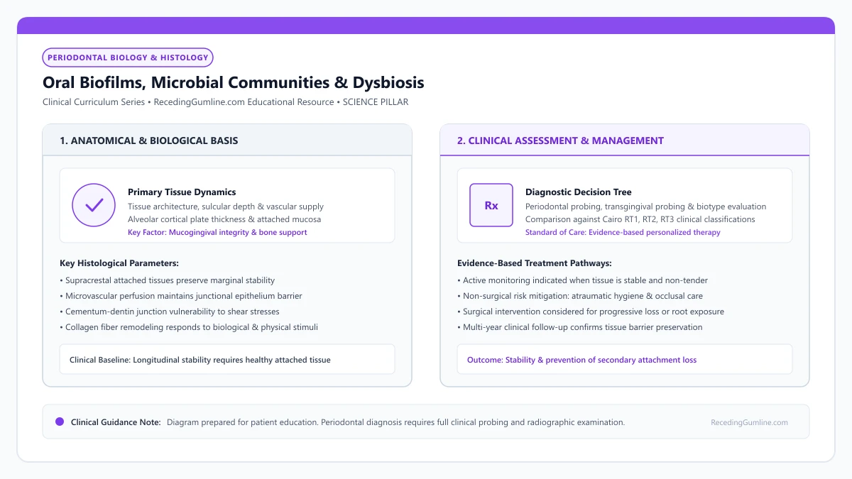
Educational illustration: Oral Biofilms, Microbial Communities & Dysbiosis. Clinical management requires comprehensive periodontal evaluation rather than isolated self-assessment.
Source: RecedingGumline.com Clinical Editorial Team (Proprietary educational diagram for RecedingGumline.com)
Biofilm Architecture & The Extracellular Polymeric Substance (EPS)
Bacteria within the oral cavity do not exist as free-floating (planktonic) single cells; they live embedded within highly organized, surface-attached communities known as biofilms. Dental plaque biofilm develops through an orderly succession: salivary glycoproteins form an acquired pellicle on the tooth within minutes of cleaning.
Early colonizers—predominantly Streptococcus and Actinomyces species—bind to pellicle receptors. These commensals synthesize an Extracellular Polymeric Substance (EPS) matrix composed of polysaccharides, proteins, lipids, and extracellular DNA. This EPS matrix acts as a protective shield, encasing bacterial microcolonies and providing structural stability.
The biofilm matrix establishes complex micro-environments with distinct nutrient gradients, pH zones, and anaerobic niches. Furthermore, the EPS matrix acts as a physical diffusion barrier, rendering bacteria within mature biofilms up to 1,000 times more resistant to antimicrobial mouthwashes and systemic antibiotics than their planktonic counterparts. Physical mechanical disruption remains the mandatory clinical requirement for biofilm elimination.
Key Scientific Insights
- Bacteria organize into structured biofilms shielded by an Extracellular Polymeric Substance (EPS).
- The EPS matrix protects bacterial colonies from host immune cells and antimicrobial chemicals.
- Biofilm bacteria exhibit up to 1,000-fold higher antibiotic resistance, requiring mechanical removal.
Socransky Microbial Complexes: From Symbiosis to Red Complex Pathogens
In 1998, Sigmund Socransky and colleagues at the Forsyth Institute published a landmark classification of subgingival plaque bacteria, grouping species into color-coded microbial complexes based on their ecological association with periodontal health versus disease.
The Yellow, Blue, Green, and Purple complexes represent early and secondary colonizers associated primarily with clinical periodontal health and early gingivitis. As plaque accumulates without mechanical disruption, oxygen levels within the subgingival sulcus plunge, creating an anaerobic, nutrient-rich environment.
This permits the emergence of the Orange Complex (including Fusobacterium nucleatum and Prevotella intermedia), which serves as a molecular bridge, facilitating the attachment of the virulent Red Complex. The Red Complex comprises three obligate anaerobic pathogens: Porphyromonas gingivalis, Tannerella forsythia, and Treponema denticola. The appearance of the Red Complex strongly correlates with bleeding on probing, deep periodontal pockets, and active alveolar bone loss.
Key Scientific Insights
- Socransky categorized subgingival bacteria into color-coded ecological complexes.
- Early colonizers (Yellow, Blue, Green) maintain periodontal health and mild gingivitis.
- The virulent Red Complex (P. gingivalis, T. forsythia, T. denticola) drives severe bone destruction.
The Keystone-Pathogen Hypothesis & Polymicrobial Synergy
Contemporary periodontology has evolved beyond the classical infectious disease model (one specific microbe causing one specific disease). In 2012, George Hajishengallis and colleagues introduced the Keystone-Pathogen Hypothesis to explain how periodontal dysbiosis occurs.
This paradigm identifies Porphyromonas gingivalis as a "keystone pathogen." Even when present in very low relative abundance (less than 0.1% of the total microbial population), P. gingivalis secretes potent cysteine endopeptidases known as gingipains. Gingipains cleave host complement component C5 into C5a and degrade antimicrobial peptides.
This targeted immune subversion uncouples bacterial killing from inflammatory recruitment. The host immune response is paralyzed, allowing the entire resident microbial community to overgrow and transition into a destructive dysbiotic state. It is this dysbiotic polymicrobial community—not P. gingivalis alone—that drives sustained host-mediated periodontal tissue breakdown and recession.
Key Scientific Insights
- P. gingivalis acts as a keystone pathogen, subverting host immune defenses even in tiny numbers.
- Gingipains cleave complement factors, paralyzing neutrophil antimicrobial killing capacity.
- Immune subversion transforms the entire commensal microbiota into a destructive dysbiotic biofilm.
Disrupting Dysbiosis: Mechanical Debridement vs. Chemical Adjuncts
Because the biofilm EPS matrix shields bacteria from chemical agents, therapeutic management requires methodical physical debridement. Professional scaling and root planing mechanically dislodges subgingival biofilm and calculus, resetting the ecological clock and allowing beneficial aerobic commensal species to recolonize the root surface.
Chemical adjuncts—such as chlorhexidine rinses, essential oils, or subgingival delivery of minocycline microspheres (Arestin)—are utilized strictly as secondary adjuncts to mechanical debridement. They help suppress bacterial re-growth during the post-instrumentation healing window.
At home, daily disruption of the supragingival biofilm with the Modified Bass brushing technique and interdental cleaning prevents subgingival ecological shifts, preserving the health of marginal gingival tissues and preventing the onset of inflammatory recession.
Key Scientific Insights
- Physical mechanical debridement via ultrasonic and hand instruments is mandatory to rupture biofilms.
- Chemical antimicrobials function solely as adjuncts to suppress post-cleaning bacterial recolonization.
- Daily atraumatic home plaque control maintains subgingival symbiosis and prevents disease recurrence.
Salivary Ecology & Microbiome Equilibrium
The oral microbiome exists in continuous dynamic equilibrium with the host salivary flow. Saliva supplies essential nutrients, antimicrobial immunoglobulins (secretory IgA), and buffer systems that maintain physiological oral pH between 6.7 and 7.3. When salivary flow is impaired by systemic medications or autoimmune conditions (xerostomia), this critical ecological buffer is lost.
In a dry oral environment, acidification of the oral cavity suppresses beneficial commensals while favoring aciduric and proteolytic anaerobes. The lack of salivary flushing allows biofilms to stagnate and mature undisturbed along the cervical gingival margin, dramatically accelerating the onset of inflammatory attachment loss and root caries.
Therapeutic protocols that stimulate natural salivary function and maintain neutral pH are vital components of long-term microbiome stabilization.
Key Scientific Insights
- Salivary flow provides constant antimicrobial immunoglobulins (sIgA) and buffering capacity.
- Salivary hypofunction accelerates subgingival biofilm maturation and tissue breakdown.
- Maintaining salivary flow and neutral oral pH preserves healthy microbial symbiosis.
Subgingival Plaque Biofilm Ecology in Periodontal Gum Recession
In the human mouth, hundreds of bacterial species coexist in complex biological communities known as biofilms. In a state of health, this oral microbiome lives in harmonious symbiosis with the host. However, when plaque is left undisturbed beneath the gingival margin, the microbial ecosystem undergoes a profound ecological shift—termed dysbiosis—transforming into a virulent anaerobic community that destroys periodontal bone and tissue.
• Subgingival plaque is an organized, multicellular biofilm protected by an extracellular polymeric substance (EPS) matrix.
• Socransky's microbial complexes categorize bacteria by pathogenic virulence, culminating in the virulent "Red Complex."
• The Red Complex—Porphyromonas gingivalis, Treponema denticola, and Tannerella forsythia—is strongly linked to advanced pocketing and attachment loss.
• P. gingivalis acts as a "keystone pathogen," orchestrating dysbiosis and remodeling benign biofilms into destructive pathogens.
The Biofilm Fortress: The Extracellular Polymeric Substance Matrix
Bacteria in the subgingival pocket do not float as solitary individuals; they construct an intricate, multicellular micro-city known as a biofilm. The bacteria secrete a slime-like extracellular polymeric substance (EPS) matrix made of polysaccharides, extracellular DNA, and proteins.
This EPS matrix acts like armor, shielding the bacteria from the host immune system (white blood cells) and rendering them up to 1,000 times more resistant to systemic antibiotics and antiseptic mouthwashes than planktonic bacteria. Water channels within the matrix circulate nutrients and remove waste, functioning as an internal circulatory system.
Dental plaque is not a random accumulation of bacteria, but a highly structured polymicrobial biofilm embedded within an extracellular polysaccharide matrix. As biofilm matures and migrates subgingivally, the micro-environment transitions from aerobic gram-positive cocci to strictly anaerobic gram-negative motile rods.
Key Scientific Insights
- Bacteria live in structured multicellular biofilms protected by an extracellular polymeric matrix
- Matrix shields pathogens from host antibodies and makes them 1,000x resistant to antibiotics
- Microscopic fluid channels circulate nutrients and chemical signals between bacterial species
Socransky's Microbial Complexes: The Deadly Red Complex
In the late 1990s, Dr. Sigmund Socransky and researchers at the Forsyth Institute organized subgingival bacteria into color-coded microbial complexes based on their association with health or disease. Yellow, green, and purple complexes consist of early colonizers that are generally benign.
However, as pockets deepen, the Orange Complex (including Fusobacterium nucleatum) bridges the gap, allowing the lethal Red Complex to emerge. The Red Complex consists of three obligate anaerobes: Porphyromonas gingivalis, Treponema denticola, and Tannerella forsythia. Their presence is virtually universal in sites undergoing active, destructive bone resorption and clinical attachment loss.
Key periodontal pathogens comprising Socransky’s red complex—Porphyromonas gingivalis, Treponema denticola, and Tannerella forsythia—dominate the deep subgingival pocket niches. These virulent organisms produce toxic proteases, hemolysins, and lipopolysaccharides that trigger severe tissue destruction.
Key Scientific Insights
- Color-coded complexes track ecological succession from early health to late disease
- Orange Complex acts as a biological bridge, facilitating colonizing anaerobes
- Red Complex (P. gingivalis, T. denticola, T. forsythia) drives active tissue destruction
The Keystone Pathogen Theory: P. Gingivalis and Dysbiosis
Modern periodontal microbiology, pioneered by Dr. George Hajishengallis, established the "Keystone Pathogen Hypothesis." Research proved that Porphyromonas gingivalis does not need to be present in massive quantities to destroy tissue; even in tiny numbers, it acts as a master manipulator.
P. gingivalis produces specialized cysteine proteases called gingipains. Gingipains cleave host complement proteins (C5a), paralyzing white blood cells and dismantling the immune response. By subverting host defenses, P. gingivalis allows the entire benign microbial community to overgrow into a hyper-virulent dysbiotic state that triggers massive bone destruction.
The physical architecture of the biofilm shields resident pathogens from systemic antibiotics and host immunological defenses. Effective clinical therapy requires physical mechanical disruption of the biofilm matrix through ultrasonic scaling and curettage, as chemical agents alone cannot penetrate mature biofilm.
Key Scientific Insights
- P. gingivalis acts as a master manipulator, remodeling benign biofilms into pathogens
- Gingipain enzymes disarm white blood cells and disable host immune defenses
- Drives the transition from symbiotic health to destructive periodontal dysbiosis
Microbial Ecology: Socransky's Complexes & Dysbiotic Shift
The subgingival microenvironment exists in a state of dynamic equilibrium between host immune defenses and bacterial biofilms. In 1998, Sigmund Socransky organized subgingival microorganisms into colored complexes based on their association with health or disease.
In healthy sites, yellow, green, and blue complexes (dominated by Streptococcus and Actinomyces species) form a stable, commensal biofilm. However, poor hygiene, mechanical stagnation, or local inflammation induces ecological dysbiosis.
The pioneer species are gradually displaced by the orange complex (Fusobacterium nucleatum, Prevotella intermedia), which creates an anaerobic, nutrient-rich environment that facilitates colonization by the virulent "red complex": Porphyromonas gingivalis, Treponema denticola, and Tannerella forsythia.
Key Scientific Insights
- Healthy subgingival sulci are colonized by commensal yellow, green, and blue bacterial complexes.
- Ecological dysbiosis shifts the microenvironment from aerobic commensals to pathogenic anaerobes.
- The red complex (P. gingivalis, T. denticola, T. forsythia) is the primary driver of destructive periodontitis.
The Extracellular Polymeric Substance (EPS) Matrix & Biofilm Recalcitrance
Subgingival bacteria do not exist as free-floating (planktonic) cells; they are encased within a self-produced Extracellular Polymeric Substance (EPS) matrix composed of exopolysaccharides, extracellular DNA (eDNA), proteins, and lipids.
This gelatinous EPS matrix provides structural stability, coordinates bacterial cell-to-cell communication (quorum sensing), and acts as a physical barrier that restricts the penetration of host antibodies, complement proteins, and systemic antibiotics.
Bacteria embedded within a mature subgingival biofilm can be up to 1,000 times more resistant to antimicrobial agents than their planktonic counterparts. Consequently, effective periodontal therapy mandates mechanical disruption via scaling and root planing; chemical mouthwashes alone cannot penetrate the EPS barrier.
Key Scientific Insights
- The EPS matrix encases subgingival bacteria in a protective mesh of polysaccharides, eDNA, and proteins.
- Biofilms exhibit up to 1,000-fold higher resistance to antimicrobial agents than free-floating bacteria.
- Mechanical instrumentation (scaling and root planing) is essential to physically disrupt the EPS matrix.
Clinical Reality Check
Antimicrobial mouthwashes and antibiotics cannot arrest periodontitis or halt progressive recession on their own. Without physical, mechanical removal of the subgingival biofilm matrix, chemical agents cannot penetrate the protective EPS shield.
Questions to Ask Your Dentist or Periodontist
- Do my periodontal probing depths indicate the presence of deep, anaerobic bacterial biofilms?
- Would subgingival microbial testing or localized antibiotic therapy (Arestin) be beneficial in my case?
- How does my current oral hygiene routine impact the balance of my oral microbiome?
- What specific therapeutic mouthwash do you recommend to maintain healthy microbial balance?
- How quickly do pathogenic bacteria recolonize my gum pockets after a deep cleaning?
- Do my periodontal probing depths indicate the presence of deep, anaerobic subgingival pockets?
- How does scaling and root planing physically disrupt this subgingival bacterial biofilm?
- What specific daily home hygiene tools (interdental brushes, water flossers) will reach into these microbial areas?
- Why can't I just take a course of antibiotics to kill the gum disease bacteria?
Unsure What Your Gum Changes Mean?
Take our free, evidence-based Gum Recession Assessment — approximately 3 minutes. Identify potential risk factors, evaluate symptoms, and receive personalized discussion questions for your dentist or periodontist.
Non-diagnostic educational triage. Private, secure, completed in your browser.
Related Educational Topics
Scientific Literature & Clinical Guidelines
4sources · Hide ▲
- Löe H, Theilade E, Jensen SB (1965).
"Experimental gingivitis in man." The Journal of Periodontology.
Clinical relevance: Classic experimental gingivitis study demonstrating that withdrawal of oral hygiene leads to bacterial plaque accumulation and reversible marginal gingival inflammation within 10 to 21 days, establishing the microbial etiology of gingival inflammation. It serves as foundational evidence for plaque-induced gingivitis, not modern comprehensive models of periodontitis or gingival recession.
- Chapple ILC, Mealey BL, Van Dyke TE, Bartold PM, Dommisch H, Eickholz P, et al. (2018).
"Periodontal health and gingival diseases and conditions on an intact and a reduced periodontium: Consensus report of workgroup 1 of the 2017 World Workshop on the Classification of Periodontal and Peri-Implant Diseases and Conditions." Journal of Clinical Periodontology.
Clinical relevance: Consensus report establishing diagnostic criteria for periodontal health and gingivitis across intact and reduced periodontia, defining clinical gingival health as <10% bleeding on probing without attachment loss and strictly differentiating gingivitis from periodontitis.
- Tonetti MS, Greenwell H, Kornman KS (2018).
"Staging and grading of periodontitis: Framework and proposal of a new classification and case definition." Journal of Clinical Periodontology.
Clinical relevance: Consensus framework establishing the multidimensional staging (severity and extent of periodontal tissue breakdown) and grading (biological rate of disease progression, incorporating smoking and diabetes as grade modifiers) for periodontitis. It addresses periodontitis diagnosis and staging, not the classification of localized gingival recession defects.
- Pihlstrom BL, Michalowicz BS, Johnson NW (2005).
"Periodontal diseases." The Lancet.
Clinical relevance: Peer-reviewed Lancet seminar review synthesizing global periodontal epidemiology, microbial etiology, host immunopathology, and systemic interactions, emphasizing prevention, biofilm disruption, and early risk factor modification.
Important Medical Notice
The contents of RecedingGumline.com, including text, graphics, self-assessment calculators, and other materials, are intended solely for educational and informational purposes. This content is not intended to replace professional dental examination, diagnosis, or treatment. Always seek the advice of a qualified dentist, periodontist, or other licensed oral healthcare provider with any questions you may have regarding a medical or dental condition. Never disregard professional medical advice or delay seeking it because of something you read on this website.