Gingival Bleeding on Brushing or Flossing as a Gum Recession Sign
Gingival bleeding upon toothbrushing or flossing is one of the most widespread clinical signs of active periodontal inflammation (see our gum recession). While frequently dismissed by patients as a minor consequence of "brushing too hard," healthy gingiva never bleeds under physiological pressure. In the biological context of gum recession, bleeding on probing signifies micro-ulceration of the sulcular epithelium, active immune-inflammatory engagement with subgingival bacterial biofilms, and potential vulnerability to an emerging receding gumline.
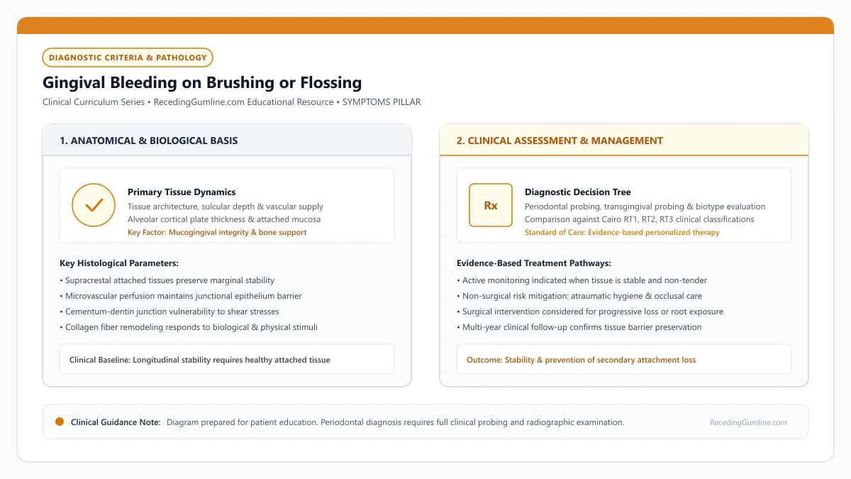
Educational illustration: Gingival Bleeding on Brushing or Flossing. Clinical management requires comprehensive periodontal evaluation rather than isolated self-assessment.
Source: RecedingGumline.com Clinical Editorial Team (Proprietary educational diagram for RecedingGumline.com)
Microscopic Sulcular Ulceration: The Source of Bleeding
To understand why gingival bleeding occurs, one must examine the microscopic histology of the gingival sulcus. In a state of periodontal health, the sulcular and junctional epithelium form a continuous, intact cellular barrier that seals underlying vascular connective tissue from the oral cavity. However, when bacterial biofilms (plaque) accumulate along the cervical margin, bacteria release metabolic endotoxins and proteolytic enzymes that penetrate the intercellular junctions.
In response, the host immune system initiates a rapid vascular inflammatory reaction. Capillaries within the supraperiosteal vascular plexus become dilated, hyperemic, and engorged with blood. Neutrophils migrate into the sulcular crevice, releasing lysosomal enzymes and reactive oxygen species to combat bacteria.
This intense inflammatory skirmish causes necrosis and fragmentation of the sulcular epithelial cells, creating microscopic ulcerations. When physical pressure from a toothbrush bristle, interdental floss, or a periodontal probe touches these ulcerated sites, the engorged, fragile capillary loops rupture immediately, resulting in visible bleeding.
Key Clinical Insights
- Bacterial endotoxins breach the sulcular barrier, triggering intense capillary vasodilation.
- Inflammatory micro-ulcerations expose fragile, engorged capillaries directly to the crevice.
- Mechanical contact from brushing or probing ruptures exposed vessels, producing immediate bleeding.
Diagnostic Boundary: Reversible Gingivitis vs. Destructive Periodontitis
A fundamental clinical distinction must be established when evaluating bleeding gums: determining whether the bleeding represents reversible gingivitis or active, destructive periodontitis. As established in the landmark experimental gingivitis studies by Harald Löe and colleagues (1965), plaque accumulation on healthy teeth induces measurable marginal gingivitis and bleeding within 10 to 21 days. Crucially, this inflammation is entirely confined to the soft gingiva; the underlying periodontal ligament and alveolar bone remain completely untouched.
If meticulous plaque removal is reinstated, gingivitis completely resolves without permanent structural tissue loss. However, if left unchecked in susceptible individuals, chronic gingivitis can transition into periodontitis. In periodontitis, the inflammatory front extends apically, activating osteoclasts that resorb alveolar bone crests and severing the collagenous Sharpey's fibers that anchor tooth roots to bone.
When recession occurs in conjunction with periodontitis, bleeding reflects active, progressive bone destruction that threatens long-term tooth retention unless professionally arrested through scaling and root planing.
Key Clinical Insights
- Löe's classic 1965 study demonstrated that biofilm induces reversible gingivitis within 10 to 21 days.
- Gingivitis involves soft-tissue inflammation without loss of underlying alveolar bone support.
- Periodontitis extends inflammation into bone and ligament, transforming bleeding into a sign of attachment loss.
The Dangerous Paradox: Masked Bleeding in Tobacco Users
While bleeding is an exceptionally sensitive indicator of inflammation, its absence does not guarantee periodontal health. The most striking clinical paradox occurs in individuals who smoke or use nicotine products.
Nicotine is a potent peripheral vasoconstrictor that stimulates local and systemic catecholamine release, severely constricting terminal capillary loops in the marginal gingiva. Consequently, smokers with severe, rapid periodontitis and advanced gingival recession frequently demonstrate little or no bleeding on brushing or probing.
This vasoconstrictive masking gives patients a false sense of security, delaying critical periodontal intervention until teeth become visibly elongated, hypermobile, or structurally compromised. Clinicians must rely on radiographic bone evaluation and clinical attachment level measurements rather than bleeding indices when evaluating tobacco users.
Key Clinical Insights
- Nicotine-induced vasoconstriction drastically reduces gingival blood flow and masks bleeding.
- Smokers frequently exhibit advanced periodontal bone loss and recession without overt bleeding.
- The absence of bleeding in a tobacco user must never be mistaken for clinical periodontal stability.
Clinical Evaluation: Bleeding on Probing (BOP) as a Prognostic Tool
In modern dental practices, Bleeding on Probing (BOP) is quantified using standardized periodontal probes (such as the UNC-15 probe) applied with a controlled light force of 0.20 to 0.25 Newtons. The clinician gently introduces the probe tip into the sulcus at six sites per tooth (mesiobuccal, mid-buccal, distobuccal, mesiolingual, mid-lingual, distolingual).
The clinical utility of BOP lies in its negative predictive value. Classic longitudinal periodontal research by Lang and colleagues demonstrated that a site exhibiting zero bleeding on probing over consecutive maintenance recalls has an over 98% probability of remaining biologically stable without attachment loss.
Conversely, sites exhibiting persistent BOP at consecutive visits demonstrate an elevated risk for progressive attachment loss and deepening recession, signaling the urgent need for localized subgingival debridement and antimicrobial therapy.
Key Clinical Insights
- Standardized probing applies a controlled light pressure (0.25 N) across 6 sites per tooth.
- Absence of BOP carries a 98% negative predictive value for periodontal attachment stability.
- Persistent bleeding at sequential recall visits identifies sites at high risk for progressive tissue retreat.
Resolution Protocols: Atraumatic Biofilm Control & Therapeutic Care
Halting gingival bleeding requires eliminating the subgingival microbial trigger while avoiding mechanical trauma to the inflamed margins. Patients should adopt the Modified Bass brushing technique using extra-soft bristles, angling the filaments at 45 degrees into the sulcular crevice to dislodge plaque without shredding fragile ulcerated tissue.
Interdental cleaning is non-negotiable. Because periodontal inflammation almost always initiates in the protected interproximal spaces beneath the contact points, daily interdental brushing or correct C-shaped flossing is essential to resolve papillary bleeding.
For acute flare-ups, short-term adjunctive chemical plaque control with therapeutic mouthrinses (such as 0.12% chlorhexidine digluconate or essential-oil formulations) suppresses bacterial load. Once professional scaling removes mineralized calculus deposits, gingival micro-ulcerations heal within 7 to 14 days, completely eliminating bleeding.
Key Clinical Insights
- The Modified Bass technique cleans the inflamed sulcus without inflicting abrasive shear trauma.
- Interdental brushes dislodge interproximal biofilms where bleeding and recession frequently initiate.
- Professional calculus debridement allows microscopic ulcerations to re-epithelialize within 1 to 2 weeks.
Sensitivity to Sweet & Acidic Foods from Receding Gums
While cold temperature is the most recognized trigger of sensitive teeth, many individuals with receding gums experience sharp, painful winces when eating sweets (chocolate, candy) or consuming acidic foods and drinks (citrus, vinegar, wine). Understanding the osmotic and chemical mechanisms behind sweet and acidic sensitivity clarifies why exposed root surfaces react violently to ordinary dietary choices.
• Sweet foods trigger pain through an osmotic gradient, drawing fluid outward through open dentinal tubules.
• Acidic foods and beverages chemically dissolve mineral tubule plugs, widening tubule apertures.
• Enamel resists osmotic shifts; sensitivity to sugar is almost exclusively a sign of exposed dentin or decay.
• Neutralizing oral acids with water rinses and using stannous fluoride dentifrices helps restore the mineral seal.
The Osmotic Mechanism: How Concentrated Sugar Draws Fluid
Tooth enamel is virtually impermeable to sugar molecules. However, when gum recession exposes root dentin, the open dentinal tubules act as microscopic fluid conduits directly connected to the dental pulp.
When you eat a concentrated sweet food (such as caramel or chocolate), the sugar creates a hypertonic solution at the tooth surface—meaning its solute concentration is vastly higher than the fluid inside your dentinal tubules. Following the laws of osmosis, water rapidly rushes outward through the tubules toward the higher sugar concentration.
Hypertonic sucrose solutions and acidic citrus beverages generate rapid osmotic fluid shifts across exposed dentinal tubules toward the tooth surface. This sudden osmotic pressure differential directly stimulates pulpal nociceptors, producing intense, sharp cervical twinges.
Key Clinical Insights
- Enamel blocks sugar; exposed root dentin allows direct osmotic fluid contact
- Hypertonic sweet solutions generate a powerful osmotic pull
- Dentinal fluid rushes outward at high velocity, triggering A-delta sensory nerve impulses
Acidic Dissolution: How Citrus, Soda & Wine Strip Root Plugs
Acidic foods and drinks—including lemon juice, sports drinks, carbonated sodas, balsamic vinegar, and white wine—operate through chemical demineralization. Hydroxyapatite crystals in dentin begin dissolving at a critical pH of 6.2 to 6.7 (compared to 5.5 for enamel), making roots far more acid-vulnerable.
When dietary acids touch an exposed root, they dissolve the microscopic mineral plugs and salivary pellicle that naturally shield tubule openings. Within seconds, tubule diameters widen, allowing greater fluid movement and multiplying sensitivity to all subsequent stimuli.
Frequent consumption of acidic sports drinks, sodas, or carbonated waters strips away the protective salivary smear layer that naturally plugs open tubules. Without a salivary smear layer, tubule apertures remain wide open and hyper-sensitive to ordinary dietary foods.
Key Clinical Insights
- Root dentin dissolves at a higher pH (6.5) than crown enamel (5.5)
- Dietary acids dissolve the microscopic smear plugs that naturally block tubules
- Widened tubule apertures leave the tooth hyper-reactive to temperature and touch
Protective Protocols: Buffering Acids and Remineralizing Roots
Managing chemical and osmotic sensitivity requires dietary timing adjustments. Immediately after eating sweets or drinking acidic beverages, do not brush your teeth. Acid-softened dentin is easily scrubbed away by bristles. Instead, rinse thoroughly with plain tap water or a baking soda rinse (1/2 teaspoon baking soda in a cup of water) to neutralize the pH.
Incorporate stannous fluoride or arginine-calcium carbonate toothpastes into your daily routine. Stannous fluoride creates an insoluble, acid-resistant tin-fluoride mineral barrier over tubule openings, shielding them against future osmotic and chemical challenges.
Patients are advised to rinse thoroughly with neutral tap water immediately following acidic intake rather than brushing vigorously with abrasive pastes. Incorporating remineralizing amorphous calcium phosphate gels helps re-establish protective intratubular mineral plugs.
Key Clinical Insights
- Never brush immediately after acidic foods; wait at least 30 minutes for saliva to re-harden dentin
- Rinse with plain water or a mild baking soda solution to neutralize oral acidity instantly
- Use stannous fluoride toothpastes to deposit acid-resistant protective mineral layers
Osmotic Dynamics: Hypertonic Solutions & Outward Fluid Siphonage
Sensitivity triggered by concentrated sweets (such as chocolate, candy, or honey) operates through an osmotic mechanism rather than thermal contraction. Concentrated sugars create a hypertonic environment in the oral cavity relative to the physiological fluid within dentinal tubules.
Through basic osmotic principles, water molecules are drawn rapidly from areas of lower solute concentration (the dental pulp and tubules) toward areas of higher concentration (the oral cavity). This sudden outward fluid siphonage exerts mechanical traction on pulpal odontoblasts, firing pain signals.
Acidic foods and beverages (citrus fruits, sodas, salad dressings, and wines) act through a dual mechanism: their low pH dissolves the superficial smear layer that plugs tubule openings, while hypertonic fruit acids simultaneously trigger osmotic fluid shifts.
Key Clinical Insights
- Concentrated sugary foods create hypertonic environments that pull tubular fluid outward.
- Osmotic fluid siphonage exerts mechanical tension on odontoblastic processes in the pulp.
- Dietary acids dissolve protective smear plugs, exposing fresh dentin to osmotic shifts.
Protecting the Acquired Pellicle: Dietary Timing & Remineralization
The primary biological defense against osmotic and acidic sensitivity is the acquired salivary pellicle—an acellular, protective protein film derived from saliva that coats exposed tooth surfaces within minutes. The pellicle acts as an organic diffusion barrier, moderating fluid flow across tubules.
However, frequent consumption of acidic beverages strips away the pellicle and erodes superficial root dentin. Dentists counsel patients to consume acidic foods exclusively during main meals, when stimulated salivary flow provides rich bicarbonate buffering capacity.
Following acidic consumption, patients should immediately rinse thoroughly with plain water or a neutralizing sodium bicarbonate solution. Brushing must be deferred for at least 30 to 45 minutes to permit salivary calcium and phosphate to remineralize the softened dentin surface.
Key Clinical Insights
- The salivary pellicle acts as a natural protein barrier, buffering osmotic fluid movement.
- Acidic foods should be confined to mealtimes when stimulated salivary buffering is highest.
- Rinsing with water and delaying brushing for 30 minutes prevents abrading softened root dentin.
Wedge-Shaped Notches at the Gumline: Cervical Lesions and Tissue Recession
Many patients run their fingernail along the gumline and discover a sharp, deep groove or V-shaped notch carved into the neck of the tooth. These physical defects—medically classified as Non-Carious Cervical Lesions (NCCLs)—are frequently accompanied by receding gums. Understanding how mechanical friction, occlusal clenching, and chemical erosion create these notches reveals why they require targeted dental care.
• Non-carious cervical lesions (NCCLs) are physical notches grooved into the tooth neck that do not originate from bacterial decay.
• Cervical notches present with sharp, well-defined internal angles (wedge-shaped) or smooth dish-shaped contours.
• Heavy biting forces (abfraction), horizontal scrubbing (abrasion), and dietary acids (erosion) act together to carve the defect.
• Restoring deep notches with flexible tooth-colored resin protects tooth structure and prevents fracture.
Clinical Morphology: Wedge-Shaped vs. Saucer-Shaped Lesions
Dentists classify cervical notches into two primary morphological shapes: wedge-shaped and saucer-shaped defects. Wedge-shaped defects exhibit sharp internal line angles, resembling a notch cut by an axe into the side of a tree. These sharp angles are classic indicators of tensile stress concentration from nocturnal clenching (abfraction).
Saucer-shaped defects, in contrast, display smooth, shallow, rounded contours with ill-defined borders. These smooth concavities are typical of frictional toothbrush abrasion or dietary acid erosion, where fluid and bristles have polished the surface over years.
Cervical wedge defects, known clinically as non-carious cervical lesions or abfractions, manifest as deep sharp V-shaped grooves at the cementoenamel junction. These notches result from a destructive combination of heavy occlusal flexure forces and aggressive horizontal toothbrush abrasion.
Key Clinical Insights
- Wedge-shaped notches feature sharp, acute internal angles typical of occlusal stress flexure
- Saucer-shaped concavities feature smooth, rounded borders typical of brushing abrasion and acids
- Premolars and canines exhibit the highest clinical prevalence of cervical notching
Structural Risks: Nerve Proximity and Tooth Fracture
While a shallow notch under 0.5 mm is primarily an aesthetic and hygiene concern, progressive notches pose serious structural threats to the tooth. As the notch deepens toward the center of the tooth, it approaches the dental pulp chamber.
A deep notch creates a mechanical stress riser—a weak point where biting forces concentrate. In severe cases, an un-restored notch can result in the entire clinical crown shearing off at the gumline during normal eating, requiring complex post-and-core surgery or extraction.
As masticatory clenching flexes the crown, microcrystalline enamel rods and root dentin fracture away along the cervical fulcrum line. The sharp notched margins trap food debris and plaque biofilm, creating a high-risk micro-environment for root caries and structural tooth fracture.
Key Clinical Insights
- Deep cervical notches can penetrate to within a millimeter of the vascular pulp
- Acts as a structural weak point that predisposes the tooth to horizontal crown fracture
- Sharp internal ledges trap plaque and food particles, accelerating root breakdown
Restorative Management: When and How to Fill Cervical Notches
When a cervical notch exceeds 1.0 mm in depth, causes persistent cold sensitivity, or weakens tooth integrity, restorative filling is indicated. Dentists clean the defect and restore it using resin-modified glass ionomer (RMGI) or microfilled flowable composite.
RMGI materials are particularly favored because they bond chemically to dentin, release protective fluoride into the root, and possess a low modulus of elasticity that allows the restoration to flex with the tooth during chewing without breaking loose.
Restorative treatment involves meticulously beveling the enamel borders and bonding flexible microfilled composite resins that can flex alongside natural tooth movements. An occlusal nightguard is routinely prescribed to neutralize the nocturnal clenching forces driving lesion formation.
Key Clinical Insights
- Restoration is indicated when notches exceed 1 mm depth or harbor chronic sensitivity
- Resin-modified glass ionomer (RMGI) releases fluoride and bonds strongly to root dentin
- Flexible composites absorb tooth flexure forces to prevent restoration debonding
Biomechanical Etiology: Lateral Tooth Flexure & Cervical Fulcrums
Cervical notching—often manifesting as sharp, wedge-shaped V-defects along the gumline—represents Non-Carious Cervical Lesions (NCCLs). While historically attributed solely to abrasive toothbrushing, modern dental biomechanics recognizes the primary role of occlusal micro-flexure, termed abfraction.
During nocturnal clenching or chewing with eccentric tooth interferences, lateral bending forces are applied to the tooth crown. Because the tooth is anchored within the rigid alveolar bone socket, the cervical margin at the cementoenamel junction (CEJ) acts as a mechanical fulcrum.
Tensile and compressive stresses concentrate along this fulcrum. Brittle enamel crystals and hydroxyapatite prisms within the thin cervical collar fracture under cyclical fatigue, detaching from the underlying dentin and leaving a distinct angular notch.
Key Clinical Insights
- Wedge-shaped cervical notches are driven by mechanical tooth flexure under bite overload.
- The cervical cementoenamel junction acts as a biomechanical fulcrum during lateral forces.
- Cyclical stress fractures brittle enamel prisms, producing sharp angular V-shaped notches.
Restorative Principles: Material Flexibility & Modulus of Elasticity
Restoring deep cervical wedge defects requires careful consideration of material physical properties. Conventional highly rigid restorative composites have a high modulus of elasticity; when placed into an abfraction defect in a bruxing patient, ongoing tooth flexure inevitably breaks the adhesive bond, causing the filling to dislodge.
To prevent restoration failure, clinicians utilize materials with a low modulus of elasticity that can flex harmoniously with the tooth. Micro-filled flowable composites and resin-modified glass ionomer cements (RMGIs) provide ideal flexural compliance.
RMGIs offer the additional advantage of chemical bonding to dentin and continuous fluoride release, protecting the vulnerable cervical margin from recurrent root decay while sealing sensitive dentinal tubules against fluid movement.
Key Clinical Insights
- Rigid composites pop out of cervical notches because they cannot flex with the tooth.
- Low-modulus materials (flowable composites, RMGIs) flex harmoniously during biting forces.
- Resin-modified glass ionomers provide chemical adhesion and continuous protective fluoride release.
Receding Gums on Front Teeth: Aesthetic Impact, Causes & Conservative vs Surgical Care
Receding gums on the front teeth (the maxillary anterior "smile zone") carries intense cosmetic, emotional, and psychological weight for patients experiencing a receding gumline. Unlike recession on hidden molar teeth, front-tooth recession is immediately visible when smiling, laughing, or speaking. Understanding the unique aesthetic dynamics, high surgical root coverage predictability, and conservative options helps patients restore smile harmony.
• Front-tooth recession is in the high-visibility aesthetic zone, causing teeth to appear uneven, elongated, or discolored.
• Maxillary front teeth frequently present with intact interdental bone (Cairo RT1), offering the highest clinical predictability for 100% complete root coverage.
• Subepithelial connective tissue grafts combined with coronally advanced flaps provide optimal aesthetic color and texture matching.
• Minimally invasive vestibular tunneling techniques (VISTA) allow front-tooth grafting without visible surface incisions.
Aesthetic Dynamics: High Lip Lines and Gingival Zenith Symmetry
In dental aesthetics, the gingival zenith is the most apical point of the gumline contour on each tooth. Under ideal symmetry, the zeniths of the two central incisors should be at the exact same height, with the lateral incisors sitting about 1 mm lower, and the canines matching the central incisors.
When recession strikes a front tooth—even by just 1 to 2 millimeters—this balance is destroyed. In patients with a high smile line (showing gums when smiling), the receded tooth looks noticeably longer and darker, creating an asymmetrical, prematurely aged appearance.
Gum recession on visible maxillary incisors creates significant aesthetic distress, often described by patients as elongated or uneven teeth. The loss of symmetrical gingival scallop contours can compromise the natural harmony of the smile line during social interactions.
Key Clinical Insights
- Gingival zenith symmetry is essential for a harmonious, balanced smile
- Recession of even 1 to 2 mm on an incisor creates prominent visual asymmetry
- Patients with high smile lines experience the greatest cosmetic and emotional distress
High Surgical Predictability: Why Front Teeth Heal Exceptionally Well
Fortunately, front teeth are anatomically favorable candidates for surgical repair. Because front-tooth recession is frequently caused by toothbrush abrasion or orthodontic alignment rather than destructive periodontitis, the interdental bone between the teeth is usually 100% intact (Cairo RT1).
When interdental bone and papillae are intact, periodontists can predictably achieve 90% to 100% complete root coverage. The intact lateral papillae provide robust collateral blood supply that nourishes the newly transplanted connective tissue graft as it heals across the root.
Anterior recession defects are frequently accompanied by localized root prominence and paper-thin labial cortical bone plates. Even minor episodes of aggressive flossing or vigorous brushing can rapidly shear through the delicate anterior attached tissue collar.
Key Clinical Insights
- Front teeth usually have intact interdental bone (Cairo RT1), maximizing coverage success
- Collateral blood supply from adjacent papillae nourishes the transplanted tissue
- Achieving 100% complete root coverage is realistic and clinically routine in RT1 cases
Aesthetic Techniques: Connective Tissue Grafts and Tunneling
In the smile zone, choice of surgical technique is critical. Free gingival grafts are strictly avoided because they heal with a pale, whitish "patch" appearance. Instead, periodontists use subepithelial connective tissue grafts (SCTG) or allograft matrices.
Surgeons often employ minimally invasive tunneling techniques (such as the VISTA procedure). Instead of cutting open the gums with visible vertical incisions, the surgeon creates a microscopic tunnel beneath the gums from a hidden access point, slides the graft into place, and anchors it with delicate sutures, leaving zero visible scars.
Surgical root coverage utilizing coronally advanced flaps combined with subepithelial connective tissue grafting achieves high aesthetic predictability in anterior sites. Restoring natural tissue thickness and harmonious marginal contours protects anterior roots from progressive exposure.
Key Clinical Insights
- Subepithelial connective tissue grafts blend seamlessly with natural gum color and texture
- Tunneling techniques (VISTA) eliminate vertical surface incisions and scarring
- Micro-sutures ensure invisible healing and smooth, natural tissue margins
Aesthetic Zone Dynamics: High Smile Lines & Papillary Scalloping
Gingival recession occurring in the aesthetic zone (maxillary incisors and canines) carries profound cosmetic and psychological implications. Patients with a "high smile line" expose several millimeters of gingival architecture during natural speech and smiling, making even 1.0 mm of localized margin retraction immediately conspicuous.
Anterior teeth exhibit pronounced architectural scalloping, where delicate interdental papillae rise several millimeters above the facial margin. The underlying facial bone plate over central and lateral incisors is exceptionally thin—often less than 0.5 mm thick.
When recession develops in the aesthetic zone, preserving the height and vascular integrity of the adjacent interdental papillae is paramount. Loss of papillae produces unsightly "black triangles" that are far more difficult to reconstruct surgically than the facial recession itself.
Key Clinical Insights
- High smile lines expose anterior gingival margins, making minor recession highly noticeable.
- Anterior incisors possess delicate, highly scalloped margins over paper-thin facial bone.
- Preserving interdental papillae is critical to prevent cosmetic "black triangle" defects.
Minimally Invasive Tunneling Approaches for Aesthetic Root Coverage
In the anterior aesthetic zone, conventional periodontal flap designs that utilize vertical releasing incisions carry a high risk of visible scar tissue formation and compromised blood supply to the papillae. Modern aesthetic periodontics relies on the minimally invasive tunneling technique (tunnel flap).
Through microscopic intrasulcular incisions, the periodontist undermines the facial soft tissue, creating a continuous subgingival tunnel connecting adjacent teeth without incising the interdental papillae. A subepithelial connective tissue graft is guided through this tunnel and positioned over the denuded roots.
The entire mucosal complex is advanced coronally and anchored with micro-sutures suspended around tooth contacts. This flapless tunneling approach preserves all native blood vessels, avoids scarring, and yields natural, harmonious aesthetic contours.
Key Clinical Insights
- Vertical releasing incisions in the aesthetic zone risk unsightly permanent scar tissue.
- The tunneling technique undermines tissues intrasulcularly without incising interdental papillae.
- Tunnel connective tissue grafting maximizes blood supply and produces natural cosmetic contours.
Receding Gums on Lower Front Teeth: Thin Cortical Plate, Calculus & Frenum Factors
The mandibular central and lateral incisors (lower front teeth) are the single most common site for advanced, progressive gingival recession in the human mouth. Positioned where salivary glands deposit mineralized tartar and where facial lip muscles exert continuous traction, these small, delicate teeth possess paper-thin bone plates that make them extraordinarily susceptible to attachment loss.
• Lower incisors feature the thinnest alveolar bone plates and narrowest bands of attached gingiva in the dentition.
• Submandibular and sublingual salivary ducts exit directly behind lower incisors, causing heavy, rapid calculus accumulation.
• High mentalis muscle attachments and labial frenums exert continuous tension, pulling the margin downward.
• Free gingival grafting or connective tissue grafting is frequently indicated to establish an impenetrable barrier of attached tissue.
The Tripartite Vulnerability: Bone, Frenum & Saliva
Lower front teeth face a "perfect storm" of biological vulnerabilities. First, the facial cortical bone plate over mandibular incisors is razor-thin—often under 0.3 mm—and frequently contains developmental dehiscences caused by crowded eruption.
Second, the lower labial frenum and mentalis muscle insert directly into the movable mucosa near the tooth margin, pulling on the gums every time you talk or chew. Third, the salivary ducts (Wharton's and Rivinus' ducts) empty directly behind these teeth, saturating the area with calcium and phosphate that turns soft plaque into rock-hard calculus within 48 hours.
Mandibular anterior incisors possess the thinnest alveolar bone plates and the narrowest bands of keratinized gingiva in the entire human dentition. Due to gravity and salivary pooling, dense subgingival calculus forms rapidly on the lingual surfaces of lower incisors, driving localized inflammation.
Key Clinical Insights
- Cortical bone over lower incisors is the thinnest in the mouth (< 0.3 mm)
- Lower labial frenum and mentalis muscle fibers exert direct downward pull
- Sublingual salivary ducts promote rapid calculus deposition that harbors bacteria
Clinical Consequences: From Root Exposure to Tooth Mobility
Because mandibular incisor roots are exceptionally narrow and short compared to molars or canines, losing 4 or 5 millimeters of attachment represents a catastrophic loss of total periodontal support. As recession extends through the narrow band of attached gingiva into the alveolar mucosa, the tissue margin separates from the root.
Once bacterial plaque penetrates the apical third of the root, alveolar bone support is lost circumferentially. The tooth begins to exhibit Class I or Class II mobility (looseness), signaling imminent tooth loss if clinical intervention is not promptly rendered.
High lingual and labial frenum attachments frequently insert directly into the marginal gingiva of lower central incisors. Continuous muscular pull from the lower lip combined with thin bone dehiscences can cause rapid, deep recession within a matter of months.
Key Clinical Insights
- Narrow incisor roots have less total surface area; 4 mm of recession represents over 40% attachment loss
- Loss of attached tissue allows movable mucosa to pull away, creating deep pockets
- Progressive bone loss leads to tooth looseness (mobility) and eventual extraction
Surgical Stabilization: Creating an Impenetrable Tissue Band
The primary treatment objective on lower front teeth is not cosmetic beauty, but structural survival. The gold-standard treatment is the Free Gingival Graft (FGG). The surgeon takes a dense, keratinized tissue graft from the palate and stitches it at the base of the receded incisors.
This graft heals into a wide, tough, thick band of attached gingiva that buffers the tooth against muscle pull and toothbrush friction. Because the lower lip covers these teeth during smiling, the slightly paler cosmetic appearance of a free gingival graft is completely hidden, making it the ideal functional solution.
Periodontists often perform free gingival grafts or connective tissue grafts on lower incisors to create a robust collar of dense keratinized tissue. Thickening this tissue band halts progressive mucogingival migration and protects fragile mandibular teeth from premature loss.
Key Clinical Insights
- Free gingival graft creates a dense, thick collar of attached keratinized tissue
- Buffers the delicate tooth margins against downward mentalis muscle traction
- Pale tissue appearance is hidden behind the lower lip, making FGG the ideal choice
Anatomical Vulnerability: Paper-Thin Cortical Plates & Shallow Vestibules
Mandibular incisors represent the single most common site for progressive gingival recession in the human dentition. This extreme vulnerability is the direct consequence of unique regional anatomy.
The labial cortical bone plate covering lower incisor roots is naturally paper-thin and frequently features developmental fenestrations and dehiscences. Furthermore, the anterior mandibular vestibule is characteristically shallow, and the insertion of the mentalis muscle and inferior labial frenum is positioned immediately adjacent to the gingival margin.
Every time the patient speaks, chews, or swallows, dynamic muscle contraction pulls on the delicate mucosal margin. In the absence of a wide band of thick attached gingiva, this perpetual muscular pull causes rapid apical stripping of the gum tissue.
Key Clinical Insights
- Lower incisors are the most common site for progressive recession due to anatomical constraints.
- Labial cortical bone over mandibular incisors is paper-thin or congenitally absent.
- Shallow vestibular depth and mentalis muscle pull exert continuous mechanical tension on the margin.
Surgical Decision: Free Gingival Grafting vs. Tunnel CTG in the Mandible
Selecting the appropriate surgical modality for lower incisor recession depends on whether the primary clinical goal is functional tissue augmentation or cosmetic root coverage. Because lower incisors are rarely visible during normal smiling, functional stabilization is typically the primary objective.
When the zone of keratinized tissue is completely absent and high muscle tension is present, the Free Gingival Graft (FGG) is the treatment of choice. The FGG creates a dense, fibrous, immovable band of attached tissue that permanently resists muscle pull, effectively deepening the vestibule and halting progression.
However, if the patient exposes lower incisors during speech and demands aesthetic root coverage, a coronally advanced tunnel graft combined with a connective tissue graft may be considered, provided interdental bone is fully preserved (Cairo RT1).
Key Clinical Insights
- Functional tissue stabilization is the primary goal for lower incisors due to low aesthetic visibility.
- Free gingival grafts widen keratinized tissue, deepen the vestibule, and permanently resist muscle pull.
- Tunnel connective tissue grafting is reserved for cases requiring aesthetic root coverage.
Receding Gums on Canine Teeth: Root Prominence and Toothbrush Angulation
The canine teeth (often called eyeteeth or cuspids) are the cornerstone of the human dental arch. Possessing the longest and most massive roots in the mouth, canines sit directly at the corner where the dental arch transitions from the front to the sides. This anatomical prominence makes canines the number one target for aggressive toothbrushing friction and mechanical gumline recession.
• Canines occupy the corner of the dental arch, creating a prominent outward root bulge.
• The outer cortical bone plate over canines is frequently paper-thin or naturally dehiscenced.
• Hand movements naturally accelerate and apply maximum scrubbing force at the arch corner.
• Root coverage grafting on canines has high success rates when interdental bone is preserved.
Arch Geometry: The "Cornerstone" Prominence
The canine tooth is situated at the exact intersection where the flat anterior dental arch turns 90 degrees to form the posterior arch. To anchor this vital tooth, the canine root is exceptionally long, conical, and bulky.
This massive root creates a pronounced outward ridge in the facial jawbone known as the canine eminence. Because the root protrudes so far facially, the cortical bone plate covering its outer surface is paper-thin, and in many individuals, developmental alveolar dehiscences leave the cervical third covered only by soft tissue.
Maxillary and mandibular canines form the prominent architectural corners of the dental arch, featuring thick roots that project buccally into the facial vestibule. Because they occupy the corner turning point, canines absorb the greatest lateral frictional forces during manual toothbrushing strokes.
Key Clinical Insights
- Canines sit at the 90-degree turning corner of the dental arch
- Bulky root creates an outward structural ridge called the canine eminence
- Facial cortical bone over the canine eminence is naturally thin or absent
Brushing Mechanics: The Apex of Hand Acceleration
When people brush their teeth, their arm and wrist movements follow a predictable biomechanical path. As the brush sweeps across the front teeth and prepares to turn along the side teeth, the hand naturally pivots at the canine.
This pivot point causes the toothbrush bristles to smash directly into the prominent canine root with maximum velocity and compressive force. Horizontal scrubbing acts like sandpaper over the prominent root bulge, rapidly scouring away the delicate gingival margin and exposing the underlying cementum.
During lateral chewing excursions, canine-guided occlusion concentrates intense mechanical stress across the cervical root concavity. The combination of root prominence, thin facial bone, and heavy canine guidance makes these teeth the most frequent site of severe localized recession.
Key Clinical Insights
- Wrist movement naturally accelerates and pivots at the canine corner
- Bristles hit the protruding canine root with maximum mechanical friction
- Horizontal scrubbing selectively abrades canines faster than neighboring teeth
Surgical Repair: Achieving Predictable Canine Coverage
Because canine recession is usually driven by mechanical abrasion in patients with healthy interdental bone, it almost always presents as a Cairo RT1 defect. This means surgical root coverage has an exceptionally high success rate (85% to 95% complete coverage).
Periodontists treat receded canines using a coronally advanced flap combined with a subepithelial connective tissue graft (CAF + SCTG) or tunneling procedure. The transplanted tissue creates a thick, resilient cushion over the canine eminence that permanently shields the root from future brushing wear.
Managing canine recession requires adjusting premature occlusal contacts during lateral excursions alongside periodontal plastic surgical coverage. Connective tissue grafting restores thick attached gingiva that resists ongoing shearing stresses during normal mastication.
Key Clinical Insights
- High predictability for 90% to 100% root coverage due to intact interdental bone (RT1)
- Coronally advanced flap plus connective tissue graft provides ideal thickness and aesthetics
- Surgically thickened tissue permanently buffers the prominent root against brushing wear
Arch Prominence & Canine Guidance Biomechanics
Maxillary and mandibular canines occupy the "cornerstone" position of the dental arch, situated at the transition between the anterior and posterior segments. Because canines have the longest and most prominent roots in the mouth, their facial root surfaces naturally protrude outward beyond the skeletal base.
This natural prominence leaves the overlying labial bone plate exceptionally thin. Furthermore, in ideal occlusion, canines provide "canine guidance"—bearing the entire lateral shear load when the jaw moves sideways, protecting the premolars and molars from destructive lateral forces.
When patients grind or clench their teeth (bruxism), the canines endure heavy lateral friction and cyclical tooth flexure. This intense occlusal loading, combined with their anatomical vulnerability, makes canines a prime target for severe abfraction and gingival recession.
Key Clinical Insights
- Canines occupy the arch corner with prominent roots that naturally protrude through thin bone.
- Canine guidance subjects these teeth to high lateral shear forces during jaw movement.
- Heavy occlusal load combined with prominent anatomy accelerates abfraction and gum recession.
Management: Occlusal Adjustment, Nightguards & Surgical Coverage
Managing canine recession requires coordinating bite equilibration with soft-tissue reconstruction. Prior to surgical grafting, the periodontist and restorative dentist evaluate the patient's lateral excursions to ensure lateral forces are distributed safely across the arch or cushioned by an occlusal nightguard.
Surgically, prominent canines are prime candidates for coronally advanced flaps with connective tissue grafts. Because the interdental papillae between the lateral incisor and first premolar are typically robust and well-vascularized, canine defects offer excellent recipient beds for graft revascularization.
If an abfraction notch is present at the canine CEJ, the deep notch is smoothed and re-contoured to create a smooth, biologically receptive root surface before the connective tissue graft is secured.
Key Clinical Insights
- Occlusal equilibration and nightguards neutralize destructive lateral forces on canines.
- Robust adjacent interdental papillae provide an excellent vascular bed for graft healing.
- Smoothing cervical notches creates an optimal root bed for connective tissue reattachment.
Receding Gums and Persistent Bad Breath: Subgingival Biofilm Reservoirs
Persistent bad breath (chronic halitosis) that does not resolve after brushing, flossing, or using commercial mouthwash is a frequent and distressing symptom in individuals with receding gums. When gum tissue detaches from tooth roots, it creates subgingival pockets that serve as oxygen-deprived biological incubators for virulent anaerobic bacteria that produce foul-smelling sulfur gases.
• Chronic halitosis in periodontal disease is driven by Volatile Sulfur Compounds (VSCs)—principally hydrogen sulfide and methyl mercaptan.
• Subgingival pockets accompanying receding gums create anaerobic sanctuaries where oxygen cannot penetrate.
• Commercial breath sprays and cosmetic mouthwashes only mask odor for 20 minutes without disrupting deep bacterial colonies.
• Mechanical subgingival debridement (scaling and root planing) eliminates the microbial reservoirs causing persistent odor.
The Chemistry of Odor: Volatile Sulfur Compounds (VSCs)
The unpleasant odor associated with gum disease is not caused by rotting food particles; it is the metabolic waste product of specific anaerobic bacteria. Bacteria such as Porphyromonas gingivalis, Treponema denticola, and Fusobacterium nucleatum feed on proteins found in saliva, dead epithelial cells, and bleeding pocket fluid.
As these bacteria digest sulfur-containing amino acids (cysteine and methionine), they excrete Volatile Sulfur Compounds (VSCs). The two primary culprits are hydrogen sulfide (which smells like rotten eggs) and methyl mercaptan (which smells like decaying cabbage and feces).
Deep periodontal pockets and exposed root concavities associated with receding gums create stagnant anaerobic niches that trap food debris and biofilm. Colonizing anaerobic bacteria, such as Porphyromonas gingivalis, metabolize sulfur-containing amino acids into volatile sulfur compounds (VSCs).
Key Clinical Insights
- Anaerobic bacteria metabolize sulfur-rich proteins in blood and tissue exudate
- Hydrogen sulfide (rotten egg smell) and methyl mercaptan (decay smell) are excreted
- Methyl mercaptan is highly toxic and accelerates periodontal collagen breakdown
The Subgingival Incubator: Why Toothbrushes Cannot Reach the Odor
In healthy mouths, the gingival sulcus is shallow (1 to 3 mm) and well-oxygenated. However, when periodontal disease causes attachment loss and gum recession, deep pockets (4 to 8 mm) form between the root and the loose tissue collar.
These deep pockets become anaerobic chambers where atmospheric oxygen cannot penetrate. Toothbrush bristles and dental floss can only reach 2 to 3 mm subgingivally, leaving the bottom 5 millimeters of bacterial sludge completely undisturbed. This is why patients can brush five times a day and still have persistent halitosis.
These volatile sulfur compounds, including hydrogen sulfide and methyl mercaptan, produce distinct, persistent oral malodor that cannot be masked by mouthwashes. The presence of bleeding sulci provides bacterial pathogens with iron-rich hemoglobin, fueling accelerated anaerobic proliferation.
Key Clinical Insights
- Deep pockets (> 4 mm) create zero-oxygen environments where virulent anaerobes flourish
- Toothbrush bristles and floss penetrate only 2 to 3 mm beneath the gumline
- Deep bacterial reservoirs remain untouched by normal home brushing and mouthwash
Clinical Elimination: Subgingival Debridement and Pocket Reduction
Eliminating periodontal bad breath requires professional intervention. The dentist or periodontist performs scaling and root planing (deep cleaning) to mechanically flush out subgingival calculus, bacterial colonies, and endotoxins using ultrasonic cavitation tips.
Once the root surfaces are cleaned, the pocket tissues shrink and readapt to the tooth, collapsing the anaerobic incubator. Patients should support healing with tongue scrapers (as 50% of oral bacteria reside on the posterior tongue) and therapeutic antimicrobial rinses prescribed by their dental team.
Eradicating halitosis linked to gum recession requires thorough professional subgingival debridement to decontaminate root surfaces and pocket environments. Daily interdental cleaning using correctly sized interproximal brushes physically removes the bacterial colonies producing foul-smelling sulfur gases.
Key Clinical Insights
- Ultrasonic scaling mechanically dislodges subgingival bacterial colonies and toxins
- Collapsing deep pockets re-introduces oxygen, killing strict anaerobic pathogens
- Tongue scraping and interdental cleaning maintain low microbial counts long-term
Biochemical Pathophysiology: Volatile Sulfur Compounds (VSCs)
Chronic halitosis (bad breath) accompanying receding gums is predominantly driven by the metabolic byproducts of anaerobic gram-negative periodontal bacteria residing in deep subgingival pockets and exposed root niches. Pathogens such as Porphyromonas gingivalis, Treponema denticola, and Fusobacterium nucleatum thrive in oxygen-depleted subgingival environments.
These proteolytic microorganisms metabolize sulfur-containing amino acids (cysteine and methionine) derived from gingival crevicular fluid, blood, and desquamated epithelial cells. This enzymatic breakdown produces Volatile Sulfur Compounds (VSCs)—principally hydrogen sulfide (H2S), methyl mercaptan (CH3SH), and dimethyl sulfide.
Methyl mercaptan is exceptionally cytotoxic; in addition to producing a pungent malodor, it increases mucosal permeability and stimulates host collagenase activity, directly accelerating periodontal attachment loss and recession.
Key Clinical Insights
- Halitosis accompanying recession stems from subgingival anaerobic bacteria in deep pockets.
- Pathogens metabolize sulfur-rich amino acids to produce volatile sulfur compounds (VSCs).
- Methyl mercaptan creates severe oral malodor while simultaneously degrading periodontal tissues.
Therapeutic Elimination: Subgingival Debridement & Zinc Formulations
Resolving halitosis associated with gum recession cannot be achieved with superficial breath mints or alcohol-based mouthwashes, which dry oral tissues and exacerbate bacterial stagnation. Treatment requires thorough professional subgingival scaling and root planing to disrupt anaerobic biofilms.
Once subgingival colonies are debrided, home care protocols incorporate interdental micro-brushes and oral irrigators to cleanse the enlarged interproximal embrasures created by receding gums. Cleaning the dorsum of the tongue with a mechanical tongue scraper removes a major reservoir of VSC-producing bacteria.
Therapeutic oral rinses containing zinc acetate or zinc chloride neutralize volatile sulfur compounds on contact. Zinc ions bind with sulfur precursors to form non-volatile, odorless zinc sulfide precipitates, providing lasting breath freshness while periodontal tissues heal.
Key Clinical Insights
- Alcohol-based mouthwashes dry tissues and worsen halitosis; professional SRP is required.
- Interdental micro-brushes and oral irrigators flush bacteria from enlarged recession embrasures.
- Zinc-containing oral rinses convert volatile sulfur compounds into odorless precipitates.
Signs Your Gum Recession Is Getting Worse: Subtle Indicators of Active Disease
Gingival recession is often an insidious, slow-moving process. Because tissue detachment typically occurs without acute pain, patients frequently assume their condition is stable when in fact active, destructive attachment loss is quietly progressing. When treating a receding gumline, recognizing the subtle clinical indicators of worsening recession empowers patients to seek timely intervention before teeth become loose.
• Documented increases in visible yellow root height across photographic records signal active recession.
• New or escalating cold and brushing sensitivity indicates freshly exposed dentinal tubules.
• Bleeding on flossing or probing confirms ongoing inflammatory breakdown of the junctional epithelium.
• Widening black triangles between teeth demonstrate progressive interdental bone loss.
Visual and Tactile Clues: The Expanding Root Margin
The most obvious sign of progressing recession is an expanding zone of exposed root. Root cementum has a distinct dull, yellowish, or darker hue compared to translucent, reflective white enamel. If you notice the boundary line (the cementoenamel junction) sitting further away from the gumline in comparison to older photos, active recession is occurring.
Another tactile indicator is running your fingernail or dental floss along the margin. If you detect a new ledge, groove, or notch that was not present six months ago, mechanical brushing friction or tooth flexure (abfraction) is actively carving into your newly exposed root dentin.
Subtle visual warning signs of active recession progression include increasing root exposure, loosening of previously tight crown margins, and deepening cervical notches. Patients may notice teeth appearing progressively longer or feel a distinct sharp ridge along the gumline with their tongue.
Key Clinical Insights
- Expanding yellow or darker root surface visible below the white crown enamel
- Comparison with baseline intraoral photographs reveals apical margin movement
- Emergence of new cervical ledges or grooves detectable by touch or dental floss
Sensory and Inflammatory Signals: Sensitivity and Bleeding
Sensory changes frequently announce recent tissue migration. When gum tissue recedes to expose fresh dentin that has never been exposed to saliva, the unoccluded dentinal tubules react violently to cold drinks or toothbrush contact. A sudden flare-up of sharp sensitivity on a specific tooth strongly suggests recent tissue loss.
Concurrently, pay close attention to your dental floss. If flossing around a receded tooth consistently draws blood or reveals pink saliva, the sulcular epithelial lining is ulcerated. Bleeding on probing (BOP) is a definitive biological sign that inflammatory cytokines are actively breaking down connective tissue fibers.
Regular clinical documentation tracking clinical attachment levels in millimeters provides definitive scientific evidence of progression. An increase of 1 mm or more in attachment loss over consecutive annual exams signals an active, uncontrolled pathological process requiring immediate intervention.
Key Clinical Insights
- Sudden onset of sharp cold sensitivity indicates freshly exposed, unoccluded tubules
- Bleeding when flossing signals active inflammatory breakdown of junctional epithelium
- Swelling, redness, or rolled tissue margins indicate active chronic periodontitis
Objective Clinical Verification: The 6-Month Probing Comparison
While self-checks are valuable, the definitive clinical test of progression is comparative periodontal charting. At every dental checkup, your hygienist or dentist records millimeter measurements of probing depth and recession distance for every tooth.
If your clinical attachment level (CAL) increases by 1 to 2 millimeters between visits, active disease progression is confirmed. This quantitative jump serves as the decisive trigger for clinical intervention—indicating that conservative monitoring has failed and that scaling, grafting, or bite adjustment is immediately required.
Ignoring progressive recession warning signs can lead to extensive alveolar bone loss, tooth mobility, and secondary root canal infections as the recession reaches lateral canals. Early periodontal consultation allows conservative intervention before complex regenerative surgeries are needed.
Key Clinical Insights
- Sequential periodontal probing charts provide definitive quantitative proof of progression
- An increase in CAL ≥ 1 to 2 mm confirms active, destructive attachment loss
- Triggers immediate transition from conservative monitoring to active clinical intervention
Clinical Surveillance: CEJ-to-Margin Probing & 3D Optical Scanning
Accurately identifying active progression in gum recession requires systematic clinical measurement rather than visual estimation. Periodontists measure the distance from the fixed, anatomical cementoenamel junction (CEJ) to the crest of the soft-tissue margin using a calibrated periodontal probe.
Recording this measurement at four- to six-month intervals provides an objective longitudinal record. An increase in recession depth of 1.0 mm or more between visits confirms active attachment loss.
Modern practices increasingly utilize intraoral 3D optical scanners. By superimposing digital surface meshes captured at different appointments, specialized software computes volumetric color-coded heat maps that detect marginal loss as subtle as 0.1 millimeters, alerting clinicians to active recession long before it becomes clinically obvious.
Key Clinical Insights
- Calibrated probing from the CEJ to the margin establishes an objective longitudinal record.
- A 1.0 mm increase in recession depth between recalls confirms active tissue breakdown.
- 3D intraoral scan superimposition detects micro-volumetric tissue loss as small as 0.1 mm.
The Clinical Tipping Point: When Conservative Monitoring Transitions to Surgery
A critical decision in periodontal therapy is identifying the exact "tipping point" where conservative monitoring is no longer appropriate and surgical intervention becomes mandatory. Structured clinical criteria guide this determination.
Surgery is indicated when: (1) sequential recall visits document progressive apical movement of the margin; (2) the remaining band of attached keratinized tissue drops below 1.0 mm, creating an unstable mucosal margin; or (3) root exposure causes debilitating thermal sensitivity that resists desensitizing agents.
Intervening at this tipping point—while the interproximal bone remains intact (Cairo RT1)—guarantees the highest probability of 100% root coverage and prevents the defect from degenerating into an irreversible RT2 or RT3 condition.
Key Clinical Insights
- Documented progressive apical movement is a primary indicator for surgical intervention.
- A remaining keratinized tissue band under 1.0 mm creates an unstable, vulnerable margin.
- Surgical intervention while interdental bone is intact (RT1) ensures optimal coverage predictability.
Stillman Clefts and McCall Festoons in Gum Recession: Slits and Rolled Margins in Tissue
When examining receding gums, clinicians occasionally identify distinctive tissue deformities that deviate from typical smooth, curved recession. First described in early 20th-century dental literature, Stillman's clefts (narrow, slit-like vertical fissures in the gum margin) and McCall's festoons (thickened, rolled, doughnut-shaped margin collars) represent classic anatomical reactions to traumatic forces and localized inflammation.
• A Stillman cleft is a narrow, vertical slit-like fissure extending from the gingival margin apically along the root.
• A McCall festoon is an exaggerated, rolled, lifesaver-shaped enlargement of the marginal gingiva.
• Historically attributed solely to occlusal bite trauma, modern periodontics recognizes them as multifactorial reactions to trauma and plaque.
• Stillman clefts can deepen rapidly, exposing roots and requiring surgical flap or graft correction.
Stillman Clefts: The "Paper Cut" Fissure in Gum Tissue
Described by Dr. Paul Stillman in 1921, a Stillman cleft is a narrow, vertical, slit-like indentation extending from the marginal gingiva down toward the root apex. It often looks as if the gum has been sliced cleanly with a razor blade or paper cut.
Clefts can be simple (a single straight vertical split) or compound (where the fissure branches or exhibits ragged edges). They most frequently occur on the facial surfaces of canines, premolars, and lower incisors. If left unmanaged, the cleft deepens until it penetrates through the mucogingival junction, exposing the root completely.
Stillman’s clefts present as narrow, vertical slit-like fissures extending apically from the gingival margin along the root surface. McCall’s festoons manifest as exaggerated, life-preserver-like rolled enlargements of the marginal gingiva circling the cervical neck of the tooth.
Key Clinical Insights
- Presents as a sharp, vertical, slit-like fissure extending apically from the margin
- Commonly triggered by aggressive horizontal brushing, fingernail picking, or occlusal stress
- Can rapidly progress into severe, full-thickness root exposure if left untreated
McCall Festoons: Enlarged, Rolled Marginal Collars
Described by periodontist J.O. McCall, a McCall festoon is an exaggerated, rounded, lifesaver-shaped enlargement of the marginal gingiva. Instead of tapering to a fine, knife-edged margin against the enamel, the gum tissue rolls into a distinct, swollen rim.
Festoons most commonly affect the facial surfaces of mandibular premolars and canines. While they may appear inflamed and red, festoons in health can be pale pink and fibrous. They represent the body's attempt to reinforce a gingival collar subjected to chronic mechanical friction or heavy biting forces.
Both clefting and festooning represent localized soft-tissue reactions to chronic mechanical trauma, such as aggressive fingernail picking or severe occlusal trauma. The localized micro-tears disrupt normal microcirculation, creating characteristic indented clefts or thickened fibrous collars.
Key Clinical Insights
- Presents as an exaggerated, rolled, doughnut-shaped thickening of the marginal tissue
- Most prevalent on mandibular canines and premolars
- Represents compensatory fibrous tissue proliferation in response to mechanical strain
Etiological Triad and Treatment Strategies
Historical periodontics blamed clefts and festoons almost exclusively on "occlusal trauma" (bite interference). Modern periodontal science recognizes a multifactorial triad: biomechanical bite forces create micro-strain at the cervical margin, which is then exacerbated by aggressive toothbrush scrubbing and bacterial plaque accumulation.
Treating Stillman clefts requires removing the causative trauma: adjusting high bite interferences, breaking fingernail picking habits, and switching to ultra-soft brushes. For deep, open clefts, periodontal plastic surgery—such as a connective tissue graft or coronally advanced flap—is performed to close the fissure and restore smooth marginal continuity.
Resolution requires completely eliminating the underlying traumatic stimulus through occlusal adjustment or habit cessation therapy. Once mechanical trauma is removed, shallow clefts often resolve spontaneously, while deep clefts may be surgically repaired using soft-tissue grafting.
Key Clinical Insights
- Multifactorial cause involving bite forces, toothbrush trauma, and plaque inflammation
- Bite adjustment (equilibration) relieves heavy traumatic forces on the affected tooth
- Periodontal plastic surgery (gum grafting) reconstructs smooth, continuous tissue margins
Diagnostic Features: Stillman's Clefts vs. McCall's Festoons
Stillman's clefts and McCall's festoons are classic clinical morphological manifestations of localized mucogingival pathology. A Stillman's cleft is a narrow, slit-like vertical indentation or fissure extending from the gingival margin apically along the root surface.
In contrast, McCall's festoons appear as distinct, doughnut-shaped or rolled, exaggerated enlargements of the marginal gingiva, most commonly observed around the labial surfaces of canines and premolars. Both lesions are often triggered by heavy occlusal trauma combined with aggressive horizontal toothbrushing.
Under excessive mechanical force, the marginal vascular plexus undergoes localized thrombosis. In thin phenotypes, the ischemic tissue collapses into a sharp cleft, whereas in thicker phenotypes, the tissue responds with hyperplastic fibrous enlargement (festooning).
Key Clinical Insights
- Stillman's clefts are narrow, slit-like vertical fissures running apically along the root.
- McCall's festoons are rolled, doughnut-shaped fibrous enlargements of the marginal gumline.
- Ischemic vascular thrombosis from trauma causes clefting in thin tissue and festoons in thick tissue.
Clinical Protocols: Occlusal Equilibration, Debridement & Grafting
Resolving Stillman's clefts and McCall's festoons requires identifying and eliminating the underlying mechanical trauma before attempting soft-tissue surgery. When occlusal interferences or clenching habits are detected, precision occlusal adjustment and custom nightguard therapy are implemented immediately.
Simultaneously, the patient's brushing technique is adjusted to gentle sulcular cleansing with extra-soft filaments. In early or mild cases, eliminating trauma allows the cleft to spontaneously re-epithelialize and shallow over several months.
However, if the cleft persists, extends to the mucogingival junction, or causes plaque stagnation, surgical connective tissue grafting is performed to re-establish a continuous, harmonious marginal architecture and seal the exposed root.
Key Clinical Insights
- Eliminating occlusal trauma and aggressive brushing must precede any surgical repair.
- Early Stillman's clefts frequently re-epithelialize and resolve spontaneously once trauma ceases.
- Persistent clefts extending past the mucogingival junction require connective tissue grafting.
Clinical Reality Check
Bleeding gums should never be treated by avoiding brushing. Halting oral hygiene allows bacterial biofilms to mature and multiply, rapidly worsening inflammation and accelerating tissue destruction. Gentle, atraumatic brushing combined with professional debridement is required.
Questions to Ask Your Dentist or Periodontist
- Does my gum bleeding indicate superficial gingivitis or underlying periodontal bone loss?
- What percentage of my mouth exhibited bleeding on probing during today's examination?
- Are my receded teeth showing active bleeding in the pockets, or are the margins stable?
- What specific size of interdental brush is best suited to clean between my receded teeth without causing trauma?
- How soon should we re-evaluate my bleeding scores following my professional deep cleaning?
- Is my sweet sensitivity caused by simple open dentinal tubules or does that receded root have an early cavity?
- Do you see evidence of acid erosion (biocorrosion) on my exposed root surfaces?
- Can you recommend an acid-resistant remineralizing paste or varnish for my receded teeth?
- How long after eating acidic foods should I wait before it is safe to brush my teeth?
- Is my cervical notch deep enough to threaten the nerve or risk tooth fracture?
- Does the notch have sharp wedge angles indicating that I am clenching or grinding my teeth?
- Should we restore this notch with a tooth-colored filling or evaluate it for a gum graft?
- What type of flexible filling material will you use to make sure it doesn't pop out when I bite?
- Is my front-tooth recession classified as a Cairo RT1 defect with 100% coverage potential?
- Do you recommend a coronally advanced flap, a tunneling technique, or a pinhole procedure for my smile?
- Will the grafted tissue blend perfectly with my natural gum color and texture?
- What temporary modifications should I make to my speech, chewing, and brushing during front-tooth healing?
- How many millimeters of attached keratinized tissue remain on my lower front teeth?
- Are my lower incisors showing any measurable mobility or bone loss between the roots?
- Do you recommend a free gingival graft or a connective tissue graft to stabilize these teeth?
- Is my lower lip frenum pulling on the receded margin, and should it be released with a frenectomy?
- Is my canine recession caused primarily by root prominence and my brushing path?
- Do my canines have intact interdental bone that makes them good candidates for a gum graft?
- Can you show me how to angle my toothbrush to avoid hitting the canine corners so hard?
- Would a connective tissue graft permanently cover this exposed root and stop my cold sensitivity?
- Are my receded gums accompanied by deep periodontal pockets that could be causing bad breath?
- Did my probing examination show bleeding or suppuration (pus) in any specific areas?
- Will scaling and root planing help eliminate the foul taste and odor in my mouth?
- What specific therapeutic mouthrinse or tongue cleaner do you recommend for my bacterial profile?
- How do my current recession and probing depth measurements compare to my chart from one year ago?
- Are there specific teeth showing active clinical attachment loss since my last checkup?
- Can you take standardized intraoral photographs today to establish an exact visual baseline?
- Is my recession currently at a stage where a gum graft is recommended, or is it safe to continue monitoring?
- Is the slit in my gum tissue a classic Stillman cleft, and how deep does it extend?
- Does my bite show excessive pressure or interference on this specific tooth?
- Do you see signs of a rolled McCall festoon around my premolars or canines?
- Will this cleft heal on its own if I stop brushing it hard, or does it require a gum graft?
Unsure What Your Gum Changes Mean?
Take our free, evidence-based Gum Recession Assessment — approximately 3 minutes. Identify potential risk factors, evaluate symptoms, and receive personalized discussion questions for your dentist or periodontist.
Non-diagnostic educational triage. Private, secure, completed in your browser.
Related Educational Topics
Scientific Literature & Clinical Guidelines
15sources · Hide ▲
- Löe H, Theilade E, Jensen SB (1965).
"Experimental gingivitis in man." The Journal of Periodontology.
Clinical relevance: Classic experimental gingivitis study demonstrating that withdrawal of oral hygiene leads to bacterial plaque accumulation and reversible marginal gingival inflammation within 10 to 21 days, establishing the microbial etiology of gingival inflammation. It serves as foundational evidence for plaque-induced gingivitis, not modern comprehensive models of periodontitis or gingival recession.
- Chapple ILC, Mealey BL, Van Dyke TE, Bartold PM, Dommisch H, Eickholz P, et al. (2018).
"Periodontal health and gingival diseases and conditions on an intact and a reduced periodontium: Consensus report of workgroup 1 of the 2017 World Workshop on the Classification of Periodontal and Peri-Implant Diseases and Conditions." Journal of Clinical Periodontology.
Clinical relevance: Consensus report establishing diagnostic criteria for periodontal health and gingivitis across intact and reduced periodontia, defining clinical gingival health as <10% bleeding on probing without attachment loss and strictly differentiating gingivitis from periodontitis.
- Tonetti MS, Greenwell H, Kornman KS (2018).
"Staging and grading of periodontitis: Framework and proposal of a new classification and case definition." Journal of Clinical Periodontology.
Clinical relevance: Consensus framework establishing the multidimensional staging (severity and extent of periodontal tissue breakdown) and grading (biological rate of disease progression, incorporating smoking and diabetes as grade modifiers) for periodontitis. It addresses periodontitis diagnosis and staging, not the classification of localized gingival recession defects.
- Lang NP, Löe H (1972).
"The relationship between the width of keratinized gingiva and gingival health." Journal of Periodontology.
Clinical relevance: Landmark clinical investigation observing that gingival sites with less than 2 mm of keratinized gingiva (corresponding to less than 1 mm of attached gingiva) frequently exhibited clinical signs of persistent marginal inflammation despite plaque control, historically establishing the 2 mm keratinized tissue reference point.
- Brännström M (1966).
"Sensitivity of dentine." Oral Surgery, Oral Medicine, Oral Pathology.
Clinical relevance: Foundational paper formulating the hydrodynamic theory of dentin hypersensitivity: rapid fluid displacement within patent dentinal tubules physically deforms intradental nerve endings at the pulp-dentin boundary, explaining thermal, mechanical, and evaporative root sensitivity.
- West NX, Seong J, Davies M (2015).
"Management of dentine hypersensitivity: efficacy of professionally and self-administered agents." Journal of Clinical Periodontology.
Clinical relevance: Systematic review evaluating professionally and self-administered desensitizing agents; found evidence supporting tubule-occluding dentifrices and potassium-based nerve desensitizers in providing transient to moderate symptom relief, with substantial heterogeneity across clinical trials.
- Pihlstrom BL, Michalowicz BS, Johnson NW (2005).
"Periodontal diseases." The Lancet.
Clinical relevance: Peer-reviewed Lancet seminar review synthesizing global periodontal epidemiology, microbial etiology, host immunopathology, and systemic interactions, emphasizing prevention, biofilm disruption, and early risk factor modification.
- Grippo JO, Simring M, Coleman TA (2012).
"Abfraction, abrasion, biocorrosion, and the enigma of noncarious cervical lesions: a 20-year perspective." Journal of Esthetic and Restorative Dentistry.
Clinical relevance: Authoritative review defining and clarifying cervical hard tissue lesions: stress-induced abfraction, frictional abrasion from dentifrices/brushes, and chemical biocorrosion (acidic erosion), explaining how multi-factorial mechanisms accelerate cervical notching once gingival recession exposes root dentin.
- Bader JD, Levitch LC, Shugars DA, Heymann HO, McClure F (1993).
"How dentists classified and treated non-carious cervical lesions." The Journal of the American Dental Association.
Clinical relevance: Multicenter clinical investigation evaluating non-carious cervical lesions (NCCLs); found that lesion prevalence and depth correlate significantly with patient age, mechanical brushing frequency, and occlusal wear facets, supporting a multifactorial interaction of abrasion and abfraction at the exposed root margin.
- Chambrone L, Salinas Ortega MA, Sukekava F, Rotundo R, Kalemaj Z, Buti J, Pini Prato GP (2018).
"Root coverage procedures for treating localised and multiple recession-type defects." Cochrane Database of Systematic Reviews.
Clinical relevance: Cochrane systematic review evaluating root-coverage procedures for localized and multiple recession-type defects. While subepithelial connective tissue grafts (SCTG) combined with coronally advanced flaps demonstrated higher rates of complete root coverage and keratinized tissue gain compared to flap advancement alone, evidence quality varied across outcomes and clinical decisions must balance donor site morbidity and patient-reported outcomes.
- Cairo F, Nieri M, Cincinelli S, Mervelt J, Pagliaro U (2011).
"The interproximal clinical attachment level to classify gingival recessions and predict root coverage outcomes: an explorative and reliability study." Journal of Clinical Periodontology.
Clinical relevance: Exploratory and reliability study establishing the Cairo classification based on interdental clinical attachment level (CAL): RT1 (no interproximal attachment loss; complete root coverage is clinically predictable), RT2 (interproximal attachment loss <= buccal loss; partial coverage predictable), and RT3 (interproximal loss exceeds buccal recession; complete coverage not predictable).
- Zucchelli G, Mounssif I (2015).
"Periodontal plastic surgery." Periodontology 2000.
Clinical relevance: Peer-reviewed review of periodontal plastic surgery modalities, detailing flap design, coronally advanced flaps, autogenous connective tissue grafting, tunneling techniques, and anatomical factors governing aesthetic and functional root coverage.
- Kassab MM, Cohen RE (2003).
"The etiology and prevalence of gingival recession." The Journal of the American Dental Association.
Clinical relevance: Comprehensive epidemiological and etiological review establishing that gingival recession affects more than 50% of adults aged 18 to 64 and over 88% of individuals aged 65 and older, driven by interactions between predisposing anatomical factors and precipitating mechanical or inflammatory stimuli.
- Jepsen S, Caton JG, Albandar JM, Bissada NF, Bouchard P, Cortellini P, et al. (2018).
"Periodontal manifestations of systemic diseases and developmental and acquired conditions: Consensus report of workgroup 3 of the 2017 World Workshop on the Classification of Periodontal and Peri-Implant Diseases and Conditions." Journal of Periodontology.
Clinical relevance: Consensus report defining mucogingival conditions, gingival phenotype (replacing biotype), non-carious cervical lesions, and the multifactorial etiology of gingival recession; emphasizes that recession can occur without periodontitis and classifies recession by interdental clinical attachment loss.
- Wennström JL (1987).
"Lack of association between width of attached gingiva and development of soft tissue recession. A 5-year longitudinal study." Journal of Clinical Periodontology.
Clinical relevance: Clinical study demonstrating that in the presence of meticulous plaque control, an extremely narrow zone or absence of attached keratinized gingiva does not inevitably lead to soft-tissue breakdown or recession progression, qualifying historical mandatory width dogmas.
Important Medical Notice
The contents of RecedingGumline.com, including text, graphics, self-assessment calculators, and other materials, are intended solely for educational and informational purposes. This content is not intended to replace professional dental examination, diagnosis, or treatment. Always seek the advice of a qualified dentist, periodontist, or other licensed oral healthcare provider with any questions you may have regarding a medical or dental condition. Never disregard professional medical advice or delay seeking it because of something you read on this website.