Tooth Mobility & Shifting in Periodontal Gum Recession
Among the most critical signs of a receding gums care, tooth mobility and shifting represent advanced clinical warning signs indicating that the structural foundation supporting the tooth has been compromised. In health, teeth possess a minute, physiological degree of mobility (less than 0.2 mm) mediated by the viscoelastic suspension of the periodontal ligament (PDL). When extensive periodontal bone loss, severe gingival recession, or traumatic occlusal forces exceed the attachment apparatus's capacity, pathological hypermobility and progressive tooth migration occur.
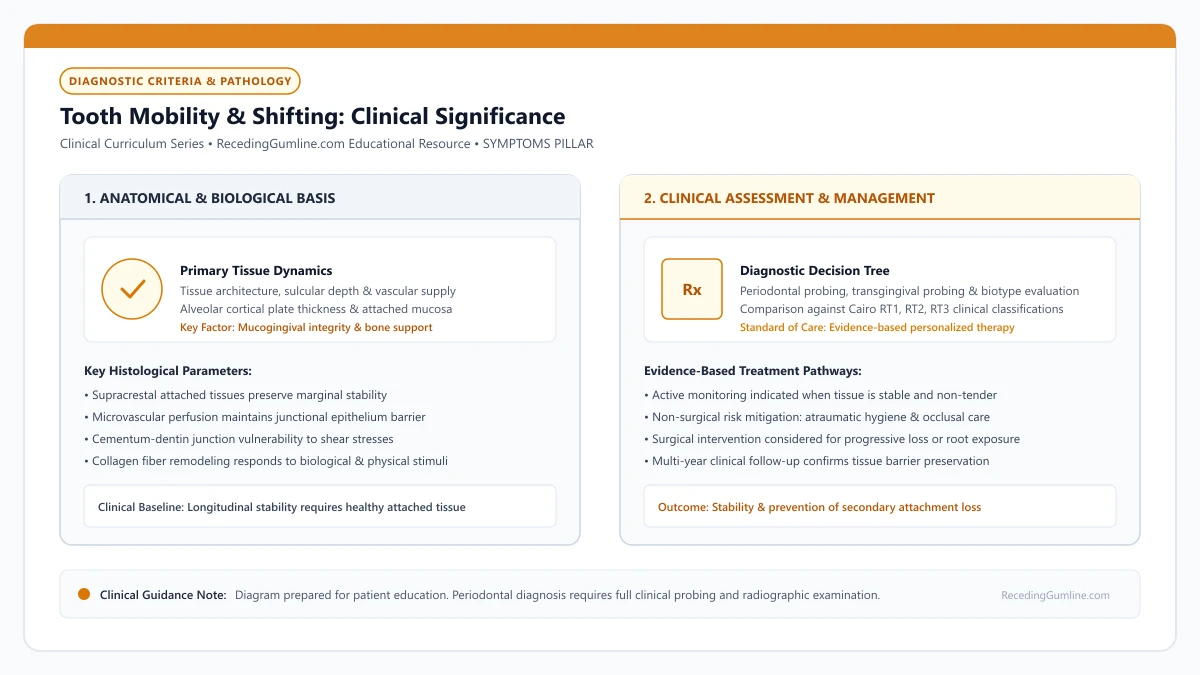
Educational illustration: Tooth Mobility & Shifting: Clinical Significance. Clinical management requires comprehensive periodontal evaluation rather than isolated self-assessment.
Source: RecedingGumline.com Clinical Editorial Team (Proprietary educational diagram for RecedingGumline.com)
Periodontal Ligament Biomechanics & Physiological Mobility
Human teeth are not rigidly fused to the jawbone; instead, each tooth is suspended within its alveolar socket by the Periodontal Ligament (PDL)—a specialized, vascular connective tissue space measuring approximately 0.15 to 0.38 mm in width. The PDL consists of dense collagen fiber bundles (Sharpey's fibers) that anchor the root cementum to the alveolar cortical bone.
The PDL acts as a sophisticated hydraulic shock absorber. When biting forces are applied, interstitial fluid within the ligament space is compressed, dampening mechanical impact and distributing forces evenly across the alveolar bone walls. This hydraulic mechanism permits a physiological mobility of 0.05 to 0.20 mm in horizontal directions, which is completely imperceptible to the patient.
Pathological tooth mobility develops through two distinct biological pathways: quantitative reduction in bone height (where severe recession and periodontitis destroy 50% or more of the root's bony support) and qualitative changes in ligament width (where active inflammation or traumatic forces widen the PDL space, creating noticeable laxity).
Key Clinical Insights
- The Periodontal Ligament acts as a hydraulic shock absorber, allowing minor physiological movement.
- Quantitative bone loss reduces the root surface area anchored within the supporting socket.
- Inflammatory degradation and occlusal trauma widen the PDL space, producing perceptible mobility.
Clinical Evaluation: The Miller Mobility Classification
Periodontists evaluate tooth mobility using two rigid instrument handles (such as the metal backs of a dental mirror and periodontal probe) placed on the buccal and lingual surfaces of the tooth. Testing with fingers is inaccurate because the soft, compressible finger pads yield, masking true movement.
The degree of mobility is categorized according to the universally accepted Miller Mobility Classification (1950), as refined in modern periodontology: Class 0 represents physiological mobility within normal limits. Class I represents barely perceptible horizontal movement, measuring up to 1.0 mm in a buccolingual direction.
Class II represents moderate horizontal movement exceeding 1.0 mm buccolingually, but without vertical displacement. Class III represents severe mobility characterized by extensive horizontal movement accompanied by vertical depression (the tooth can be visibly pushed downward into its socket). Class III mobility indicates a severely compromised prognosis and high risk of imminent tooth loss.
Key Clinical Insights
- Mobility must be tested using two rigid instrument handles rather than soft finger pads.
- Miller Class I (< 1 mm horizontal) and Class II (> 1 mm horizontal) represent reversible or manageable states.
- Miller Class III involves vertical depressibility, indicating a guarded to poor long-term prognosis.
Primary vs. Secondary Occlusal Trauma
A fundamental diagnostic milestone is distinguishing primary occlusal trauma from secondary occlusal trauma. Primary occlusal trauma occurs when excessive, abnormal biting forces—such as severe nocturnal bruxism, clenching, or a high dental filling—act upon a tooth with normal, healthy alveolar bone height.
In primary trauma, the periodontal ligament space widens symmetrically to dissipate the heavy forces, causing transient mobility. Crucially, primary trauma does not cause apical migration of the junctional epithelium or recession in a clean environment; once the bite is adjusted or a nightguard is worn, the mobility completely reverses.
In contrast, Secondary Occlusal Trauma occurs when normal, everyday biting and chewing forces act upon a tooth that has already lost substantial alveolar bone support due to past periodontitis and advanced recession. Because the crown-to-root ratio is severely compromised, normal physiological loads overwhelm the weakened remaining foundation, causing progressive, permanent tooth drifting, flaring, and mobility.
Key Clinical Insights
- Primary occlusal trauma involves excessive force on normal bone; mobility is fully reversible.
- Secondary occlusal trauma involves normal chewing force on severely reduced alveolar bone.
- Secondary trauma accelerates attachment loss and causes teeth to splay outward (pathological migration).
Pathological Tooth Migration & Anterior Flaring
When anterior teeth suffer substantial bone loss and gingival recession, patients frequently observe that their front teeth are drifting apart, rotating, or splaying outward toward the lip (anterior flaring). This condition is termed Pathological Tooth Migration (PTM).
In health, tooth position is maintained in a delicate dynamic equilibrium known as the "neutral zone"—the balanced space where resting inward pressure from the lips is exactly counterbalanced by outward pressure from the tongue.
When advanced recession and bone loss destroy the apical periodontal anchor, the tooth can no longer resist normal swallowing forces from the tongue (which exerts up to 1.5 to 2.0 kilograms of pressure thousands of times per day). The teeth flare labially, creating wide interdental spaces, progressive elongation, and bite collapse.
Key Clinical Insights
- The neutral zone maintains tooth alignment through balanced forces between the lips and tongue.
- Weakened bone support permits normal tongue pressure to push anterior teeth labially (flaring).
- Pathological tooth migration leads to open spaces, bite collapse, and progressive aesthetic distortion.
Stabilization Protocols: Occlusal Adjustment & Periodontal Splinting
Managing mobile and shifting teeth requires a structured, multi-disciplinary protocol. The absolute first priority is controlling active periodontal infection: scaling and root planing must eliminate subgingival biofilms and resolve inflammatory edema within the periodontal ligament.
Next, occlusal therapy is implemented: occlusal equilibration (gentle reshaping of premature biting contacts) eliminates traumatic interferences, and a custom hard acrylic nightguard is fabricated to absorb nocturnal clenching forces.
For teeth with persistent Class II mobility that impairs comfortable chewing, Periodontal Splinting provides immediate functional stabilization. Using high-strength braided polyethylene fiber ribbons (such as Ribbond) bonded to the lingual surfaces with flowable composite, several teeth are joined into a rigid, unified block. Splinting distributes chewing forces across multiple teeth, providing remarkable functional comfort and enabling stable long-term tooth retention.
Key Clinical Insights
- Resolving subgingival infection is mandatory before attempting mechanical tooth stabilization.
- Occlusal adjustment removes premature bite contacts, relieving traumatic hyperfunction.
- Bonded fiber-composite periodontal splints join mobile teeth into a rigid unit, restoring comfortable chewing.
Clinical Reality Check
Periodontal splinting stabilizes loose teeth and improves chewing comfort, but it does not regrow lost alveolar bone. Splinted teeth require meticulous interdental hygiene with specialized threaders or water flossers to prevent subgingival plaque stagnation along the bonded wire.
Questions to Ask Your Dentist or Periodontist
- What Miller mobility grade do my loose teeth have, and what is their realistic long-term prognosis?
- Is my tooth looseness caused by active infection, heavy night grinding, or past bone loss?
- Would a bonded fiber splint stabilize my teeth and make chewing comfortable again?
- Do I need a custom nightguard to protect my loose teeth from ongoing nocturnal clenching forces?
- What specific flossing tools do I need to keep my splinted teeth clean and infection-free?
Unsure What Your Gum Changes Mean?
Take our free, evidence-based Gum Recession Assessment — approximately 3 minutes. Identify potential risk factors, evaluate symptoms, and receive personalized discussion questions for your dentist or periodontist.
Non-diagnostic educational triage. Private, secure, completed in your browser.
Related Educational Topics
Scientific Literature & Clinical Guidelines
4sources · Hide ▲
- Pihlstrom BL, Michalowicz BS, Johnson NW (2005).
"Periodontal diseases." The Lancet.
Clinical relevance: Peer-reviewed Lancet seminar review synthesizing global periodontal epidemiology, microbial etiology, host immunopathology, and systemic interactions, emphasizing prevention, biofilm disruption, and early risk factor modification.
- Tonetti MS, Greenwell H, Kornman KS (2018).
"Staging and grading of periodontitis: Framework and proposal of a new classification and case definition." Journal of Clinical Periodontology.
Clinical relevance: Consensus framework establishing the multidimensional staging (severity and extent of periodontal tissue breakdown) and grading (biological rate of disease progression, incorporating smoking and diabetes as grade modifiers) for periodontitis. It addresses periodontitis diagnosis and staging, not the classification of localized gingival recession defects.
- Chapple ILC, Mealey BL, Van Dyke TE, Bartold PM, Dommisch H, Eickholz P, et al. (2018).
"Periodontal health and gingival diseases and conditions on an intact and a reduced periodontium: Consensus report of workgroup 1 of the 2017 World Workshop on the Classification of Periodontal and Peri-Implant Diseases and Conditions." Journal of Clinical Periodontology.
Clinical relevance: Consensus report establishing diagnostic criteria for periodontal health and gingivitis across intact and reduced periodontia, defining clinical gingival health as <10% bleeding on probing without attachment loss and strictly differentiating gingivitis from periodontitis.
- Ramfjord SP, Caffesse RG, Morrison EC, Burgett FG, Nissle RR, Shick RA (1987).
"4 modalities of periodontal treatment compared over 5 years." Journal of Clinical Periodontology.
Clinical relevance: Longitudinal clinical trial comparing four periodontal treatment modalities over 5 years; established that rigorous professional maintenance at 3-month intervals successfully maintains clinical attachment levels and prevents further periodontal tissue breakdown regardless of the initial surgical or non-surgical modality used.
Important Medical Notice
The contents of RecedingGumline.com, including text, graphics, self-assessment calculators, and other materials, are intended solely for educational and informational purposes. This content is not intended to replace professional dental examination, diagnosis, or treatment. Always seek the advice of a qualified dentist, periodontist, or other licensed oral healthcare provider with any questions you may have regarding a medical or dental condition. Never disregard professional medical advice or delay seeking it because of something you read on this website.