Alveolar Bone Dehiscence & Fenestration in Gum Recession
What are alveolar bone dehiscences and fenestrations, and how do they cause receding gums?
The stability of the gingival margin is directly governed by the skeletal architecture of the alveolar process (see our receding gumline factors). In many individuals, the outer cortical bone plate covering tooth roots contains natural anatomical defects known as dehiscences (cleft-like gaps in the crestal bone) and fenestrations (isolated bone windows). Understanding these skeletal variations explains why receding gums can develop even in the absence of active periodontal infection.
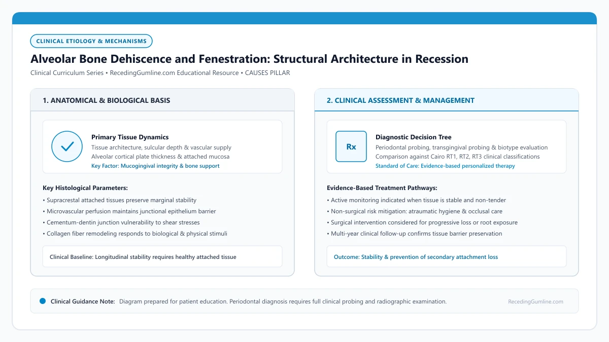
Educational illustration: Alveolar Bone Dehiscence and Fenestration: Structural Architecture in Recession. Clinical management requires comprehensive periodontal evaluation rather than isolated self-assessment.
Source: RecedingGumline.com Clinical Editorial Team (Proprietary educational diagram for RecedingGumline.com)
Key Clinical Distinctions & Diagnostic Boundaries
- A dehiscence involves the marginal bone crest, whereas a fenestration leaves an intact bridge of bone at the marginal crest.
- Dehiscences can be developmental (congenital) or acquired through orthodontic expansion and chronic inflammation.
- A tooth with a dehiscence can have normal probing depths (1 to 2 mm) despite extensive structural bone absence.
- Soft-tissue grafting over a dehiscence provides a thick fibrous pad that prevents further apical migration, even though the bone plate itself is not regenerated.
Anatomical Anatomy: Dehiscence vs. Fenestration
The alveolar bone forms the sockets (alveoli) that support the teeth. Under ideal anatomical conditions, the facial cortical plate extends to within 1.5 to 2.0 mm of the cementoenamel junction, providing continuous circumferential bone support.
However, developmental variations frequently occur. A dehiscence is an isolated cleft or dip where the marginal alveolar crest is completely missing, exposing the root surface in a continuous V-shape. In contrast, a fenestration is an isolated "window" of missing bone where the root penetrates the cortex, but an intact bridge of crestal bone remains above it.
An alveolar bone dehiscence represents an isolated V-shaped or U-shaped absence of cortical bone along the coronal facial aspect of a root. Without underlying cortical bone support, the overlying gingival margin depends entirely on suprabony soft-tissue attachment to maintain its vertical position.
Clinical Considerations:
- Dehiscence: Continuous V-shaped loss of marginal crestal bone exposing the cervical root
- Fenestration: Isolated window defect with an intact bridge of bone at the marginal collar
- Dehiscences directly cause the overlying gingival margin to collapse into recession
Etiology: Developmental Occurrence vs. Acquired Breakdown
Dehiscences can be developmental or acquired. Developmental dehiscences occur during tooth eruption when large tooth roots erupt through narrow alveolar ridges, common in patients with thin facial profiles, prominent canines, or crowded lower incisors.
Acquired dehiscences develop later in life. They are triggered when orthodontic arch expansion pushes roots facially through the cortical plate, or when chronic biofilm-induced inflammation resorbs the razor-thin facial bone. Once the cortical plate is gone, the overlying attached gingiva lacks blood supply from the periosteum, accelerating soft-tissue recession.
Direct trauma from aggressive horizontal scrubbing or accidental hard food impaction can easily shear through this unsupported soft-tissue sleeve. Once the thin junctional epithelium breaks down, marginal tissue rapidly collapses to the level of the existing bone crest.
Clinical Considerations:
- Developmental dehiscences arise when roots erupt through narrow alveolar ridges
- Acquired dehiscences result from orthodontic expansion or inflammatory bone loss
- Loss of underlying periosteal blood supply leaves the gingival margin fragile
Diagnostic Imaging: Why Standard Dental X-Rays Miss Dehiscences
A major source of patient confusion is why their regular dental x-rays look "completely normal" despite significant gum recession. Standard intraoral periapical and bitewing radiographs are two-dimensional projections that superimpose the thick lingual/palatal bone over the thin facial plate.
Consequently, a facial dehiscence is completely invisible on standard x-rays. Only 3D Cone-Beam Computed Tomography (CBCT) or direct visual observation during periodontal flap reflection can accurately reveal the presence and depth of an alveolar bone dehiscence.
Surgical management of extensive dehiscence defects requires specialized regenerative techniques such as guided tissue regeneration with bone grafts and collagen membranes. Rebuilding interdental bone support and thickening the overlying mucosa prevents further apical migration of the margin.
Clinical Considerations:
- Standard 2D radiographs superimpose lingual bone, masking facial dehiscences
- Cone-beam CT (CBCT) provides true 3D cross-sectional imaging of cortical bone thickness
- Treatment focuses on thick soft-tissue grafting to shield the denuded bone defect
Flapless Diagnostic Indicators & Dehiscence Identification
An alveolar bone dehiscence is a non-continuous skeletal defect characterized by the absence of the facial or lingual cortical bone plate extending apically from the marginal crest. Identifying dehiscences without raising a full-thickness mucoperiosteal flap requires careful clinical examination and tactile evaluation.
Clinicians detect underlying dehiscences by observing localized root prominence, palpable root contours through thin alveolar mucosa, and sudden tissue discoloration under lateral lighting. When combined with a shallow probing depth but significant clinical attachment loss, a bony dehiscence is virtually certain.
Because dehiscences remove the structural framework that anchors periodontal ligament Sharpey's fibers, the marginal tissue is held solely by supra-alveolar connective tissue fibers, leaving the margin fragile and prone to rapid detachment under physical challenge.
Clinical Considerations:
- Dehiscences represent a V-shaped or U-shaped absence of marginal cortical bone along the root.
- Tactile palpation and root prominence indicate bone loss even when tissue is intact.
- Absence of cortical bone deprives the marginal tissue of rigid structural support and vascular plexus supply.
Periodontal Plastic & Regenerative Considerations
Managing gingival recession occurring over a significant alveolar dehiscence requires advanced surgical consideration. Conventional flap repositioning alone frequently fails because the repositioned flap lacks a rigid osseous vascular bed against which to establish new fibrous attachment.
To overcome this anatomical hurdle, periodontists combine subepithelial connective tissue grafts with biologically active enamel matrix derivatives (EMD) or recombinant human platelet-derived growth factors (rhPDGF-BB). These biologic mediators stimulate cementogenesis and recruit osteoblasts to induce true periodontal regeneration.
While complete coronal bone regeneration over a deep dehiscence is biologically challenging, achieving thick keratinized soft-tissue coverage establishes a durable biological seal that halts further attachment loss and provides lifelong root protection.
Clinical Considerations:
- Flap repositioning over deep dehiscences requires soft-tissue grafting to overcome vascular limitations.
- Enamel matrix derivatives and growth factors enhance cellular recruitment along denuded root dentin.
- Thick soft-tissue coverage forms an effective protective biological seal even over osseous deficiencies.
Clinical Reality Check
Even though soft-tissue grafts cover roots over a dehiscence, they do not regenerate new cortical bone over the facial surface; they create a thick, dense fibrous barrier that shields the root permanently.
Questions to Ask Your Periodontist or Dentist
- Do my receded teeth exhibit signs of an underlying alveolar bone dehiscence?
- Why does my regular dental x-ray show normal bone between my teeth while my gums are receded on the front?
- Would a 3D CBCT scan be helpful to assess my bone thickness before any planned treatment?
- How does soft-tissue grafting protect a tooth root that has lost its outer facial bone?
Unsure What Your Gum Changes Mean?
Take our free, evidence-based Gum Recession Assessment — approximately 3 minutes. Identify potential risk factors, evaluate symptoms, and receive personalized discussion questions for your dentist or periodontist.
Non-diagnostic educational triage. Private, secure, completed in your browser.
Related Educational Topics
Clinical Evidence & Claim Traceability (2 Mapped Assertions)
Scientific Literature & Clinical Guidelines
3sources · Hide ▲
- Jepsen S, Caton JG, Albandar JM, Bissada NF, Bouchard P, Cortellini P, et al. (2018).
"Periodontal manifestations of systemic diseases and developmental and acquired conditions: Consensus report of workgroup 3 of the 2017 World Workshop on the Classification of Periodontal and Peri-Implant Diseases and Conditions." Journal of Periodontology.
Clinical relevance: Consensus report defining mucogingival conditions, gingival phenotype (replacing biotype), non-carious cervical lesions, and the multifactorial etiology of gingival recession; emphasizes that recession can occur without periodontitis and classifies recession by interdental clinical attachment loss.
- Pihlstrom BL, Michalowicz BS, Johnson NW (2005).
"Periodontal diseases." The Lancet.
Clinical relevance: Peer-reviewed Lancet seminar review synthesizing global periodontal epidemiology, microbial etiology, host immunopathology, and systemic interactions, emphasizing prevention, biofilm disruption, and early risk factor modification.
- Wennström JL (1987).
"Lack of association between width of attached gingiva and development of soft tissue recession. A 5-year longitudinal study." Journal of Clinical Periodontology.
Clinical relevance: Clinical study demonstrating that in the presence of meticulous plaque control, an extremely narrow zone or absence of attached keratinized gingiva does not inevitably lead to soft-tissue breakdown or recession progression, qualifying historical mandatory width dogmas.
Important Medical Notice
The contents of RecedingGumline.com, including text, graphics, self-assessment calculators, and other materials, are intended solely for educational and informational purposes. This content is not intended to replace professional dental examination, diagnosis, or treatment. Always seek the advice of a qualified dentist, periodontist, or other licensed oral healthcare provider with any questions you may have regarding a medical or dental condition. Never disregard professional medical advice or delay seeking it because of something you read on this website.