Thin Gingival Phenotype & Anatomy in Gum Recession
The anatomical architecture of the oral vestibule and mucogingival complex exerts a profound physical influence on the long-term stability of the gingival margin. As highlighted in our management of receding gums overview, an adequate band of dense, firmly bound keratinized attached gingiva shields the tooth collar from muscle forces. However, when congenital anatomical variations exist—such as a high labial frenum attachment, an abnormally shallow vestibular depth, or an absence of attached gingiva—normal muscular movements of the lips and cheeks can exert direct mechanical pull on the gumline, driving progressive recession.
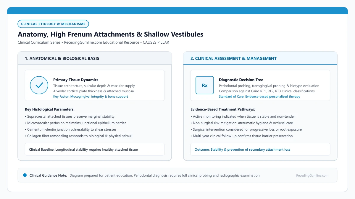
Educational illustration: Anatomy, High Frenum Attachments & Shallow Vestibules. Clinical management requires comprehensive periodontal evaluation rather than isolated self-assessment.
Source: RecedingGumline.com Clinical Editorial Team (Proprietary educational diagram for RecedingGumline.com)
Frenum Anatomy & Mucogingival Tension Mechanics
A frenum is a fold of mucous membrane containing dense collagenous connective tissue fibers, elastic fibers, and occasionally muscle slips originating from the orbicularis oris or mentalis musculature. Human oral anatomy features several prominent frena, most notably the maxillary labial frenum, the mandibular labial frenum, and the lingual frenum beneath the tongue.
Under normal anatomical conditions, the frenum inserts into the alveolar mucosa well apical to the mucogingival junction, leaving a broad, protective band of attached gingiva between the muscle attachment and the tooth margin. In aberrant anatomical configurations, however, the frenum inserts high on the attached gingiva (gingival insertion), directly into the free gingival margin (marginal insertion), or crosses the margin entirely to insert into the interdental papilla (papillary penetrating insertion).
When a frenum inserts at or near the gingival margin, every movement of the lip—during talking, eating, smiling, and laughing—transmits direct mechanical tension to the soft-tissue collar. This tension pulls the gingival margin away from the tooth surface, opening the sulcular seal, facilitating plaque biofilm ingress, and physically peeling the tissue apically.
Key Etiological Insights
- Frena are fibrous folds connecting lips and cheeks to the underlying alveolar periosteum.
- Aberrant insertions at or near the gingival margin transmit direct dynamic muscular tension.
- Dynamic muscle pull opens the gingival sulcus, accelerating localized plaque accumulation and recession.
Shallow Vestibular Depth & Keratinized Tissue Deficiency
Vestibular depth is defined as the vertical distance from the coronal margin of the gingiva to the deepest fold of the buccal fornix. An adequate vestibular depth (typically 5 mm or greater) is essential for proper oral function, facilitating saliva clearance, food deflection, and comfortable positioning of oral hygiene devices.
When an individual possesses a congenitally shallow vestibule, the movable alveolar mucosa and mentalis muscle attachments sit immediately adjacent to the cervical margin of the teeth, particularly in the mandibular anterior quadrant. This anatomical configuration results in a severe deficiency or total absence of attached keratinized gingiva.
Without a band of attached gingiva firmly bound to the underlying periosteum, the free mucosal margin lacks structural rigidity. During mastication, food boluses are forcefully driven directly into the unattached margin rather than being deflected away. Furthermore, positioning a toothbrush into a shallow vestibule causes the brush head to pinch the alveolar mucosa, prompting patients to avoid the area or scrub traumatically.
Key Etiological Insights
- A shallow vestibule (< 5 mm) lacks sufficient room for proper food deflection and hygiene access.
- Shallow vestibular depth frequently coexists with a complete absence of protective attached gingiva.
- Food impaction and mechanical toothbrush impingement rapidly accelerate tissue loss in shallow vestibules.
Clinical Diagnostic Evaluation: The Tension Test
Periodontists evaluate frenal tension using a simple, highly sensitive clinical maneuver known as the Tension Test or Blanching Test. The clinician grasps the patient's lip or cheek between the thumb and forefinger and gently stretches it outward, downward, or laterally.
If the frenal attachment exerts pathological tension on the gingival margin, the free margin will noticeably pull away from the tooth root, or the interdental papilla will blanch (turn pale white) as blood is squeezed out of the microvasculature. A positive blanching test confirms that muscle dynamics are actively contributing to gingival instability.
In addition, the clinician measures the width of keratinized tissue (KT) and attached gingiva (AG) using a periodontal probe. Classic landmark studies by Lang and Löe (1972) established that sites with less than 2 mm of keratinized tissue (including less than 1 mm of attached gingiva) frequently manifest persistent inflammation and progressive recession when oral hygiene is suboptimal.
Key Etiological Insights
- The Tension Test identifies whether lip movement pulls the gingival margin away from the tooth.
- Blanching of the papilla under lip traction proves active mechanical ischemia and tension.
- Less than 2 mm of keratinized tissue represents an anatomical risk zone for progressive recession.
Surgical Management: Frenectomy & Vestibuloplasty
When aberrant frenum pull or a shallow vestibule drives progressive recession, conservative modifications alone are rarely sufficient. Relieving the mechanical tension requires surgical intervention.
A Frenectomy involves the complete surgical or laser excision of the frenal fold, including its deep attachment to the underlying periosteum. In many cases, a simple frenectomy is combined with a Free Gingival Graft (FGG) or Vestibuloplasty. During this procedure, donor tissue harvested from the palate is secured into a surgically created recipient bed at the base of the tooth.
This dual approach achieves two vital clinical goals: it permanently relocates muscle attachments apically into a deepened vestibule, and it creates a wide, thick, immobile collar of dense keratinized attached gingiva that resists all future mechanical pull, providing permanent stability against recurrent recession.
Key Etiological Insights
- Frenectomy surgically releases the muscular and fibrous attachment pulling on the margin.
- Vestibuloplasty deepens the oral fold, creating physical clearance for oral hygiene devices.
- Free gingival grafting creates a broad, immobile zone of attached tissue that permanently halts recession.
Clinical Reality Check
Performing a frenectomy without addressing a co-existing deficiency in attached gingiva often yields incomplete clinical results. When attached gingiva is less than 1 mm, grafting should be sequenced alongside or immediately following frenum release to ensure long-term stability.
Questions to Ask Your Dentist or Periodontist
- Does the tension from my lip or frenum pull directly on my receding gums?
- How many millimeters of attached, keratinized gum tissue do I have around these vulnerable teeth?
- Would a laser or conventional frenectomy be recommended to stop my gumline from receding further?
- Do I need a soft-tissue graft in addition to a frenectomy to rebuild a protective barrier?
- Is my shallow vestibule making it difficult for me to clean these teeth properly?
Unsure What Your Gum Changes Mean?
Take our free, evidence-based Gum Recession Assessment — approximately 3 minutes. Identify potential risk factors, evaluate symptoms, and receive personalized discussion questions for your dentist or periodontist.
Non-diagnostic educational triage. Private, secure, completed in your browser.
Related Educational Topics
Scientific Literature & Clinical Guidelines
4sources · Hide ▲
- Lang NP, Löe H (1972).
"The relationship between the width of keratinized gingiva and gingival health." Journal of Periodontology.
Clinical relevance: Landmark clinical investigation observing that gingival sites with less than 2 mm of keratinized gingiva (corresponding to less than 1 mm of attached gingiva) frequently exhibited clinical signs of persistent marginal inflammation despite plaque control, historically establishing the 2 mm keratinized tissue reference point.
- Cairo F, Nieri M, Cincinelli S, Mervelt J, Pagliaro U (2011).
"The interproximal clinical attachment level to classify gingival recessions and predict root coverage outcomes: an explorative and reliability study." Journal of Clinical Periodontology.
Clinical relevance: Exploratory and reliability study establishing the Cairo classification based on interdental clinical attachment level (CAL): RT1 (no interproximal attachment loss; complete root coverage is clinically predictable), RT2 (interproximal attachment loss <= buccal loss; partial coverage predictable), and RT3 (interproximal loss exceeds buccal recession; complete coverage not predictable).
- Jepsen S, Caton JG, Albandar JM, Bissada NF, Bouchard P, Cortellini P, et al. (2018).
"Periodontal manifestations of systemic diseases and developmental and acquired conditions: Consensus report of workgroup 3 of the 2017 World Workshop on the Classification of Periodontal and Peri-Implant Diseases and Conditions." Journal of Periodontology.
Clinical relevance: Consensus report defining mucogingival conditions, gingival phenotype (replacing biotype), non-carious cervical lesions, and the multifactorial etiology of gingival recession; emphasizes that recession can occur without periodontitis and classifies recession by interdental clinical attachment loss.
- Tonetti MS, Greenwell H, Kornman KS (2018).
"Staging and grading of periodontitis: Framework and proposal of a new classification and case definition." Journal of Clinical Periodontology.
Clinical relevance: Consensus framework establishing the multidimensional staging (severity and extent of periodontal tissue breakdown) and grading (biological rate of disease progression, incorporating smoking and diabetes as grade modifiers) for periodontitis. It addresses periodontitis diagnosis and staging, not the classification of localized gingival recession defects.
Important Medical Notice
The contents of RecedingGumline.com, including text, graphics, self-assessment calculators, and other materials, are intended solely for educational and informational purposes. This content is not intended to replace professional dental examination, diagnosis, or treatment. Always seek the advice of a qualified dentist, periodontist, or other licensed oral healthcare provider with any questions you may have regarding a medical or dental condition. Never disregard professional medical advice or delay seeking it because of something you read on this website.