Prominent Tooth Roots & Labial Bone Dehiscence in Gum Recession
Why do prominent, protruding teeth experience more gum recession than properly aligned teeth?
The three-dimensional position of a tooth root within the dental arch plays a decisive role in soft-tissue stability (see our gum recession overview). When a tooth erupts too far facially (buccally) or tilts outward, its prominent root stretches the overlying cortical bone and gingival tissue, creating an anatomical hot spot for isolated, localized gum recession.
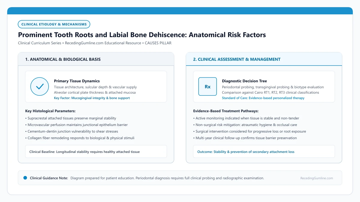
Educational illustration: Prominent Tooth Roots and Labial Bone Dehiscence: Anatomical Risk Factors. Clinical management requires comprehensive periodontal evaluation rather than isolated self-assessment.
Source: RecedingGumline.com Clinical Editorial Team (Proprietary educational diagram for RecedingGumline.com)
Key Clinical Distinctions & Diagnostic Boundaries
- Recession on prominent teeth is primarily driven by anatomical position and mechanical abrasion, not systemic periodontitis.
- Prominent roots often lack facial cortical bone completely from the time of eruption (developmental dehiscence).
- The receded margin on a prominent tooth often has zero bleeding or pocketing, presenting as clinically pink and firm.
- Attempting surgical grafting on a root that severely protrudes from the arch has lower predictability unless the root is first repositioned.
The Arch Trajectory: How Eruption Creates Prominent Contours
The dental arch has a curved, parabolic shape. Certain teeth—most notably maxillary canines, upper premolars, and lower central incisors—frequently erupt at the corners or outer contours of this curve. When dental crowding occurs, these teeth are often squeezed facially outside the main line of the alveolar bone.
As a consequence of this outward position, the facial cortical bone plate fails to form over the prominent root convexities, creating a developmental alveolar dehiscence. Instead of being encased in solid bone, the outer root surface is covered only by a paper-thin layer of periosteum and mucosa.
Teeth that are positioned buccally outside the primary curvature of the dental arch exhibit prominent root profiles that stretch the overlying soft tissue. This anatomical displacement leaves little to no vascularized cancellous bone covering the facial root convexities.
Clinical Considerations:
- Crowding pushes canines and incisors facially outside the alveolar bone envelope
- Cortical bone fails to develop over prominent root convexities
- Leaves the root vulnerable with only soft tissue coverage and no skeletal support
The "Speed Bump" Effect: Concentrated Brushing Abrasion
When an individual brushes their teeth using horizontal scrubbing strokes, prominent roots act like speed bumps in the road. The toothbrush bristles hit the protruding root contour with significantly greater force and friction than the adjacent, receded, or normally positioned teeth.
Over months and years, this concentrated mechanical wear strips away the thin gingival margin. Because there is no underlying bone to resist the force, the tissue rapidly recedes apically until it reaches a level where bone is present, exposing the yellow root surface.
During daily oral hygiene, standard toothbrush bristles exert disproportionately higher shearing forces against protruding root surfaces compared to well-aligned adjacent teeth. Over years of vigorous brushing, the thin tissue band stretched over these convexities recedes rapidly.
Clinical Considerations:
- Protruding roots receive disproportionate mechanical impact during toothbrushing
- Horizontal scrubbing selectively wears down the most prominent teeth in the arch
- Recession continues apically until it encounters a stable bone foundation
Clinical Sequencing: Orthodontics vs. Periodontal Grafting
Treating recession on a prominent tooth requires careful diagnostic planning. If a periodontist simply places a gum graft over a root that juts out 2 millimeters beyond the dental arch, the graft has no lateral vascular bed and is highly likely to fail or relapse.
The ideal interdisciplinary strategy often involves orthodontic alignment first: using braces or clear aligners to upright the tooth and torque the root lingually back inside the alveolar bone envelope. Once the root is centered in the bone, a connective tissue graft can achieve predictable, permanent root coverage.
Periodontists often coordinate with orthodontic specialists to evaluate whether minor lingual root torque can reposition protruding roots back into the center of the alveolar ridge. Bringing the root inside the cortical bone plate significantly enhances the predictability of surgical soft-tissue coverage.
Clinical Considerations:
- Grafting over severely protruding roots has reduced biological predictability
- Orthodontic root torquing moves the root back into the protective bone housing
- Subsequent grafting achieves predictable coverage and long-term attachment stability
Cone-Beam Computed Tomography (CBCT) in Prominent Root Evaluation
Evaluating teeth with prominent root contours requires imaging beyond standard two-dimensional bitewing or periapical radiographs, which cannot resolve the thin labial cortical bone plate. High-resolution Cone-Beam Computed Tomography (CBCT) provides cross-sectional volumetric visualization of the alveolar housing, revealing the exact spatial relationship between the tooth root and the buccal plate.
CBCT analyses consistently show that prominent tooth roots—frequently canines and premolars positioned buccally in the arch—often possess labial bone plates thinner than 0.5 millimeters or exhibit complete congenital dehiscences. In such sites, the root surface is covered exclusively by soft tissue and periosteum, rendering it vulnerable to rapid marginal retraction if exposed to friction or chronic plaque.
This radiographic insight is pivotal before undergoing restorative dentistry, periodontal surgery, or orthodontic tooth movement, enabling clinicians to identify non-visible osseous deficiencies and tailor treatment parameters accordingly.
By analyzing cross-sectional bone dimensions prior to intervention, clinicians can identify whether root prominence requires orthodontic movement or phenotype modification before attempting soft tissue coverage.
Clinical Considerations:
- CBCT cross-sectional imaging reveals cortical bone plates thinner than 0.5 mm that conventional x-rays miss.
- Prominent roots often lack buccal bone coverage, existing with congenital dehiscences under the soft tissue.
- Preoperative 3D imaging prevents surgical surprises and guides protective restorative planning.
Orthodontic Decompensation & Root Centering Strategies
When prominent root contours contribute to localized recession, interdisciplinary orthodontic and periodontal planning is essential. Moving a prominent tooth root back into the center of the alveolar envelope—a process known as orthodontic decompensation—can re-establish protective osseous architecture around the root prominence.
Orthodontists utilize controlled continuous light forces with precise torque mechanics to guide the root lingually without compromising the apical neurovascular bundle. Clinical evidence indicates that centering the root within the cancellous bone creates a more favorable anatomical bed, significantly increasing the success rate of subsequent soft-tissue coverage procedures.
Attempting soft-tissue grafting over a severely displaced or prominent root without first addressing its three-dimensional position frequently yields incomplete root coverage, as the avascular root surface remains outside the biological protective envelope.
Clinical Considerations:
- Torque-controlled orthodontic movement centers prominent roots within the cancellous alveolar housing.
- Centering the root creates an optimal vascular bed for subsequent soft-tissue grafting procedures.
- Grafting over prominent, uncentered roots carries higher rates of partial failure due to lack of osseous support.
Clinical Reality Check
When an isolated receded tooth is positioned further facially than adjacent teeth, modifying brushing mechanics is essential, but orthodontic repositioning of the tooth root may be required for predictable surgical coverage.
Questions to Ask Your Periodontist or Dentist
- Is my receded tooth positioned noticeably further outward in the dental arch than the adjacent teeth?
- Does my clinical exam indicate an alveolar bone dehiscence over this specific root?
- Should we consider orthodontic alignment to move the root inward before attempting a gum graft?
- What modifications should I make to my brushing path to avoid hitting this prominent tooth so hard?
Unsure What Your Gum Changes Mean?
Take our free, evidence-based Gum Recession Assessment — approximately 3 minutes. Identify potential risk factors, evaluate symptoms, and receive personalized discussion questions for your dentist or periodontist.
Non-diagnostic educational triage. Private, secure, completed in your browser.
Related Educational Topics
Clinical Evidence & Claim Traceability (2 Mapped Assertions)
Scientific Literature & Clinical Guidelines
3sources · Hide ▲
- Jepsen S, Caton JG, Albandar JM, Bissada NF, Bouchard P, Cortellini P, et al. (2018).
"Periodontal manifestations of systemic diseases and developmental and acquired conditions: Consensus report of workgroup 3 of the 2017 World Workshop on the Classification of Periodontal and Peri-Implant Diseases and Conditions." Journal of Periodontology.
Clinical relevance: Consensus report defining mucogingival conditions, gingival phenotype (replacing biotype), non-carious cervical lesions, and the multifactorial etiology of gingival recession; emphasizes that recession can occur without periodontitis and classifies recession by interdental clinical attachment loss.
- Wennström JL (1987).
"Lack of association between width of attached gingiva and development of soft tissue recession. A 5-year longitudinal study." Journal of Clinical Periodontology.
Clinical relevance: Clinical study demonstrating that in the presence of meticulous plaque control, an extremely narrow zone or absence of attached keratinized gingiva does not inevitably lead to soft-tissue breakdown or recession progression, qualifying historical mandatory width dogmas.
- Zucchelli G, Mounssif I (2015).
"Periodontal plastic surgery." Periodontology 2000.
Clinical relevance: Peer-reviewed review of periodontal plastic surgery modalities, detailing flap design, coronally advanced flaps, autogenous connective tissue grafting, tunneling techniques, and anatomical factors governing aesthetic and functional root coverage.
Important Medical Notice
The contents of RecedingGumline.com, including text, graphics, self-assessment calculators, and other materials, are intended solely for educational and informational purposes. This content is not intended to replace professional dental examination, diagnosis, or treatment. Always seek the advice of a qualified dentist, periodontist, or other licensed oral healthcare provider with any questions you may have regarding a medical or dental condition. Never disregard professional medical advice or delay seeking it because of something you read on this website.