Gum Graft Surgery: Techniques, Indications & Considerations
Gingival graft surgery encompasses a spectrum of microsurgical periodontal procedures designed to augment deficient attached keratinized tissue, prevent progressive tissue retreat, and achieve root coverage over exposed, sensitive root surfaces. When non-surgical modifications cannot halt periodontal recession guide or when patients experience severe root hypersensitivity and esthetic compromise, soft-tissue grafting represents the established, evidence-based clinical benchmark for restoring the anatomical barrier of the periodontium.
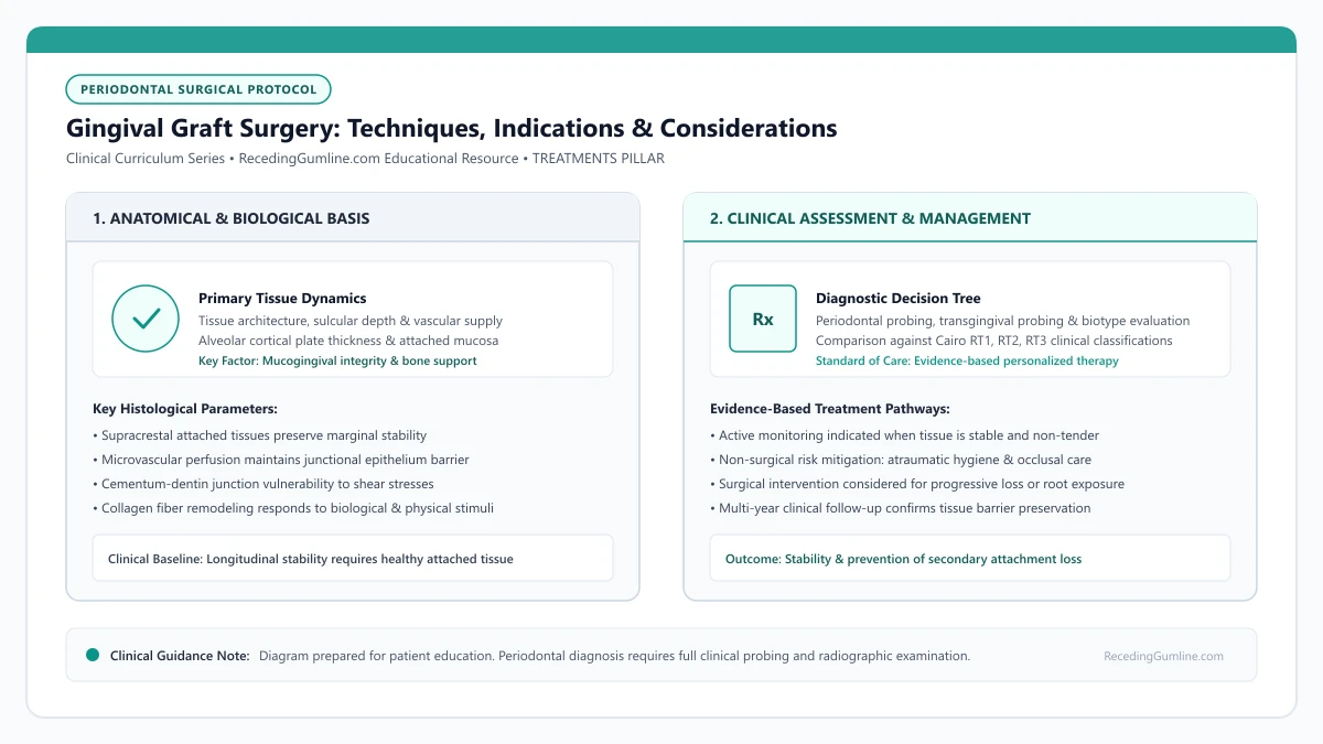
Educational illustration: Gingival Graft Surgery: Techniques, Indications & Considerations. Clinical management requires comprehensive periodontal evaluation rather than isolated self-assessment.
Source: RecedingGumline.com Clinical Editorial Team (Proprietary educational diagram for RecedingGumline.com)
Surgical Modalities: CTG vs. FGG vs. Allograft
Periodontists utilize several distinct grafting modalities depending on whether the clinical objective is root coverage, keratinized tissue augmentation, or a combination of both. The Subepithelial Connective Tissue Graft (CTG) represents the established clinical benchmark for root coverage in esthetically sensitive areas. Harvested from beneath the epithelial surface of the hard palate, the connective tissue graft is placed over the denuded root and covered by a coronally advanced flap (CAF) or inserted via a microsurgical tunnel.
The Free Gingival Graft (FGG) involves harvesting both the epithelium and underlying lamina propria from the palate. The graft is sutured directly onto an exposed periosteal bed apical to the recession defect. While the FGG creates a dense, tough band of keratinized tissue that halts muscle pull and stabilizes the margin, it heals with a paler, "tire-patch" color mismatch, making it ideal for mandibular anterior teeth but less suited for the maxillary esthetic zone.
For patients desiring to avoid a secondary palatal donor harvest, Acellular Dermal Matrix (ADM) allografts (derived from processed human donor tissue) or porcine-derived collagen xenografts provide proven alternatives. While allografts eliminate palatal wound discomfort and provide unlimited graft volume for multiple teeth, autogenous CTG consistently achieves greater long-term tissue thickness and higher percentages of complete root coverage.
Key Clinical Insights
- Connective Tissue Grafts (CTG) deliver optimal esthetic integration and predictable root coverage.
- Free Gingival Grafts (FGG) excel at widening attached keratinized tissue in non-esthetic zones.
- Allografts avoid palatal donor harvesting but achieve slightly lower complete coverage rates.
Palatal Donor Anatomy & Surgical Safety Zones
Harvesting autogenous donor tissue requires thorough knowledge of palatal anatomy to avoid complications. The primary anatomical landmark is the Greater Palatine Foramen and its anterior neurovascular bundle, which runs along the vault of the hard palate.
The safe distance from the gingival margins of the maxillary teeth to the greater palatine artery varies according to palatal vault shape: high (U-shaped) vaults provide 12 to 17 mm of vertical clearance, whereas flat (shallow) vaults offer only 7 to 10 mm. Periodontists utilize precise single-incision or trap-door techniques between the canine and first molar to harvest subepithelial tissue without violating the primary artery.
Postoperative palatal management has advanced significantly with modern techniques. Placing collagen sponges, cyanoacrylate tissue adhesives, or custom-fabricated thermoplastic palatal stents shields the donor site during eating, substantially reducing postoperative discomfort and accelerating primary closure.
Key Clinical Insights
- The Greater Palatine Artery represents the critical anatomical landmark during palatal tissue harvesting.
- High palatal vaults offer greater vertical safety zones than shallow or flat anatomical vaults.
- Protective palatal stents and specialized suturing significantly minimize post-surgical donor discomfort.
Prognostic Predictability: The Cairo Classification System
The predictability of surgical root coverage is governed by the height of the adjacent interproximal bone and papilla. In 2011, Cairo and colleagues introduced an evidence-based classification system that replaced the classic Miller (1985) system, establishing objective prognostic benchmarks.
Cairo Recession Type 1 (RT1) defects feature facial recession with zero loss of interproximal bone or papilla height. RT1 defects possess an exceptional surgical prognosis, with clinical trials demonstrating that 100% complete root coverage (CRC) can be routinely achieved using a CTG paired with a coronally advanced flap.
Cairo Recession Type 2 (RT2) defects feature interproximal attachment loss that is less than or equal to the buccal recession. Partial root coverage is highly predictable in RT2, but complete coverage is anatomically limited by the interdental bone height. Cairo Recession Type 3 (RT3) defects exhibit severe interproximal bone loss exceeding the buccal recession; in RT3, root coverage is unpredictable, and grafting is performed solely to augment attached tissue and halt progressive tooth loss.
Key Clinical Insights
- Cairo RT1 (intact interproximal bone) offers the highest predictability for 100% complete root coverage.
- Cairo RT2 allows partial root coverage limited by the height of adjacent interdental bone crests.
- Cairo RT3 defects cannot predictably achieve root coverage and are managed for structural stability.
Biological Revascularization & Postoperative Healing Stages
A soft-tissue graft begins as an avascular piece of donor tissue placed over an avascular mineralized root surface. Its survival depends entirely on a sequence of biological healing stages. During the initial 0 to 3 days (plasmatic circulation), the graft survives purely through passive diffusion of nutrients from the capillaries of the surrounding recipient bed.
Between days 4 and 7 (revascularization phase), capillary buds from the recipient periosteum and flap margin anastomose with pre-existing vascular channels in the graft, establishing true functional blood perfusion. By day 14, organic connective tissue attachment forms across the root via a thin junctional epithelium and dense collagen fiber adaptation.
Complete tissue maturation and collagen remodeling continue for 6 to 12 months post-surgery. During this period, "creeping attachment"—the slow coronal migration of the healed margin—can occasionally provide an additional 0.5 to 1.0 mm of root coverage over time.
Key Clinical Insights
- During the first 72 hours, the graft survives solely via passive diffusion of plasmatic fluid.
- Functional capillary revascularization establishes between days 4 and 7 post-surgery.
- Full collagen remodeling and tissue maturation require 6 to 12 months of undisturbed healing.
What to Expect at a Gum Graft Consultation: Diagnostic Assessment & Surgical Planning
When evaluating options for moderate to severe gum recession, a gum graft consultation is a specialized clinical evaluation where a periodontist determines whether surgical soft-tissue augmentation is indicated. The appointment involves comprehensive diagnostic charting, evaluation of donor tissue availability, discussion of surgical techniques, and establishing realistic anatomical expectations for root coverage.
• The surgeon evaluates your gingival phenotype, width of keratinized tissue, and interproximal bone levels to establish surgical feasibility.
• Palatal donor tissue thickness is assessed if autogenous grafting (connective tissue graft) is considered.
• Surgical goals may focus primarily on halting progressive recession, thickening thin tissue, or achieving cosmetic root coverage.
• A thorough consultation clarifies post-operative restrictions, recovery timelines, and total financial estimates before scheduling.
Diagnostic Measurements Performed During the Visit
During your consultation, the periodontist performs detailed measurements beyond a standard dental checkup. They measure the vertical depth and horizontal width of recession on each affected tooth, record the width of remaining keratinized attached gingiva, and probe adjacent teeth to verify interdental papilla height.
High-resolution clinical photographs and periapical radiographs are captured to document baseline conditions. These records allow the clinician to identify whether the tooth root has a cervical notch (abfraction) or prominent contour that could alter root coverage predictability.
The clinician also examines occlusal interferences and excessive lateral forces that may exacerbate cervical stress concentrations. Evaluating the opposing dentition ensures that post-surgical healing will not be disturbed by premature functional contacts during mastication.
Key Clinical Insights
- Direct millimeter documentation of recession depth and width
- Evaluation of remaining attached keratinized tissue band
- Assessment of interdental bone height on diagnostic periapical radiographs
Evaluating Donor Tissue Options: Palate vs. Biologic Allograft
If surgical grafting is recommended, the clinician examines the roof of your mouth (the hard palate). The height and vault shape of the palate, as well as soft-tissue thickness, dictate how much autogenous connective tissue can be safely harvested without compromising the greater palatine neurovascular bundle.
If palatal tissue is exceptionally thin, if multiple teeth across multiple quadrants require treatment, or if the patient prefers to avoid a palatal wound, the surgeon will present acellular dermal matrix (allograft) or xenogeneic collagen matrices as alternatives, discussing the trade-offs in recovery time and coverage predictability.
Patient preferences regarding surgical morbidity, recovery time, and aesthetic priorities are carefully weighed against biological predictability. A transparent discussion outlines the exact structural differences between autogenous palatal tissue and allogeneic acellular dermal matrices.
Key Clinical Insights
- Palatal tissue thickness determines feasibility of autogenous graft harvesting
- Palatal anatomy is evaluated to protect the greater palatine neurovascular structures
- Allograft materials eliminate the palatal donor site but exhibit slightly lower complete coverage rates in advanced defects
Establishing Realistic Surgical Goals and Anatomical Ceilings
An essential role of the consultation is establishing honest expectations. If your recession is classified as Cairo RT1 (no interdental bone loss), the periodontist can aim for complete root coverage. However, if interproximal bone resorption has already taken place (Cairo RT2 or RT3), full coverage is biologically unattainable.
In advanced situations, the realistic clinical objective shifts from complete aesthetic root coverage to augmenting the protective zone of keratinized tissue, thickening the biotype, and arresting progressive apical migration of the margin.
A complete medical history review identifies systemic factors such as unmanaged diabetes, osteopenia, or cardiovascular medications that could compromise microvascular anastomosis. Smoking cessation protocols are established well in advance to prevent microcirculatory graft failure.
Key Clinical Insights
- Cairo RT1 defects offer predictable complete root coverage
- Cairo RT2 and RT3 defects have biological ceilings limiting coverage to interdental bone height
- Tissue augmentation remains successful when it achieves stability even without 100% root coverage
Pre-Operative Health Risk Assessment & Medical Screening
During the initial surgical consultation, periodontists conduct comprehensive medical and periodontal risk evaluations to ensure patient safety and optimize healing capacity. Systemic conditions that compromise microvascular circulation—such as poorly controlled Type 2 diabetes (HbA1c > 7.0%) or active tobacco and nicotine use—significantly impair graft revascularization.
Patients taking antiplatelet agents (aspirin, clopidogrel) or anticoagulants (warfarin, apixaban) require careful hematologic coordination with their managing physician. While minor soft-tissue procedures can often proceed under established therapeutic INR ranges, surgical protocols must incorporate advanced local hemostatic measures at palatal donor sites.
The consultation also screens for nocturnal bruxism, untreated malocclusion, and gastric reflux, ensuring that predisposing mechanical and chemical forces are identified and managed before surgical intervention.
Key Clinical Insights
- Systemic vascular conditions and active nicotine use compromise graft revascularization.
- Anticoagulant and antiplatelet therapy require preoperative coordination and local hemostatics.
- Bruxism and chemical erosion risk factors must be identified and managed prior to surgery.
Establishing Realistic Expectations: Aesthetics vs. Tissue Stability
A vital objective of the periodontal consultation is aligning patient expectations with biological realities. Patients frequently assume that gum grafting will restore their gumline to its original youthful appearance across all receded teeth.
The periodontist educates the patient regarding anatomical limitations. In Cairo RT1 defects with intact interdental bone, 100% complete root coverage is a realistic clinical objective. However, in RT2 and RT3 defects with interdental bone loss, complete coverage is biologically unachievable; the primary surgical goal shifts to stopping progression and thickening the tissue band.
Clear photographic documentation and 3D visual models allow patients to visualize anticipated postoperative root coverage and understand the distinction between cosmetic enhancement and functional tissue stabilization.
Key Clinical Insights
- Intact interdental bone (RT1) allows predictable complete root coverage.
- Interdental bone loss (RT2/RT3) limits coverage; surgery focuses on halting progression.
- Preoperative photographic counseling establishes realistic aesthetic and functional expectations.
Gum Graft Recovery Timeline: Day-by-Day Healing Milestones & Tissue Maturation
For patients undergoing surgical correction of receding gums, recovery following soft-tissue grafting involves distinct biological phases spanning several months. Understanding the day-by-day healing milestones—from initial plasmatic circulation and capillary ingrowth to complete collagen remodeling—helps patients adhere to critical postoperative protocols and protect delicate graft revascularization.
• Days 1 to 3 rely entirely on plasmatic imbibition for graft nutrition; mechanical disturbance must be strictly avoided.
• Days 4 to 10 feature active capillary revascularization and vascular anastomosis between recipient bed and donor tissue.
• Weeks 2 to 4 allow suture removal and gradual resumption of gentle plaque removal with ultra-soft brushes.
• Full histological maturation and keratinization of the grafted tissue requires 8 to 12 weeks.
Days 1 to 3: The Critical Plasmatic Imbibition Phase
During the first 48 to 72 hours, the transplanted tissue has no direct blood supply. It survives through plasmatic imbibition—a passive diffusion process wherein nutrients, fluid, and oxygen pass from the recipient periosteal vascular bed through a micro-thin fibrin clot into the graft.
Any mechanical movement during this phase, such as tongue manipulation, chewing firm foods near the site, or pulling the lip to inspect the stitches, can rupture the fibrin network. Rupture severs this nutrient lifeline, leading to partial or complete graft necrosis.
Microvascular perfusion begins establishing capillary buds into the donor connective tissue within 48 to 72 hours through plasmatic circulation. Patients must strictly avoid disturbing this fragile biological fibrin seal through physical palpation, tongue exploration, or premature visual inspection.
Key Clinical Insights
- Graft relies entirely on fluid diffusion from the underlying recipient bed
- Fibrin clot must remain completely undisturbed by muscle pull or friction
- Soft, cool foods and zero mechanical toothbrush contact on the surgical site are mandatory
Days 4 to 14: Capillary Anastomosis and Superficial Desquamation
Between days 4 and 7, capillary buds from the recipient bed sprout and anastomose (connect) with pre-existing vascular channels within the connective tissue graft. By day 7 to 10, true blood circulation is established throughout the core of the graft.
Patients frequently become alarmed around day 5 to 7 because the surface of the graft appears white, grey, or yellowish. This does not represent an infection or graft failure; rather, it reflects normal desquamation of the ischemic superficial epithelium while the deep connective tissue core remains vital and firmly attached.
During the second week post-procedure, collagen fiber synthesis accelerates as immature granulation tissue transitions into organized mucosal architecture. Gentle plaque control adjacent to the site is maintained without direct mechanical contact from standard bristle brushes.
Key Clinical Insights
- New capillary ingrowth establishes active blood perfusion by days 7 to 10
- Whitish or sloughing surface tissue represents normal epithelial turnover
- Sutures are typically evaluated and gently removed between days 10 and 14
Weeks 2 to 12: Tissue Maturation, Creeping Attachment & Remodeling
Once sutures are removed, the newly formed epithelium proliferates across the graft surface. Over weeks 4 to 8, collagen bundles reorient and anchor to the root surface via a junctional epithelial seal and dense connective tissue adaptation.
Between 2 and 6 months, patients may experience what clinicians call creeping attachment—a gradual coronal migration of the gingival margin over previously exposed root surfaces, particularly in narrow Cairo RT1 defects. Full histological maturity and natural color blending are achieved by month 3.
Full maturation of the grafted tissue, including final color matching and tissue thickness blending, continues for up to six to nine months. Long-term tissue stability requires sustained elimination of the original etiological factors that prompted recession initially.
Key Clinical Insights
- Gradual coronal tissue migration (creeping attachment) may occur over 6 months
- Color and contour blend naturally with adjacent gingiva by week 12
- Periodontal probing of the grafted site is strictly contraindicated until 6 months post-op
Cellular Healing Kinetics: From Plasmatic Circulation to Maturation
The biological healing of a soft-tissue graft follows a precisely coordinated sequence of cellular events. During the first 0 to 48 hours (plasmatic circulation phase), the donor graft has no direct blood supply and survives entirely by passive diffusion of nutrients and oxygen from the host recipient bed fluid.
Between days 3 and 14 (revascularization phase), capillary sprouts from the recipient bed and overlying flap anastomose with pre-existing vessels within the donor connective tissue. Fibroblasts migrate into the interface, synthesizing an immature procollagen matrix that physically bonds the graft to the denuded root surface and adjacent periosteum.
From week 4 through month 6 (maturation and remodeling phase), collagen fibers organize into dense, highly ordered bundles, and the overlying epithelium achieves full keratinization. Complete histologic tissue maturation and marginal creeping attachment continue for up to 12 months postoperatively.
Key Clinical Insights
- Days 0-2: Graft survives solely via passive plasmatic fluid diffusion.
- Days 3-14: Direct capillary anastomosis and collagen matrix synthesis anchor the graft.
- Months 1-6: Tissue undergoes structural remodeling, keratinization, and potential creeping attachment.
Milestone-Based Activity Resumption & Postoperative Guidelines
Successful graft healing requires adhering to milestone-based physical restrictions that protect developing microvascular networks from mechanical disruption. During the initial 72 hours, patients must avoid all vigorous cardiovascular exercise, heavy lifting, and head-down postures to prevent blood pressure spikes that could induce palatal bleeding or hematoma formation.
Dietary progression proceeds from strictly cold liquids and purees (yogurt, smoothies, lukewarm broth) during days 1 to 7, advancing to soft, non-chew foods (scrambled eggs, pasta, mashed vegetables) during weeks 2 and 3. Hard, crunchy, or acidic foods are strictly prohibited until week 4.
Any mechanical contact with the surgical site—including toothbrushes, floss, or exploratory tongue movements—is strictly forbidden for the first two to three weeks; plaque control is maintained solely via prescribed antimicrobial rinses.
Key Clinical Insights
- Strenuous physical exertion is prohibited for 72 hours to prevent hematoma and bleeding.
- Diet progresses from cool liquids (week 1) to soft foods (weeks 2-3) before normal diet resumes.
- Zero mechanical tooth contact is permitted on the graft site for the first 2-3 weeks.
Managing Discomfort After Gum Graft Surgery: Palatal Protection & Analgesic Protocols
Pain after gum graft surgery is overwhelmingly concentrated at the palatal donor site rather than the recipient gum tissue. Understanding how to manage this donor area using protective palatal stents, multimodal pain regimens, and dietary adjustments ensures a smooth, manageable recovery without unneeded reliance on heavy narcotics.
• The recipient gum graft site typically produces mild pressure or tenderness, while the palatal donor site behaves similarly to a superficial burn.
• A custom clear acrylic palatal stent or surgical collagen dressing dramatically minimizes donor site irritation during eating and speaking.
• Combining scheduled acetaminophen and ibuprofen provides greater analgesic efficacy compared to opioid monotherapy for dental pain.
• Cold, smooth liquids and pureed foods for the first 72 hours prevent mechanical disruption of the palatal blood clot.
Why the Palate Hurts: Understanding Donor Site Wound Biology
When an autogenous graft is taken, connective tissue is harvested from the vault of the palate between the canine and second molar. In subepithelial connective tissue grafting, clinicians typically use a "single incision" or "envelope" approach that allows primary closure of the palate with sutures, leaving minimal exposed raw tissue.
In contrast, a free gingival graft removes the superficial epithelium along with the connective tissue, creating an open secondary wound resembling a severe burn from hot pizza. This open wound takes 14 to 21 days to completely re-epithelialize, explaining why donor site protection is critical.
Custom clear acrylic palatal stents or thermoplastic surgical templates provide physical insulation against tongue friction and direct contact with masticated food boluses. Wearing the appliance consistently during the initial 48 hours dramatically lowers perceived donor-site pain scores.
Key Clinical Insights
- SCTG harvests leave a closed incision with minimal discomfort
- FGG procedures leave a secondary wound requiring 2 to 3 weeks to re-epithelialize
- Tongue friction and hard food contact are the primary triggers of palatal pain
Palatal Stents and Surgical Dressings: Mechanical Shielding
A custom palatal stent—a clear, thin acrylic tray fabricated before surgery to fit over your upper teeth and roof of mouth—is one of the most effective tools for eliminating donor site pain. Wearing the stent prevents your tongue, food particles, and beverages from irritating the raw palatal tissue.
Most periodontists recommend wearing the stent continuously for the first 24 to 48 hours, removing it only for gentle water rinses. Surgeons may also place biological dressings such as gelatin sponges, oxidized cellulose, or cyanoacrylate adhesives over the donor site to seal exposed capillaries.
Alternating acetaminophen with nonsteroidal anti-inflammatory agents like ibuprofen creates synergistic peripheral and central analgesia without requiring opioid prescriptions. Cold compress applications to the external facial cheek for 20-minute intervals reduce post-operative inflammatory edema.
Key Clinical Insights
- Custom acrylic stents mechanically shield the palatal wound from tongue movement and food
- Stents are worn continuously for the first 24 to 48 hours for maximum stabilization
- Hemostatic gelatin sponges or surgical tissue adhesives aid clotting and pain relief
Evidence-Based Analgesic Protocols: NSAIDs vs. Opioids
Modern dental pain research demonstrates that a multimodal analgesic strategy—combining ibuprofen (400 to 600 mg) with acetaminophen (500 mg) taken concurrently or in an alternating schedule every 6 hours—provides profound relief by targeting both peripheral prostaglandin synthesis and central pain processing.
This non-opioid combination consistently outperforms narcotics while avoiding side effects such as nausea, dizziness, constipation, and cognitive impairment. Patients should take their first dose while the surgical local anesthesia is still numb to prevent "pain breakthrough."
Patients are instructed to consume chilled nutrient-dense liquid and pureed foods such as Greek yogurt, smoothies, and lukewarm broths. Avoiding citrus, hot spices, and sharp crispy foods prevents mechanical disruption of stabilizing hemostatic gelatin sponges placed in the palate.
Key Clinical Insights
- Combining ibuprofen and acetaminophen provides synergistic analgesic relief
- First dose should be administered before local anesthesia completely wears off
- Aspirin must be strictly avoided to prevent postoperative palatal bleeding
Palatal Wound Protection: Vacuum-Formed Stents & Hemostatic Biomaterials
When autogenous connective tissue is harvested from the hard palate, managing the donor site is the primary determinant of postoperative patient comfort. Unprotected palatal donor wounds are subjected to continuous friction from the tongue, speech articulation, and food bolus movement.
To shield this vulnerable site, periodontists fabricate a custom vacuum-formed acrylic palatal stent prior to surgery. Worn continuously for the first 24 to 48 hours, the clear stent provides rigid mechanical protection, preventing food contact and dramatically reducing sensory irritation.
Within the donor wound, surgeons place hemostatic collagen sponges, oxidized regenerated cellulose, or cyanoacrylate tissue adhesives secured with cross-mattress sutures. These biomaterials accelerate clot stabilization and suppress pain receptor exposure.
Key Clinical Insights
- Vacuum-formed acrylic palatal stents physically shield the donor wound from tongue and food trauma.
- Continuous stent wear for 24-48 hours provides dramatic reduction in donor site discomfort.
- Collagen sponges and cross-mattress sutures stabilize blood clots and protect sensory nerve endings.
Evidence-Based Multimodal Analgesia: Optimizing Pain Control
Contemporary periodontal surgical protocols prioritize multimodal, non-opioid pain management regimens that achieve effective analgesia while avoiding opioid-related side effects. An established clinical protocol combines an NSAID with acetaminophen in staggered dosing schedules.
Administering ibuprofen (600 mg) combined with acetaminophen (500 mg) every six hours establishes synergistic anti-inflammatory and central antipyretic analgesia. This combination blocks peripheral prostaglandin synthesis at the surgical site while dampening central nervous system nociceptive processing.
Long-acting local anesthetics (such as bupivacaine with 1:200,000 epinephrine) administered at the conclusion of surgery provide 6 to 9 hours of profound local numbness, allowing patients to travel home, begin oral analgesics, and rest comfortably before surgical sensation returns.
Key Clinical Insights
- Staggered ibuprofen (600 mg) and acetaminophen (500 mg) provides effective non-opioid analgesia.
- Synergistic action blocks both peripheral prostaglandin production and central pain signaling.
- Long-acting bupivacaine local anesthesia delivers 6-9 hours of extended postoperative numbness.
Gum Graft Surgery Risks, Complications & Graft Failure Signs
While periodontal soft-tissue grafting boasts high success rates in experienced hands, it remains a delicate micro-surgical procedure subject to biological and mechanical complications. Understanding potential risks—including persistent bleeding, flap dehiscence, infection, and partial graft necrosis—equips patients to detect issues early and seek prompt intervention.
• True complete graft failure is uncommon, but partial graft necrosis or incomplete root coverage can occur due to anatomical limits or mechanical trauma.
• Post-operative bleeding from the palatal donor site is the most frequent acute complication, typically manageable with direct pressure.
• Infection occurs in fewer than 2% of cases when strict sterile protocols and postoperative antimicrobial rinses are utilized.
• Flap dehiscence (separation of the incision margins) within the first week significantly reduces final root coverage percentage.
Acute Postoperative Complications: Hemorrhage and Hematoma
The most immediate post-surgical concern is bleeding, almost always originating from the palatal donor site. The hard palate contains the greater palatine artery and its branches. Minor oozing during the first 12 to 24 hours is common and causes pink-tinged saliva, but active, continuous red bleeding requires attention.
Patients can usually control donor site bleeding by applying firm, direct pressure with a damp gauze pad or a moistened caffeinated black tea bag for 20 to 30 minutes continuously. The tannic acid in black tea acts as a natural topical vasoconstrictor and coagulant.
Total graft failure occurs when capillary revascularization fails to establish within the first 96 hours, resulting in sloughing white necrotic tissue. Maintaining meticulous surgical immobility with fine microsurgical sutures minimizes dead space and maximizes graft survival rates.
Key Clinical Insights
- Pink saliva is normal oozing; steady welling of dark red blood indicates active bleeding
- Continuous direct pressure with a damp black tea bag for 20 minutes promotes hemostasis
- Avoid spitting or rinsing vigorously, as pressure changes dislodge early blood clots
Differentiating Normal Epithelial Sloughing from True Graft Failure
A frequent cause of acute patient anxiety is the appearance of the recipient graft around days 5 to 8. The outer epithelial cells, deprived of direct circulation, undergo necrosis and turn white, grey, or yellowish before shedding. This superficial sloughing is a natural biological step in re-epithelialization.
True graft necrosis occurs when the deep collagenous connective tissue core fails to revascularize. Signs of genuine graft failure include complete disintegration of the tissue block, foul odor, spontaneous bleeding upon minimal contact, purulent drainage (pus), and the complete re-exposure of the denuded root surface.
Palatal hemorrhage from branches of the greater palatine artery is prevented through precise anatomical landmark identification during harvesting. Clinicians maintain direct digital pressure or apply local hemostatic agents like collagen plugs and oxidized regenerated cellulose if persistent oozing occurs.
Key Clinical Insights
- Superficial white/yellow film is normal dead epithelial cells sloughing off
- True graft failure involves loss of the underlying connective tissue core and persistent pus
- Periodontal surgeons evaluate tissue vitality by gentle inspection, never through probing
Long-Term Structural and Aesthetic Considerations
Longer-term complications relate to aesthetics and sensory changes. When a free gingival graft is placed on a front tooth, it retains palatal epithelial characteristics, often resulting in a "patch-like," paler color and thicker texture that clashes with adjacent gingiva. Subepithelial connective tissue grafting largely avoids this cosmetic issue.
Transient sensory alterations in the roof of the mouth—such as localized numbness, tingling, or hypersensitivity—occur in a small percentage of patients if minor terminal branches of the palatine nerve are stretched or severed. In the vast majority of cases, sensation returns fully within 3 to 6 months.
When residual root exposure occurs due to partial flap shrinkage, secondary corrective procedures may be considered only after twelve months of tissue maturation. Patients must understand that anatomical defects classified as Cairo RT3 cannot achieve 100% root coverage due to interproximal bone resorption.
Key Clinical Insights
- Free gingival grafts may heal with a paler, thicker "tire-patch" appearance
- SCTG yields refined color and contour blending with adjacent gums
- Palatal numbness or tingling is usually temporary, resolving within 3 to 6 months
Managing Early Flap Dehiscence & Graft Revascularization Failure
The most critical early complication following gum graft surgery is flap dehiscence—the premature separation of the overlying surgical flap from the recipient tissue margin, exposing the underlying donor graft. Flap dehiscence typically occurs between postoperative days 3 and 7, often triggered by excessive muscle tension, speaking trauma, or uncooperative lip retraction.
When donor tissue is exposed prematurely, its superficial layers undergo ischemic necrosis due to lack of blood supply. Clinicians recognize necrosis by the appearance of a pale white or yellowish sloughing tissue layer over the recipient bed.
Management requires strict conservative handling: the clinician avoids re-suturing the fragile, friable tissue, prescribing gentle chlorhexidine irrigation and close monitoring. In many instances, the deep portion of the graft survives, leaving adequate keratinized tissue even if total root coverage is compromised.
Key Clinical Insights
- Flap dehiscence occurs when mechanical tension separates the flap, exposing the graft.
- Ischemic necrosis appears as a white or yellowish sloughing layer over the donor tissue.
- Conservative management with antimicrobial irrigation preserves surviving basal graft layers.
Postoperative Hemorrhage: Palatal Anatomy & Emergency Protocols
Secondary hemorrhage from the palatal donor site is an uncommon but significant surgical risk. The greater palatine artery courses anteriorly along the junction of the horizontal palatine process and vertical alveolar process, approximately 7 to 17 millimeters from the gingival margin of the maxillary molars.
If a donor tissue harvest inadvertently encroaches upon the greater palatine neurovascular bundle or lacerates terminal arterial branches, delayed secondary bleeding may occur as local vasoconstrictors wear off. Patients must be educated on immediate home hemostasis protocols: applying firm, continuous pressure to the site using a moistened gauze pad or black tea bag (rich in astringent tannic acid) for 20 uninterrupted minutes.
If bleeding persists, the patient must contact the surgical team for in-office intervention, which may include local anesthetic infiltration, electrocautery, deep ligation sutures, or gelatin sponge placement.
Key Clinical Insights
- Palatal harvest sites must maintain a safe distance from the greater palatine artery.
- Immediate home management utilizes firm, uninterrupted pressure with damp gauze or tea bags for 20 minutes.
- Persistent bleeding requires clinical intervention with ligation, cautery, or hemostatic agents.
Gum Graft Aftercare Protocol: Hygiene Modification, Diet & Stent Management
The clinical success of a periodontal gum graft depends as much on patient postoperative compliance as on surgical execution. Because the newly transplanted soft-tissue relies on delicate capillary connections formed during the first two weeks, patients must adhere to strict behavioral, dietary, and hygiene modifications to safeguard the surgical site.
• Do not brush or floss the surgical recipient site for 14 to 21 days until cleared by your periodontist.
• Use prescribed 0.12% chlorhexidine gluconate as a gentle soak rather than vigorous swishing to chemically control plaque.
• Maintain a soft, non-chewing diet avoiding crunchy, hot, acidic, or sharp foods that could mechanically dislodge the graft.
• Refrain from aerobic exercise and heavy lifting for at least 3 to 7 days to prevent elevated blood pressure and bleeding.
Oral Hygiene Modifications: Chemical Biofilm Control
Direct mechanical contact with a toothbrush or floss on the surgical recipient site must be completely avoided for at least two to three weeks. Even the softest bristle can tear anchoring fibrin strands and detach the graft from the vascular root bed.
To keep the area clean, clinicians prescribe a 0.12% chlorhexidine gluconate oral rinse. Instead of swishing vigorously, take a small sip, tilt your head from side to side to bathe the surgical site for 30 to 60 seconds, and allow the liquid to gently spill out of your mouth into the sink. Brushing and flossing of all untreated teeth should continue as normal.
Patients must avoid retracting the lip or cheek to inspect the surgical site, as tensile stress can shear fragile microvessels before collateral circulation matures. Mechanical tooth brushing is suspended in the operated sextant until the surgical team confirms adequate epithelial sealing.
Key Clinical Insights
- Zero mechanical brushing on surgical teeth for 2 to 3 weeks
- Use chlorhexidine with passive head-tilting rather than vigorous swishing
- Brush and floss non-treated quadrants normally to control overall bacterial load
Dietary Guidelines: Protecting the Donor and Recipient Sites
For the first 48 to 72 hours, adhere strictly to cold or room-temperature liquid and smooth pureed foods, such as smoothies (eaten with a spoon, never a straw), yogurt, applesauce, cooled broths, and scrambled eggs. Avoid very hot foods and drinks, which cause vasodilation and increase bleeding.
From days 4 to 14, advance to soft foods requiring minimal chewing, such as well-cooked pasta, soft fish, mashed potatoes, and oatmeal. Steer clear of hard, crunchy, crusty, or seeded items (chips, nuts, popcorn, berries), as a single sharp crumb wedged under the flap can cause severe infection and graft loss.
Soft foods must be chewed exclusively on the contralateral unoperated side of the dental arch to minimize mechanical disruption. High-protein nutritional intake supports fibroblast proliferation and rapid extracellular matrix synthesis during the critical acute healing window.
Key Clinical Insights
- Cold and room-temperature purees are ideal for the first 3 days
- Never use a drinking straw, which creates suction and dislodges blood clots
- Strictly eliminate nuts, chips, popcorn, and small seeds that could lodge under the flap
Physical Activity, Elevation & Lifestyle Precautions
Elevated heart rate and blood pressure increase vascular pressure in the head and neck, easily triggering bleeding at both the palatal donor site and the recipient bed. Patients should avoid running, weightlifting, yoga inversions, and strenuous labor for at least 3 to 7 days.
When sleeping, keep your head elevated on two to three pillows for the first 3 nights to reduce head-level venous pressure, thereby minimizing swelling and morning throbbing. Smoking and alcohol consumption must be strictly avoided during the entire initial 14-day revascularization window.
Post-operative instructions include clear contact guidelines for reporting active arterial bleeding, sudden disproportionate swelling, or severe systemic fever. Timely clinical assessment within 24 hours ensures minor postoperative variations are managed before compromising surgical outcomes.
Key Clinical Insights
- Pause all strenuous exercise and heavy lifting for 3 to 7 days
- Sleep with the head elevated on multiple pillows to reduce edema and throbbing
- Zero smoking or nicotine use to protect microvascular capillary survival
Plaque Control Protocols: Phased Transition During Weeks 1 to 4
Maintaining oral hygiene during the initial month after gum graft surgery requires balancing bacterial biofilm elimination against the critical need to avoid mechanical disturbance of the fragile surgical site. Plaque accumulation on adjacent teeth introduces cytotoxic endotoxins that provoke marginal inflammation and compromise graft revascularization.
During weeks 1 and 2, patients brush and floss non-surgical areas normally but avoid the operative site entirely. The surgical site is cleansed exclusively through gentle passive bathing with 0.12% chlorhexidine digluconate mouthwash twice daily, held in the mouth for 60 seconds without vigorous swishing or spitting.
At the week 3 follow-up appointment, once initial epithelial bridging is confirmed, the periodontist introduces a post-surgical ultra-soft micro-bristle brush (diameter 0.08 mm). The patient is instructed to gently sweep the bristles coronally from the gum toward the incisal edge, avoiding sulcular scrubbing until full tissue maturation is achieved at week 6.
Key Clinical Insights
- Non-surgical teeth are brushed normally, while the graft site avoids all mechanical contact.
- Twice-daily 0.12% chlorhexidine bathing provides chemical plaque control without physical disturbance.
- Ultra-soft micro-filament brushes are introduced at week 3 with gentle coronal sweeping strokes.
Nutritional Protocols for Optimizing Connective Tissue Synthesis
Post-surgical healing and graft incorporation impose significant metabolic demands on periodontal fibroblasts, which must synthesize vast networks of new Type I and Type III collagen. Targeted nutritional support optimizes systemic wound healing capacity.
Vitamin C (ascorbic acid) is an essential cofactor for prolyl and lysyl hydroxylase enzymes, which stabilize the triple-helix collagen molecule; deficiency impairs connective tissue cross-linking and increases capillary fragility. Supplementation with 500 to 1,000 mg of Vitamin C daily supports optimal wound healing.
Adequate dietary protein intake (supplying essential amino acids glycine, proline, and hydroxyproline) along with zinc and Vitamin A provides the cellular building blocks required for rapid epithelial proliferation and capillary angiogenisis.
Key Clinical Insights
- Fibroblasts require specific metabolic cofactors to synthesize dense new collagen networks.
- Vitamin C is essential for enzymatic hydroxylation and structural collagen stabilization.
- Adequate protein, zinc, and Vitamin A intake accelerate epithelialization and microvascular angiogenesis.
Chlorhexidine Mouthrinse in Periodontal Healing: Indications, Duration & Staining
Chlorhexidine gluconate (0.12% in the United States, 0.20% in Europe) is widely regarded as the established benchmark antimicrobial agent in clinical periodontics. Prescribed following periodontal surgery or active scaling, chlorhexidine provides potent chemical plaque control when mechanical toothbrushing is temporarily impossible. However, its significant side-effect profile requires strict clinical guidance on duration and application.
• Chlorhexidine possesses unique substantivity, binding to oral tissues and releasing antimicrobial action for over 12 hours.
• It is indicated after gum graft surgery or scaling when mechanical brushing would disrupt healing tissues.
• Use should strictly be limited to 10 to 14 days to prevent extensive brown extrinsic tooth staining and taste alteration.
• Do not rinse with chlorhexidine immediately after brushing; sodium lauryl sulfate in toothpaste deactivates chlorhexidine molecules.
Mechanism of Action: The Power of Antimicrobial Substantivity
Chlorhexidine is a cationic (positively charged) bisbiguanide compound. The cell walls of oral bacteria and the mucosal tissues of your mouth carry negative electrical charges. Because of these opposite charges, chlorhexidine molecules bind tenaciously to oral surfaces.
This property is known as substantivity. Rather than washing away within minutes, chlorhexidine remains bound to oral tissues, slowly releasing bactericidal molecules over 8 to 12 hours. It ruptures bacterial cell membranes, effectively killing both gram-positive and gram-negative pathogens.
The therapeutic benefit of 0.12% chlorhexidine gluconate stems from its high substantivity, binding to oral soft and hard tissues and slowly releasing bactericidal concentrations over 12 hours. This sustained release prevents plaque biofilm accumulation when brushing is surgically contraindicated.
Key Clinical Insights
- Positively charged molecule binds strongly to negatively charged bacterial walls
- Substantivity provides continuous bacterial killing for up to 12 hours after rinsing
- Kills periodontal pathogens and prevents bacterial colonization on surgical sutures
Clinical Protocols: Proper Usage and Toothpaste Interference
Periodontists prescribe chlorhexidine primarily following soft-tissue grafting or flap surgery. During the first two weeks, toothbrush bristles cannot touch the surgical site. A 15 ml dose of chlorhexidine rinsed gently for 60 seconds twice daily provides complete plaque control.
A vital clinical rule is never using chlorhexidine immediately after brushing your teeth with standard toothpaste. Most commercial toothpastes contain Sodium Lauryl Sulfate (SLS), an anionic surfactant that binds to and completely neutralizes chlorhexidine. Always wait at least 30 minutes between brushing and rinsing.
Extrinsic brown staining of enamel margins and tongue surfaces is a common, reversible side effect of chlorhexidine use exceeding two weeks. Patients should be reassured that professional prophylaxis readily removes surface stains once surgical healing permits standard polishing.
Key Clinical Insights
- Rinse with 15 ml twice daily for 60 seconds during post-surgical healing
- Tilt head gently rather than swishing aggressively to avoid flap strain
- Wait 30 minutes after brushing with regular toothpaste to prevent neutralization by SLS
Managing Side Effects: Staining, Taste Changes & Duration Limits
The most noticeable drawback of chlorhexidine is brown extrinsic staining on teeth, tongue, and dental fillings. The rinse reacts with dietary tannins found in coffee, tea, red wine, and berries. These stains do not indicate tooth decay; they are purely surface deposits easily polished away at your next dental visit.
To minimize staining and prevent temporary blunting of taste buds, use chlorhexidine only for the 10 to 14 days prescribed by your surgeon. Never use chlorhexidine as a permanent, daily mouthwash, as long-term use promotes supragingival calculus formation and disrupts the healthy oral microbiome.
Because chlorhexidine exhibits non-specific cytotoxicity against regenerating fibroblasts at high concentrations, rinses should be used strictly for the prescribed 10 to 14 day window. Extending chlorhexidine therapy beyond two weeks may disrupt the oral microbiome and delay soft-tissue epithelialization.
Key Clinical Insights
- Brown staining is extrinsic and safely polished away during professional cleanings
- Limit consumption of coffee, tea, and red wine during use to reduce staining severity
- Strictly discontinue after 10 to 14 days unless specifically instructed otherwise
Pharmacodynamics: The 12-Hour Substantivity Mechanism
Chlorhexidine digluconate (0.12%) is widely considered the established benchmark topical antimicrobial in periodontal post-surgical care. Its clinical efficacy is derived not only from its broad-spectrum bactericidal activity against gram-positive and gram-negative bacteria, but primarily from its unique property of "substantivity."
Chlorhexidine carries a strong positive (cationic) charge. When rinsed in the mouth, it binds electrostatically to negatively charged oral surfaces: tooth enamel, pellicle proteins, salivary mucins, and oral epithelial cell walls.
Over the subsequent 8 to 12 hours, chlorhexidine is slowly released back into the oral fluids in active bactericidal concentrations, maintaining continuous suppression of subgingival plaque formation during the critical initial phases of soft-tissue graft healing.
Key Clinical Insights
- Chlorhexidine possesses broad-spectrum bactericidal activity against oral pathogens.
- Cationic molecules bind electrostatically to oral surfaces, establishing 8-12 hours of substantivity.
- Continuous slow release maintains a sterile surgical field without requiring mechanical brushing.
Managing Side Effects: Extrinsic Staining, Calculus & Taste Alteration
Despite its antimicrobial potency, chlorhexidine therapy is strictly limited to short-term post-surgical use (typically 10 to 14 days) due to well-documented side effects. The most common adverse effect is brown extrinsic staining of tooth enamel and composite restorations.
Staining occurs when chlorhexidine reacts with dietary chromogens present in coffee, tea, red wine, and tobacco. Additionally, chlorhexidine denatures salivary proteins, temporarily altering taste perception (dysgeusia)—particularly sweet and salty sensations—and accelerating supragingival calculus deposition.
Patients are advised to avoid dietary chromogens for at least one hour after rinsing and never to combine chlorhexidine with standard toothpaste, as anionic detergents like sodium lauryl sulfate (SLS) neutralize chlorhexidine's antimicrobial efficacy.
Key Clinical Insights
- Chlorhexidine use is restricted to 10-14 days to minimize staining and taste alteration.
- Extrinsic staining is accelerated by dietary chromogens (coffee, tea, red wine).
- Sodium lauryl sulfate in conventional toothpaste inactivates chlorhexidine; rinse times must be separated.
Clinical Reality Check
Root coverage cannot be predicted based solely on the depth of buccal recession; the interproximal bone level is the definitive biological determinant. Patients must be evaluated using the Cairo classification so expectations regarding complete versus partial coverage are clinically realistic.
Questions to Ask Your Dentist or Periodontist
- What is my Cairo classification (RT1, RT2, or RT3) for the receded teeth being considered for grafting?
- What percentage of root coverage can I realistically expect based on my interproximal bone levels?
- Do you recommend an autogenous connective tissue graft from my palate or an allograft donor tissue?
- What is the expected postoperative recovery timeline, and will I need a protective palatal stent?
- How long must I avoid chewing or brushing directly on the surgical site during healing?
- What Cairo or Miller classification defect do my affected teeth present with?
- Do you recommend an autograft from my palate or an allograft donor tissue, and what are the specific trade-offs for my case?
- What percentage of root coverage is biologically realistic given my current bone levels?
- How much downtime will I need from work, exercise, and normal oral hygiene activities?
- What specific day should I return for my post-operative suture check?
- When can I safely resume gentle brushing with an ultra-soft surgical brush around the treated teeth?
- Is my palatal donor site covered by a protective stent, surgical glue, or dissolvable sponge?
- What emergency signs should prompt me to contact your office immediately outside of business hours?
- Will my palatal harvest be performed using a closed single-incision technique or an open technique?
- Do you provide or recommend a custom acrylic palatal stent for me to wear after surgery?
- What exact schedule of ibuprofen and acetaminophen should I follow during the first three days?
- What should I do if the palatal site begins bleeding after I get home?
- What emergency number should I call if I experience uncontrolled bleeding over the weekend?
- Does my procedure use a technique that minimizes the risk of cosmetic color mismatch?
- Are there specific medications (such as blood thinners or supplements) I should pause before surgery?
- What is your protocol if a graft experiences partial detachment during the first week?
- How many times per day should I use the chlorhexidine mouthrinse, and for how many weeks?
- When can I transition from passive rinsing back to using a soft manual toothbrush on the surgical site?
- How many days of rest should I take before returning to my gym or workout routine?
- Can I continue using my regular fluoridated toothpaste on my non-surgical teeth?
- How many days specifically should I continue using chlorhexidine after my procedure?
- Can you recommend a sodium lauryl sulfate (SLS)-free toothpaste that won't deactivate the rinse?
- If I develop brown staining on my front teeth, will your hygienist polish it off at my follow-up visit?
- Should I dilute the rinse with water if it causes a stinging or burning sensation on my tongue?
Unsure What Your Gum Changes Mean?
Take our free, evidence-based Gum Recession Assessment — approximately 3 minutes. Identify potential risk factors, evaluate symptoms, and receive personalized discussion questions for your dentist or periodontist.
Non-diagnostic educational triage. Private, secure, completed in your browser.
Related Educational Topics
Scientific Literature & Clinical Guidelines
9sources · Hide ▲
- Cairo F, Nieri M, Cincinelli S, Mervelt J, Pagliaro U (2011).
"The interproximal clinical attachment level to classify gingival recessions and predict root coverage outcomes: an explorative and reliability study." Journal of Clinical Periodontology.
Clinical relevance: Exploratory and reliability study establishing the Cairo classification based on interdental clinical attachment level (CAL): RT1 (no interproximal attachment loss; complete root coverage is clinically predictable), RT2 (interproximal attachment loss <= buccal loss; partial coverage predictable), and RT3 (interproximal loss exceeds buccal recession; complete coverage not predictable).
- Chambrone L, Salinas Ortega MA, Sukekava F, Rotundo R, Kalemaj Z, Buti J, Pini Prato GP (2018).
"Root coverage procedures for treating localised and multiple recession-type defects." Cochrane Database of Systematic Reviews.
Clinical relevance: Cochrane systematic review evaluating root-coverage procedures for localized and multiple recession-type defects. While subepithelial connective tissue grafts (SCTG) combined with coronally advanced flaps demonstrated higher rates of complete root coverage and keratinized tissue gain compared to flap advancement alone, evidence quality varied across outcomes and clinical decisions must balance donor site morbidity and patient-reported outcomes.
- Zucchelli G, Mounssif I (2015).
"Periodontal plastic surgery." Periodontology 2000.
Clinical relevance: Peer-reviewed review of periodontal plastic surgery modalities, detailing flap design, coronally advanced flaps, autogenous connective tissue grafting, tunneling techniques, and anatomical factors governing aesthetic and functional root coverage.
- Cortellini P, Bissada NF (2018).
"Mucogingival conditions in the natural dentition: Narrative review, case definitions, and diagnostic considerations." Journal of Periodontology.
Clinical relevance: World Workshop 2017 comprehensive review defining mucogingival conditions, establishing diagnostic criteria for thin vs thick periodontal phenotypes, and detailing surgical indications including progressive recession, hypersensitivity, aesthetic dissatisfaction, and root caries vulnerability.
- Sculean A, Nikolidakis D, Schwarz F (2008).
"Regeneration of periodontal tissues: combinations of barrier membranes and grafting materials - biological foundation and preclinical evidence: a systematic review." Journal of Clinical Periodontology.
Clinical relevance: Comprehensive systematic review analyzing clinical and histological outcomes of biomaterials in periodontal regeneration; confirmed that enamel matrix derivative (EMD) and barrier membranes for guided tissue regeneration achieve histological evidence of true regeneration (new cementum, periodontal ligament, and bone).
- Kassab MM, Cohen RE (2003).
"The etiology and prevalence of gingival recession." The Journal of the American Dental Association.
Clinical relevance: Comprehensive epidemiological and etiological review establishing that gingival recession affects more than 50% of adults aged 18 to 64 and over 88% of individuals aged 65 and older, driven by interactions between predisposing anatomical factors and precipitating mechanical or inflammatory stimuli.
- Slot DE, Wiggelinkhuizen L, Rosema NA, Van der Weijden GA (2012).
"The efficacy of manual toothbrushes following a brushing exercise: a systematic review." International Journal of Dental Hygiene.
Clinical relevance: Systematic review evaluating plaque removal efficacy of manual toothbrushes following a single brushing exercise; observed that bristle design variations produce modest differences in plaque scores, while aggressive force or stiff bristles do not improve plaque removal and clinical evidence linking mechanical brushing technique directly to gingival recession remains contradictory.
- Sanz M, Herrera D, Kebschull M, Chapple ILC, Jepsen S, Beglundh T, et al. (2020).
"Treatment of stage I-III periodontitis—The EFP S3 level clinical practice guideline." Journal of Clinical Periodontology.
Clinical relevance: Evidence-based clinical practice guideline establishing the 4-step therapeutic pathway for periodontal management: Step 1 (behavioral and supragingival hygiene), Step 2 (subgingival instrumentation), Step 3 (surgical therapy where indicated), and Step 4 (supportive periodontal care at 3- to 4-month intervals).
- Tonetti MS, Greenwell H, Kornman KS (2018).
"Staging and grading of periodontitis: Framework and proposal of a new classification and case definition." Journal of Clinical Periodontology.
Clinical relevance: Consensus framework establishing the multidimensional staging (severity and extent of periodontal tissue breakdown) and grading (biological rate of disease progression, incorporating smoking and diabetes as grade modifiers) for periodontitis. It addresses periodontitis diagnosis and staging, not the classification of localized gingival recession defects.
Important Medical Notice
The contents of RecedingGumline.com, including text, graphics, self-assessment calculators, and other materials, are intended solely for educational and informational purposes. This content is not intended to replace professional dental examination, diagnosis, or treatment. Always seek the advice of a qualified dentist, periodontist, or other licensed oral healthcare provider with any questions you may have regarding a medical or dental condition. Never disregard professional medical advice or delay seeking it because of something you read on this website.