Oral Piercings & Physical Trauma Causes of Gum Recession
Direct physical trauma to the periodontium can precipitate rapid development of gum recession in the complete absence of bacterial periodontal disease. Among the most frequent physical etiologies are oral jewelry—specifically tongue barbells and labret studs—which continuously impinge upon adjacent mucosal tissues. In addition, excessive occlusal forces, parafunctional habits, and self-inflicted foreign body trauma exert localized destructive forces that compromise the integrity of the marginal attachment apparatus.
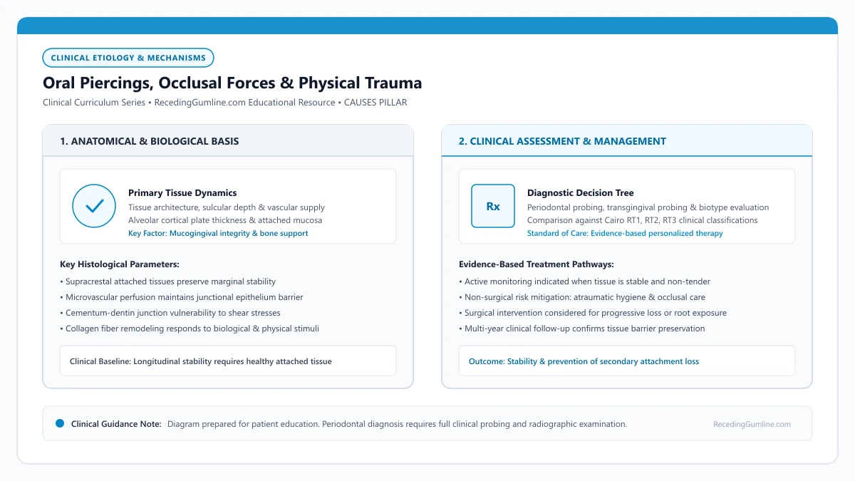
Educational illustration: Oral Piercings, Occlusal Forces & Physical Trauma. Clinical management requires comprehensive periodontal evaluation rather than isolated self-assessment.
Source: RecedingGumline.com Clinical Editorial Team (Proprietary educational diagram for RecedingGumline.com)
Oral Jewelry: Kinetic Impact & Friction Dynamics
Oral piercings represent a high-risk mechanical factor for rapid gingival attachment loss. Tongue barbells and lower lip labret studs are situated in close physical proximity to the thin lingual and labial gingival tissues of the mandibular anterior dentition. During normal physiological activities—such as speech, mastication, swallowing, and involuntary oral manipulation—the metallic or plastic jewelry repeatedly strikes and rubs against the adjacent gingival margin.
Cross-sectional clinical studies demonstrate that over 50% of individuals wearing lower lip labrets develop measurable buccal recession on mandibular central and lateral incisors within two years of insertion. The mechanism involves repetitive mechanical friction that strips away the thin sulcular epithelium, inducing localized inflammatory necrosis and progressive apical migration of the junctional epithelium.
The severity of piercing-induced recession correlates directly with barbell length, disc diameter, and duration of wear. Long tongue barbells allow the lower sphere to contact lingual mandibular gingiva, frequently producing severe lingual recession and deep vertical bone clefts that compromise tooth stability.
[object Object]
Key Etiological Insights
- Metallic oral jewelry inflicts repetitive mechanical friction on thin anterior gingival margins.
- Up to half of lip piercing wearers exhibit localized buccal recession within 24 months of placement.
- Lingual tongue jewelry can destroy lingual attached gingiva, creating challenging vertical bone defects.
Occlusal Traumatism & Non-Carious Cervical Lesions (Abfraction)
Occlusal trauma occurs when functional or parafunctional biting forces exceed the physiological adaptive capacity of the periodontal attachment apparatus. During nocturnal bruxism and chronic tooth clenching, lateral and non-axial forces subject the tooth crown to heavy bending moments. Because the tooth flexes slightly under lateral stress, tensile and compressive stresses concentrate at the fulcrum point—the cervical fulcrum near the cementoenamel junction (CEJ).
Biomechanical models propose that repetitive cervical flexure causes microscopic fatigue fractures in the crystalline enamel prisms and dentinal tubules, creating wedge-shaped Non-Carious Cervical Lesions (abfractions). While occlusal forces do not directly destroy the epithelial attachment in a clean environment, cervical tooth flexure disrupts the marginal seal, rendering the adjacent thin gingiva highly vulnerable to mechanical abrasion and localized recession.
Furthermore, secondary occlusal trauma on teeth with pre-existing attachment loss accelerates the rate of periodontal breakdown, widening the periodontal ligament space and exacerbating tooth mobility.
Key Etiological Insights
- Lateral biting forces induce tooth flexure, concentrating mechanical stresses at the cervical fulcrum.
- Cervical enamel and dentin microfractures (abfraction) disrupt the marginal gingival attachment interface.
- Severe bruxism acts as an accelerant of recession, particularly on teeth with pre-existing thin bone support.
Habitual Foreign Body Trauma & Factitious Injuries
Localized gingival recession is frequently traced to chronic, habitual foreign object trauma. Patients often engage in subconscious or factitious habits that focus mechanical pressure onto an isolated tooth margin. Common examples include chronic picking of the gingival margin with fingernails, aggressive use of wooden toothpicks or interdental wedges, chewing on pens or paperclips, and traumatic fingernail biting.
Fingernail picking (gingivitis artefacta) is especially prevalent in adolescents and individuals under psychological stress. The patient repeatedly digs a thumbnail or fingernail into the labial sulcus of a specific tooth, mechanically stripping the gingiva away from the root. This produces an isolated, V-shaped cleft (Stillman's cleft) or deep rectangular recession defect with hyperkeratotic, calloused border tissue.
Similarly, improper use of hard plastic interdental picks or forceful wedging of wooden sticks into interproximal spaces crushes the delicate interdental papilla, converting healthy col architecture into open "black triangles" and initiating interproximal recession.
Key Etiological Insights
- Subconscious habits like fingernail picking or pen biting inflict severe localized mechanical lacerations.
- Gingivitis artefacta typically manifests as an isolated, asymmetrical V-shaped gingival cleft.
- Aggressive toothpick wedging crushes interdental papillae, creating permanent black triangle defects.
Trauma Cessation, Stabilization & Surgical Recovery
The primary step in managing trauma-induced recession is the absolute cessation of the offending physical insult. Oral jewelry must be permanently removed; switching to acrylic or silicone studs reduces impact force but does not eliminate chronic friction against delicate margins. Behavioral counseling and awareness training are necessary to resolve factitious picking habits.
For occlusal trauma and bruxism, fabricating a custom hard acrylic occlusal guard (nightguard) is essential. The splint distributes biting forces evenly along the dental arch, eliminates non-axial lateral interferences, and stabilizes the cervical margins from ongoing flexural strain.
Once the mechanical cause is eliminated and the tissue is monitored for 2 to 3 months of inflammation-free healing, surgical intervention—such as a subepithelial connective tissue graft or coronally advanced flap—can be evaluated to restore lost keratinized tissue and achieve root coverage.
Key Etiological Insights
- Immediate, permanent removal of oral piercings is required to arrest ongoing soft-tissue destruction.
- Custom occlusal splints dissipate nocturnal bruxism forces, protecting cervical margins from flexure.
- Surgical soft-tissue grafting should be deferred until the physical traumatic etiology has been fully resolved.
Traumatic Oral Habits: Fingernail Picking, Foreign Object Friction & Factitious Injury
While bacterial plaque and toothbrush abrasion represent the most frequent causes of gum recession, a subset of patients experience severe, localized attachment loss driven by repetitive mechanical oral habits. Clinically termed factitious injuries or self-inflicted gingival trauma, habits such as picking the gums with fingernails, chewing on pens, or wedging toothpicks can gouge deep clefts into soft tissue.
• Factitious gingival injury involves mechanical tissue destruction caused by habitual physical trauma.
• Habitual fingernail picking is the most common self-inflicted mechanical etiology, often presenting as a deep V-shaped cleft.
• Habits involving pens, paperclips, Bobby pins, and toothpicks cause isolated, site-specific tissue tearing.
• Surgical grafting is strictly contraindicated until the traumatic behavioral habit is completely extinguished.
Clinical Recognition: The Anatomy of a Traumatic Cleft
Self-inflicted gingival trauma, medically classified as gingivitis artefacta, creates a very specific pattern of tissue loss. Unlike the smooth, parabolic recession caused by toothbrushing, factitious injuries often present as deep, narrow, slit-like clefts (Stillman clefts) or irregular, macerated tissue tears.
The defect is almost always isolated to an accessible tooth—most commonly maxillary or mandibular incisors and canines reachable by the dominant hand. The tissue margin may exhibit hyperkeratotic white calluses or persistent ulcerations from chronic fingernail scraping.
Chronic foreign object habits such as chewing on wooden toothpicks, biting pen caps, or digging fingernails into the gumline cause focal mechanical ulceration. Repetitive localized trauma tears the delicate junctional epithelium and destroys underlying periodontal ligament fibers.
Key Etiological Insights
- Presents as sharp, narrow, slit-like clefts rather than smooth recession curves
- Strictly confined to teeth easily reached by the patient's dominant hand
- Margins may exhibit whitish calloused thickening or active ulceration
Common Culprits: Fingernails, Pens, and Toothpicks
The most widespread causative habit is compulsive fingernail scratching or picking at the gingival margin. This habit frequently begins as an attempt to dislodge a perceived piece of trapped food, eventually evolving into an unconscious, repetitive anxiety-relief mechanism.
Other common foreign objects include chewing on plastic pen caps, biting paperclips, twisting hairpins (bobby pins) between teeth, and the aggressive, improper use of wooden toothpicks. Wedging wooden sticks repeatedly into interdental spaces shreds the fragile papilla, causing irreversible "black triangles."
These habitual micro-injuries often produce localized Stillman’s clefts or sharp asymmetrical recession notches confined to a single tooth. Patients are frequently unconscious of these nervous habits during periods of intense concentration or psychological stress.
Key Etiological Insights
- Fingernail picking often starts from food impaction and becomes a subconscious tick
- Chewing on pens and metal paperclips exerts heavy frictional trauma on tissue margins
- Aggressive toothpick wedging destroys the interdental papillae, creating black triangles
Management: Behavioral Extinction Must Precede Surgery
The cornerstone of managing factitious recession is non-judgmental patient awareness. Because the behavior is usually subconscious, confronting the patient gently and demonstrating how the defect matches their fingernail angle is often sufficient to help them stop.
Surgical root coverage must be postponed for at least 3 to 6 months after the habit is verified to be completely extinct. If a patient undergoes grafting while still unconsciously picking at their mouth, the newly placed donor tissue will be torn away, resulting in total surgical failure.
Behavioral modification therapy combined with custom protective occlusal appliances helps patients eliminate subconscious trauma patterns. Once traumatic habits are completely ceased, the localized defect can be evaluated for surgical root coverage or long-term conservative monitoring.
Key Etiological Insights
- Building gentle, non-judgmental patient awareness is the essential first step
- Wear thin cotton gloves or adhesive bandages on fingers at night if picking occurs during sleep
- Surgery must be delayed until 3 to 6 months of complete habit cessation is documented
Factitious Gingival Injuries & Mechanical Self-Trauma
Traumatic oral habits, clinically classified as factitious or self-inflicted gingival injuries (gingivitis artefacta), represent an underdiagnosed etiology of localized gum recession. Patients frequently engage in repetitive subconscious behaviors such as gouging the gingival margin with fingernails, toothpicks, paperclips, or pen caps.
These mechanical micro-traumas tear the attached gingiva and puncture the underlying periosteum. Over time, repeated localized lacerations destroy the supra-crestal fiber apparatus, culminating in bizarre, angular, or crescent-shaped recession defects that do not align with typical plaque or brushing patterns.
Accurate clinical diagnosis requires compassionate communication and careful forensic evaluation of defect shapes. Treating these defects requires habit cessation before any surgical reconstruction can be contemplated, as re-injury will destroy surgical results.
Key Etiological Insights
- Gingivitis artefacta involves repetitive self-inflicted mechanical injury from fingernails or tools.
- Repeated puncture wounds destroy the periosteum and supra-crestal fiber apparatus.
- Complete habit elimination is an absolute prerequisite prior to attempting surgical repair.
Interdisciplinary Behavioral Management & Tissue Stabilization
Addressing traumatic oral habits requires combining periodontal intervention with behavioral modification strategies. Clinicians work collaboratively with patients to identify emotional or situational triggers—such as high-stress periods or deep concentration—that provoke the damaging habit.
Temporary acrylic protective splints or clear vacuum-formed aligners can be fabricated to physically shield vulnerable marginal tissues from unconscious finger or object probing. This barrier approach allows traumatized tissues an opportunity to re-epithelialize without continuous disturbance.
Once habit cessation is verified over a consecutive six-month observation period, periodontists can re-evaluate the defect for soft-tissue grafting to restore aesthetic contours and protect exposed root dentin.
Key Etiological Insights
- Identifying situational triggers helps patients replace damaging motor habits.
- Custom protective oral splints physically barrier the gingiva from repetitive trauma.
- A minimum six-month habit-free verification period is required before surgical grafting.
Oral Piercings and Gingival Recession: Mechanical Trauma from Barbells and Studs
Oral piercings—including tongue barbells, labret studs, and lip rings—are well-documented etiological factors for severe, localized periodontal tissue destruction. The constant physical tapping, friction, and pressure of metallic jewelry against delicate gingival margins causes rapid recession and bone loss, often affecting young individuals with otherwise pristine oral health.
• Tongue piercings primarily damage the lingual gingiva of lower incisors, while lip piercings damage facial gingiva.
• Up to 50% of individuals wearing tongue barbells for more than two years develop measurable gingival recession.
• Repetitive mechanical impact strips away thin attached tissue and crushes underlying cortical bone.
• Replacing metal jewelry with flexible polymer/acrylic studs reduces, but does not eliminate, mechanical trauma.
Tongue Barbells: The Destructive Lingual Impact
The standard tongue piercing consists of a metallic barbell (usually titanium or surgical steel) secured with threaded balls at either end. During speech, swallowing, and chewing, the lower ball rests directly against or strikes the lingual (tongue-side) gum tissue and bone behind the mandibular central and lateral incisors.
The lingual cortical plate and overlying mucosa in this region are exceptionally delicate. The repetitive percussion of metal against bone causes pressure necrosis of the periosteum, leading to rapid, vertical lingual recession and deep bone loss that frequently exposes root surfaces within 6 to 12 months.
Metallic intraoral barbells and labret studs constantly strike against the lingual and facial surfaces of anterior mandibular incisors during speech and swallowing. This repetitive metal-on-tooth impact causes severe mechanical chipping of enamel and continuous gingival stripping.
Key Etiological Insights
- Lower barbell ball continuously impacts lingual tissue behind lower front teeth
- Causes rapid pressure necrosis of delicate lingual periosteum and bone
- Lingual recession is technically challenging to graft due to vascular limitations
Lip Rings and Labret Studs: Facial Tissue Rubbing
Piercings through the lower lip (labret piercings) feature a flat metallic backplate designed to sit flush against the inside of the lip. However, during normal facial movement, this metal disc rubs directly against the facial attached gingiva of the lower incisors and canines.
The friction of the disc continuously abrades the keratinized surface epithelium. Over time, this wear causes a distinctive circular or oval patch of recession matching the exact dimensions of the backplate. If left untreated, the recession extends through the mucogingival junction, exposing the root apex.
Studies indicate that over 50% of individuals with lower lip or tongue piercings demonstrate localized gingival recession within two years of piercing insertion. The damage is characteristically localized to the exact tooth site that contacts the metal jewelry stud.
Key Etiological Insights
- Flat labret backplates exert continuous friction on outer facial gums
- Creates circular or oval recession defects mirroring the jewelry contour
- Can cause complete loss of attached keratinized tissue down to the root tip
Harm Reduction: Material Substitution and Surgical Repair
The definitive clinical recommendation for stopping piercing-induced recession is complete and permanent removal of the jewelry. Once the mechanical trauma ceases, superficial mucosal inflammation will resolve within weeks, although receded gums and lost bone will not spontaneously regenerate.
For individuals unwilling to remove their piercing, harm-reduction strategies include downsizing to the shortest possible barbell stem to minimize movement and switching to flexible, medical-grade bioplastic (PTFE) or acrylic jewelry. If grafting is required to save affected teeth, jewelry must be removed permanently prior to surgery.
Removing oral jewelry or switching to flexible polytetrafluoroethylene (PTFE) or bioplastic studs dramatically reduces impact trauma. However, preexisting tissue recession will not spontaneously regenerate after jewelry removal and typically requires connective tissue grafting.
Key Etiological Insights
- Permanent jewelry removal is the definitive measure to arrest progressive tissue damage
- Downsizing stem length and using bioplastic/PTFE studs reduces impact force
- Soft-tissue grafting cannot be attempted until jewelry is permanently removed
Dynamic Biomechanics: Stud Friction & Lingual Flap Abrasion
Oral jewelry—particularly labret studs in the lower lip and barbells through the anterior tongue—inflicts severe dynamic mechanical trauma on the adjacent periodontal apparatus. During everyday speech, mastication, and habitual "playing" with the jewelry, metallic or plastic backings repeatedly strike and rub against the thin gingival margin.
This continuous percussion causes chronic mechanical irritation, triggering localized tissue ischemia and cellular apoptosis. The lingual and labial tissues of mandibular incisors are especially vulnerable due to their naturally thin cortical bone plate and minimal keratinized tissue band.
Longitudinal clinical studies show that over 50% of individuals with lip piercings for more than two years develop measurable gingival recession, often accompanied by localized pocketing and alarming alveolar bone loss extending to the root apex.
Key Etiological Insights
- Piercing backings subject anterior teeth to continuous repetitive percussive impact.
- Thin mandibular incisor bone plates are particularly susceptible to rapid bone loss.
- Over 50% of long-term piercing wearers develop significant clinical attachment loss.
Clinical Protocol: Removal, Stabilization & Surgical Reconstruction
When recession is detected in the vicinity of oral jewelry, the primary clinical imperative is immediate and permanent removal of the hardware. Substituting metal studs with flexible silicone or polytetrafluoroethylene (PTFE) alternatives may reduce impact forces but does not eliminate ongoing tissue friction.
Following piercing removal, a 12-week observational period is observed to allow inflammatory resolution and spontaneous soft-tissue rebound. In sites where keratinized tissue has been destroyed or deep V-shaped clefts have formed, surgical connective tissue grafting is indicated to rebuild the tissue barrier.
Patients must be informed that reinserting jewelry following successful grafting almost universally induces recurrence of the recession defect, as the newly regenerated tissue cannot withstand chronic metallic trauma.
Key Etiological Insights
- Permanent jewelry removal is the essential initial step; flexible studs do not prevent friction.
- A 12-week tissue recovery window allows baseline inflammatory resolution before surgery.
- Reinserting piercings after surgical grafting leads to rapid recurrence of recession defects.
Clinical Reality Check
Surgical root coverage grafting should never be attempted while traumatic oral jewelry or factitious picking habits persist. The newly grafted tissue has zero defense against mechanical friction, leading to immediate necrosis or recurrent recession.
Questions to Ask Your Dentist or Periodontist
- Has my lip or tongue piercing caused permanent bone loss beneath my receded gums?
- Do you see evidence of tooth grinding, clenching, or abfraction notches contributing to my gumline recession?
- Would a custom nightguard help protect my teeth from further cervical wear and recession?
- Is my receded tooth a good candidate for a connective tissue graft once the piercing is removed?
- How much natural healing can I expect after eliminating the mechanical source of trauma?
- Does the shape of my receded gum look like it was caused by physical picking or foreign object pressure?
- How can I tell if my tissue has started healing on its own once I stop touching the area?
- How long do I need to be completely habit-free before we can evaluate whether a gum graft is possible?
- Are there protective oral appliances (like a nightguard) that could prevent me from reaching this area?
- Has my tongue or lip piercing caused measurable gum recession or bone loss behind my teeth?
- If I switch to an acrylic or plastic barbell, will it prevent my recession from getting worse?
- How much bone support do my lower incisors currently have on their tongue-side surfaces?
- If I remove my piercing today, will my receded gums need a surgical graft to stay stable?
Unsure What Your Gum Changes Mean?
Take our free, evidence-based Gum Recession Assessment — approximately 3 minutes. Identify potential risk factors, evaluate symptoms, and receive personalized discussion questions for your dentist or periodontist.
Non-diagnostic educational triage. Private, secure, completed in your browser.
Related Educational Topics
Scientific Literature & Clinical Guidelines
6sources · Hide ▲
- Jepsen S, Caton JG, Albandar JM, Bissada NF, Bouchard P, Cortellini P, et al. (2018).
"Periodontal manifestations of systemic diseases and developmental and acquired conditions: Consensus report of workgroup 3 of the 2017 World Workshop on the Classification of Periodontal and Peri-Implant Diseases and Conditions." Journal of Periodontology.
Clinical relevance: Consensus report defining mucogingival conditions, gingival phenotype (replacing biotype), non-carious cervical lesions, and the multifactorial etiology of gingival recession; emphasizes that recession can occur without periodontitis and classifies recession by interdental clinical attachment loss.
- Cairo F, Nieri M, Cincinelli S, Mervelt J, Pagliaro U (2011).
"The interproximal clinical attachment level to classify gingival recessions and predict root coverage outcomes: an explorative and reliability study." Journal of Clinical Periodontology.
Clinical relevance: Exploratory and reliability study establishing the Cairo classification based on interdental clinical attachment level (CAL): RT1 (no interproximal attachment loss; complete root coverage is clinically predictable), RT2 (interproximal attachment loss <= buccal loss; partial coverage predictable), and RT3 (interproximal loss exceeds buccal recession; complete coverage not predictable).
- Tonetti MS, Greenwell H, Kornman KS (2018).
"Staging and grading of periodontitis: Framework and proposal of a new classification and case definition." Journal of Clinical Periodontology.
Clinical relevance: Consensus framework establishing the multidimensional staging (severity and extent of periodontal tissue breakdown) and grading (biological rate of disease progression, incorporating smoking and diabetes as grade modifiers) for periodontitis. It addresses periodontitis diagnosis and staging, not the classification of localized gingival recession defects.
- Grippo JO, Simring M, Coleman TA (2012).
"Abfraction, abrasion, biocorrosion, and the enigma of noncarious cervical lesions: a 20-year perspective." Journal of Esthetic and Restorative Dentistry.
Clinical relevance: Authoritative review defining and clarifying cervical hard tissue lesions: stress-induced abfraction, frictional abrasion from dentifrices/brushes, and chemical biocorrosion (acidic erosion), explaining how multi-factorial mechanisms accelerate cervical notching once gingival recession exposes root dentin.
- Kassab MM, Cohen RE (2003).
"The etiology and prevalence of gingival recession." The Journal of the American Dental Association.
Clinical relevance: Comprehensive epidemiological and etiological review establishing that gingival recession affects more than 50% of adults aged 18 to 64 and over 88% of individuals aged 65 and older, driven by interactions between predisposing anatomical factors and precipitating mechanical or inflammatory stimuli.
- Pihlstrom BL, Michalowicz BS, Johnson NW (2005).
"Periodontal diseases." The Lancet.
Clinical relevance: Peer-reviewed Lancet seminar review synthesizing global periodontal epidemiology, microbial etiology, host immunopathology, and systemic interactions, emphasizing prevention, biofilm disruption, and early risk factor modification.
Important Medical Notice
The contents of RecedingGumline.com, including text, graphics, self-assessment calculators, and other materials, are intended solely for educational and informational purposes. This content is not intended to replace professional dental examination, diagnosis, or treatment. Always seek the advice of a qualified dentist, periodontist, or other licensed oral healthcare provider with any questions you may have regarding a medical or dental condition. Never disregard professional medical advice or delay seeking it because of something you read on this website.