Visible Root Exposure & Cervical Notches in Gum Recession
Visible root exposure represents the definitive structural hallmark of receding gumline assessment. When the marginal gingiva retreats apically, the anatomical boundary of the tooth—the cementoenamel junction (CEJ)—becomes exposed to view. Patients frequently notice a distinct aesthetic transition from smooth, pearlescent crown enamel to dull, yellowish-tan root surfaces, often accompanied by sharp cervical ledges or wedge-shaped grooves known as Non-Carious Cervical Lesions (NCCLs).
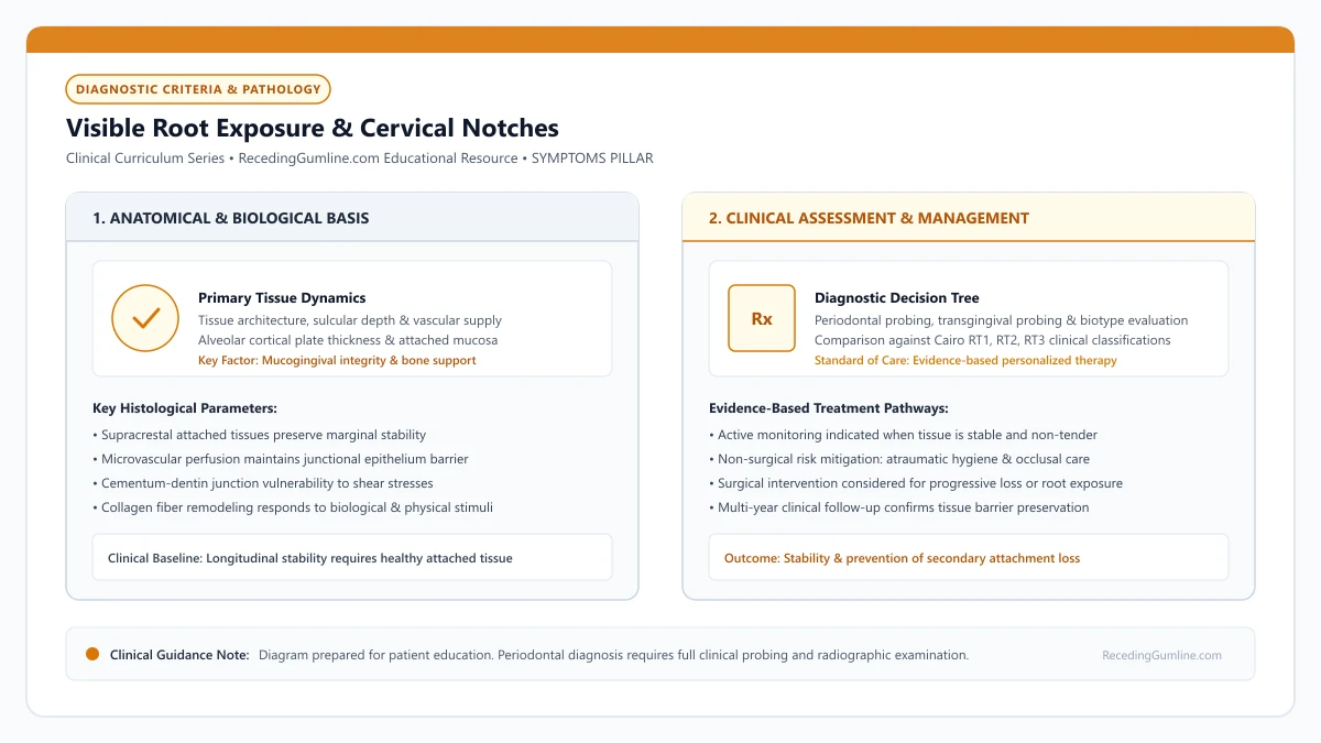
Educational illustration: Visible Root Exposure & Cervical Notches. Clinical management requires comprehensive periodontal evaluation rather than isolated self-assessment.
Source: RecedingGumline.com Clinical Editorial Team (Proprietary educational diagram for RecedingGumline.com)
Anatomical Landmarks: The Cementoenamel Junction (CEJ)
In a healthy periodontium, the free gingival margin rests approximately 1.0 to 2.0 mm coronal to the cementoenamel junction (CEJ), completely concealing the anatomical root within the gingival sulcus and alveolar housing. The CEJ represents the micro-anatomical interface where the crystalline enamel of the anatomical crown meets the cementum of the anatomical root.
Gingival recession is formally defined in clinical periodontology as the location of the marginal gingiva apical to the cementoenamel junction. Once the margin drops below this line, the root surface is permanently denuded.
Because the protective enamel cap terminates at the CEJ, the exposed root surface is covered only by an ultra-thin layer of acellular extrinsic fiber cementum, measuring a mere 20 to 50 micrometers in thickness. Once exposed to oral hygiene friction and dietary acids, this delicate cementum layer is quickly lost, exposing the softer, porous dentin beneath.
Key Clinical Insights
- The CEJ marks the anatomical boundary between enamel-covered crown and cementum-covered root.
- Recession is clinically defined as marginal gingival retreat positioned apical to the CEJ.
- Exposed root cementum is microscopically thin and wears away rapidly under daily functional friction.
Non-Carious Cervical Lesions (NCCLs): Abrasion, Abfraction & Erosion
Patients presenting with exposed roots frequently exhibit distinct notches, ledges, or deep wedge-shaped grooves at the gumline. Historically, these defects were attributed purely to aggressive toothbrushing. Modern dental science recognizes that Non-Carious Cervical Lesions (NCCLs) result from a complex, multi-factorial synergy involving three distinct mechanisms: abrasion, abfraction, and biocorrosion (erosion).
Abrasion represents the mechanical friction exerted by toothbrush bristles and abrasive dentifrices across the exposed root. Abfraction involves heavy occlusal forces—such as nocturnal bruxism and clenching—that cause the tooth crown to flex laterally under load, concentrating tensile stresses at the cervical fulcrum and inducing microfractures in the crystalline enamel and dentin.
Biocorrosion represents chemical dissolution from dietary acids (citrus, sodas, wine) or gastric acids (GERD, acid reflux). When these three factors operate concurrently, tissue destruction accelerates dramatically: acid softens the dentin matrix, occlusal flexure micro-fractures the mineral lattice, and toothbrush bristles sweep away the degraded tissue, gouging deep cervical clefts.
Key Clinical Insights
- Cervical notches (NCCLs) develop from a multi-factorial synergy of abrasion, abfraction, and erosion.
- Occlusal clenching forces flex the tooth at the cervical fulcrum, micro-fracturing dentin bonds.
- Dietary and gastric acids soften root dentin, accelerating mechanical wear from manual brushing.
Visual Aesthetics & Root Color Discrepancy
A primary complaint that prompts patients to seek professional evaluation is the noticeable change in smile aesthetics caused by exposed roots. Anatomical crown enamel is thick, highly translucent, and white, reflecting light with a characteristic pearlescent sheen.
In contrast, root surfaces are composed of dentin, which has a significantly higher organic content, lower mineral density, and a naturally warm, yellowish-tan or brownish hue. Furthermore, because exposed dentin is porous, it readily absorbs extrinsic pigments from coffee, tea, red wine, and dietary chromogens.
When recession occurs on anterior teeth or premolars within the aesthetic smile zone, the visible transition between white crown enamel and dark, yellowish root surfaces creates an asymmetrical, aged appearance that causes significant patient distress.
Key Clinical Insights
- Enamel is highly mineralized, white, and translucent; exposed root dentin is naturally yellowish-tan.
- Porous root dentin readily absorbs food and beverage pigments, causing noticeable dark staining.
- Anterior root exposure disrupts smile harmony and creates an asymmetrical, prematurely aged look.
Vulnerability to Root Caries & Structural Degradation
Beyond aesthetic concerns, exposed root surfaces introduce serious biological vulnerabilities. While enamel demineralization occurs at a critical pH of 5.5, root cementum and dentin begin dissolving at a much higher, less acidic threshold of pH 6.2 to 6.7.
This higher critical pH means that exposed roots can demineralize even in mildly acidic oral environments where enamel remains completely unaffected. When plaque biofilms accumulate on exposed root notches, cariogenic bacteria (such as Streptococcus mutans and Actinomyces species) produce organic acids that rapidly penetrate the dentinal tubules, producing soft, leathery root caries (cavities).
Root caries can spread rapidly circumferentially along the gumline, undermining coronal tooth structure and threatening pulp vitality if not diagnosed and treated promptly.
Key Clinical Insights
- Root dentin demineralizes at pH 6.2 to 6.7, making it far more vulnerable to cavities than enamel (pH 5.5).
- Bacterial biofilms on exposed roots can rapidly initiate soft, destructive root surface decay.
- Circumferential root decay undermines the anatomical crown and can rapidly compromise tooth vitality.
Clinical Treatment Decision Matrix: Graft vs. Restore vs. Monitor
Managing visible root exposure and cervical notches requires selecting the appropriate therapeutic modality based on defect anatomy. If a cervical notch is shallow (< 1 mm), asymptomatic, and accompanied by adequate attached gingiva, conservative monitoring with low-abrasivity fluoride toothpaste is indicated.
If the primary clinical issue is soft-tissue recession with intact interproximal bone (Cairo RT1), periodontal soft-tissue grafting (such as a subepithelial connective tissue graft or coronally advanced flap) represents the ideal biological treatment, restoring natural attached gingiva and covering the denuded root.
However, if the cervical notch is exceptionally deep (> 1.5 mm) with sharp undercut borders, attempting surgical grafting alone may result in flap collapse into the defect. In such cases, a combined restorative-periodontal approach is recommended: the deep portion of the notch is restored with a highly polished resin-modified glass ionomer (RMGI) or composite, and soft-tissue grafting is subsequently performed to cover the remaining apical margin.
Key Clinical Insights
- Shallow, asymptomatic root exposure with adequate attached tissue is safely managed via monitoring.
- Cairo RT1 soft-tissue defects without deep hard-tissue loss are best resolved via gum grafting.
- Deep structural notches require a combined restorative glass ionomer filling and surgical soft-tissue graft.
Cold Sensitivity from Gum Recession: Hydrodynamic Mechanism & Immediate Relief
Sharp, stabbing tooth pain when sipping iced water, eating ice cream, or breathing cold winter air is the most frequent symptom of gum recession that prompts patients to visit a dentist. When gingival margins pull back, delicate root surfaces are stripped of protective coverage, exposing thousands of microscopic fluid-filled channels directly to temperature changes.
• Cold sensitivity occurs because exposed root dentin lacks protective enamel, leaving dentinal tubules open.
• Cold stimuli cause rapid fluid movement within tubules, exciting A-delta nerve fibers in the dental pulp.
• Potassium nitrate toothpaste depolarizes pulpal nerves, requiring 2 to 4 weeks of consistent twice-daily use for relief.
• In-office varnishes and bonding agents provide immediate physical occlusion of open tubules.
Anatomy: Why Tooth Roots Have No Enamel Defense
The anatomical crown of a tooth is covered by enamel—the hardest substance in the human body, composed of 96% mineralized crystals with zero nerve endings. In contrast, tooth roots are covered only by an ultra-thin layer of cementum (less than 50 micrometers thick).
When gum recession exposes the root, daily toothbrushing or acidic saliva quickly wears away this micro-thin cementum layer. This denudes the underlying dentin, exposing up to 30,000 microscopic dentinal tubules per square millimeter directly to the oral cavity.
When the protective gingival margin recedes, the fragile cementum layer quickly wears away to expose underlying dentinal tubules. Temperature drops cause rapid hydrodynamic fluid contraction within these microscopic channels, stimulating pulp nerve endings and creating sharp, transient thermal pain.
Key Clinical Insights
- Enamel protects the crown; roots are protected only by delicate soft tissue and thin cementum
- Mechanical brushing or acid exposure strips away cementum, exposing dentinal tubules
- Each square millimeter of exposed dentin contains tens of thousands of open tubule channels
The Hydrodynamic Cascade: How Cold Triggers the Nerve
Each dentinal tubule is filled with dentinal fluid (an extracellular fluid communicating directly with the dental pulp). According to Brännström's Hydrodynamic Theory, when cold liquid or air touches an open tubule, the fluid contracts rapidly, creating sudden capillary suction.
This outward fluid displacement moves at several millimeters per second, mechanically distorting the cell bodies of odontoblasts and the unmyelinated endings of A-delta nerve fibers located at the pulp-dentin junction. The sensory nerve fires an immediate electrical impulse to the brain, perceived as sharp, piercing pain.
Daily exposure to ice water, cold weather inhalation, or chilled beverages repeatedly activates pulpal A-beta and A-delta sensory fibers. If untreated, persistent thermal irritation can induce low-grade reversible pulpitis, causing sensitivity episodes to linger longer over time.
Key Clinical Insights
- Cold causes rapid outward fluid contraction within open dentinal tubules
- Fluid movement mechanically deforms A-delta sensory nerve fibers at the pulpal border
- Triggers an immediate, sharp pain signal that dissipates once temperature normalizes
Actionable Relief: From Over-the-Counter Pastes to Professional Sealants
Relief strategies operate through two distinct mechanisms: nerve desensitization and tubule occlusion. Over-the-counter toothpastes containing potassium nitrate diffuse potassium ions down the tubules to depolarize nerve membranes, blocking pain signals. However, this requires 2 to 4 weeks of twice-daily use.
For immediate relief, professional dental treatments physically plug the open tubule mouths. Dentists apply 5% sodium fluoride varnishes, glutaraldehyde protein-coagulants (Gluma), or light-cured resin bonding agents that instantly seal the openings, preventing fluid movement entirely.
Clinical desensitization regimens target this fluid movement by utilizing stannous fluoride or potassium nitrate agents to depolarize nerve endings and precipitate microcrystalline barriers. In persistent cases, flowable composite resin bonding seals open tubule apertures permanently.
Key Clinical Insights
- Potassium nitrate toothpastes calm pulpal nerves but require 2 to 4 weeks to take effect
- Do not rinse with water immediately after brushing; let active desensitizing paste coat the roots
- In-office fluoride varnishes and resin bonding agents provide instant physical tubule seals
Hydrodynamic Fluid Movement & Intrapulpal A-Delta Nerve Excitation
Cold sensitivity associated with receding gums is explained by Brännström's hydrodynamic theory. When exposed root cementum is worn away by brushing or dissolved by acids, thousands of microscopic dentinal tubules—each communicating directly with the dental pulp—are left open to the oral environment.
Sudden exposure to cold liquids or chilled air causes the dentinal fluid within these microscopic tubules to rapidly contract. This physical volume reduction creates an immediate outward fluid flow toward the tooth surface at velocities exceeding several millimeters per second.
This rapid fluid displacement deforms the cell bodies of odontoblasts and stretches the unmyelinated terminals of intradental A-delta mechanoreceptor nerve fibers located in the subodontoblastic plexus of Raschkow, triggering an instantaneous, sharp neural pain impulse.
Key Clinical Insights
- Cold exposure causes tubular dentinal fluid to contract rapidly, inducing outward fluid flow.
- Rapid fluid movement stretches mechanoreceptor nerve terminals in the dental pulp.
- Intradental A-delta sensory fibers fire an instantaneous, sharp pain response.
Desensitizing Dentifrice Chemistry: Depolarization vs. Crystal Occlusion
Over-the-counter management of root sensitivity relies on two primary biochemical mechanisms: nerve depolarization and physical tubule occlusion. Toothpastes formulated with 5% potassium nitrate work via continuous neural depolarization.
Potassium ions diffuse down the dentinal tubules into the pulp chamber, increasing the extracellular potassium concentration around A-delta nerve endings. This prevents the normal depolarization-repolarization cycle, dampening nerve excitability. However, potassium nitrate requires 2 to 4 weeks of twice-daily continuous use before therapeutic intrapulpal concentrations accumulate.
Alternatively, occluding dentifrices containing stannous fluoride, calcium sodium phosphosilicate (NovaMin), or arginine and calcium carbonate physically precipitate insoluble mineral complexes inside the tubule orifices, sealing fluid movement within days.
Key Clinical Insights
- Potassium nitrate requires 2-4 weeks of daily use to accumulate and dampen nerve firing.
- Occluding formulations (stannous fluoride, NovaMin) form insoluble mineral plugs within tubules.
- Combining physical tubule occlusion with nerve desensitization provides optimal symptom relief.
Root Cavities and Receding Gums: Vulnerability of Exposed Cementum and Dentin
When gums recede, the biological protective barrier over the root surface is lost. While tooth enamel is exceptionally hard and acid-resistant, the exposed cementum and dentin of the root are soft, porous, and highly susceptible to dental decay. Root caries (cavities on receded roots) represent one of the most destructive and rapidly progressing complications of gum recession.
• Root dentin demineralizes at a critical pH of 6.2 to 6.7, dissolving twice as easily as tooth enamel (pH 5.5).
• Active root caries presents as a soft, sticky, leathery brown or yellowish lesion at or below the receded margin.
• Older adults and individuals with medication-induced dry mouth (xerostomia) face severe root cavity risks.
• High-potency 5,000 ppm sodium fluoride dentifrices and silver diamine fluoride (SDF) arrest early root decay.
Biochemical Vulnerability: Why Roots Melt at pH 6.5
Tooth enamel is a dense crystalline matrix composed of 96% inorganic hydroxyapatite. It remains intact until oral acidity drops to a pH of 5.5. Exposed root dentin, however, contains only 70% inorganic mineral, with the remaining 30% composed of organic collagen fibers and water.
Because of this lower mineral density, dentin begins dissolving at a much higher pH threshold of 6.2 to 6.7. Ordinary dietary carbohydrates, sweetened beverages, or mild plaque acids that would cause zero harm to enamel immediately begin demineralizing exposed root surfaces.
Root cementum and underlying radicular dentin have a significantly higher critical pH threshold (pH 6.2 to 6.7) for demineralization compared to dense enamel (pH 5.5). When recession exposes the root surface, normal dietary acids can trigger rapid biochemical demineralization.
Key Clinical Insights
- Dentin demineralizes at a critical pH of 6.2 to 6.7 (enamel dissolves at 5.5)
- Dentin contains 30% organic collagen, allowing rapid bacterial enzymatic destruction
- Mild dietary acids and plaque sugars that spare enamel actively dissolve roots
Clinical Diagnosis: Differentiating Active from Arrested Root Decay
Dentists diagnose root cavities using visual inspection and gentle tactile exploration. An active, progressing root cavity presents as a soft, mushy, or leathery depression, ranging in color from pale yellow to light brown. A dental explorer sticks in the softened collagen matrix.
In contrast, an arrested (healed) root lesion has successfully remineralized from saliva and fluoride. It appears dark brown or jet black, with a hard, shiny, glassy surface that resists the probe. Arrested lesions require no drilling; they are biological scars that should be left alone and monitored.
Root caries lesions often spread rapidly along the circumferential cementoenamel junction rather than penetrating deeply in a localized pit. Soft, yellow-brown necrotic dentin lesions can encircle the neck of the tooth, undermining the crown and jeopardizing structural integrity.
Key Clinical Insights
- Active decay: Soft, sticky, leathery, yellowish-brown surface requiring treatment
- Arrested decay: Hard, glassy, dark brown or black surface that is stable and safe
- Never drill into an arrested, hard root surface; aggressive drilling weakens the tooth
Treatment Strategies: Prescription Fluoride, SDF & Glass Ionomer
Preventing root cavities requires elevating oral fluoride concentrations. Dentists prescribe 1.1% sodium fluoride toothpaste (5,000 ppm, such as PreviDent), which hardens exposed dentin against acid attacks. Another non-invasive therapy is Silver Diamine Fluoride (SDF), an antimicrobial liquid applied chairside that instantly halts active root decay, though it stains the decay black.
When a root cavity has created a structural hole, the dentist must gently excavate the softened decay and place a restoration. Resin-Modified Glass Ionomer (RMGI) is the material of choice: it bonds chemically to root dentin in moist subgingival environments and continuously releases fluoride to prevent recurrent cavities.
Preventive protocols require high-concentration prescription 5,000 ppm sodium fluoride dentifrices and silver diamine fluoride (SDF) applications to arrest active decay. Restoring root caries requires moisture-tolerant glass ionomer cements that chemically bond to dentin and release fluoride.
Key Clinical Insights
- Prescription 5,000 ppm sodium fluoride toothpaste provides daily high-potency remineralization
- Silver Diamine Fluoride (SDF) arrests active root decay non-invasively without drilling
- Resin-modified glass ionomer (RMGI) is the premier restorative material for root cavities
Demineralization Dynamics: The Critical pH Threshold (pH 6.2 to 6.7)
When gingival recession exposes the tooth root, it bares cementum and dentin—tissues that are far more chemically vulnerable to bacterial decay than enamel. While dense crown enamel resists demineralization until oral pH drops below 5.5, root cementum and dentin demineralize at a critical pH of 6.2 to 6.7.
This narrow buffer means that even mildly acidic dietary exposures or minor bacterial plaque accumulation can initiate root demineralization. Furthermore, root dentin contains approximately 20% organic collagen matrix, which is degraded by bacterial proteolytic enzymes once the mineral crystals are dissolved.
Root caries progresses rapidly and circumferentially around the cervical collar, often invading subgingivally and undermining tooth structure beneath the remaining gumline.
Key Clinical Insights
- Root cementum and dentin demineralize at pH 6.2-6.7, compared to pH 5.5 for enamel.
- Mild dietary acidity or minor plaque easily triggers root surface demineralization.
- Bacterial proteolytic enzymes dissolve the organic collagen matrix, causing rapid circumferential decay.
Chemotherapeutic Prevention: 5,000 ppm Dentifrice & Silver Diamine Fluoride
Preventing and arresting root decay on exposed roots demands aggressive chemotherapeutic intervention beyond standard over-the-counter fluoride toothpaste. The clinical standard of care prescription is 1.1% sodium fluoride dentifrice (5,000 ppm fluoride).
Brushing nightly with 5,000 ppm fluoride creates a dense reservoir of calcium fluoride on the root surface, driving continuous remineralization and forming fluorapatite, which lowers the critical dissolution pH to 4.5.
For active, non-cavitated or cavitated root lesions, 38% Silver Diamine Fluoride (SDF) provides immediate antimicrobial and remineralizing efficacy. Silver ions penetrate dentinal tubules, killing cariogenic bacteria and inhibiting matrix metalloproteinases, while fluoride arrests lesion progression, transforming soft decay into a hardened, glass-like surface.
Key Clinical Insights
- High-concentration 5,000 ppm fluoride dentifrice significantly reduces root caries incidence.
- Fluorapatite formation lowers the critical demineralization pH of root dentin to 4.5.
- Silver Diamine Fluoride (SDF) instantly kills bacteria and hardens active root decay.
Clinical Reality Check
Placing a large composite filling over an exposed root without addressing concurrent lack of attached gingiva often fails long-term. Subgingival restoration margins attract plaque biofilms, triggering chronic marginal inflammation that drives further apical recession.
Questions to Ask Your Dentist or Periodontist
- Is my cervical notch caused primarily by toothbrush abrasion, teeth clenching, or acid erosion?
- Would this receded tooth be better treated with a gum graft or a tooth-colored filling?
- Are my exposed root surfaces currently showing any signs of early root cavities or demineralization?
- What prescription high-fluoride toothpaste or remineralizing paste should I use to protect these roots?
- Can a combined procedure—filling the deep notch and placing a gum graft—restore both tooth and gum?
- Does my cold sensitivity indicate simple exposed dentin or early nerve inflammation (pulpitis)?
- Can you apply an in-office desensitizing varnish or resin seal to these sensitive roots today?
- Which specific active ingredient in toothpaste (potassium nitrate, stannous fluoride, or arginine) will work best for me?
- Would covering this receded root with a gum graft permanently eliminate my cold sensitivity?
- Do my exposed root surfaces have active root cavities or are they hard and arrested?
- Would a prescription 5,000 ppm fluoride toothpaste help protect my receded roots from decay?
- Am I a candidate for Silver Diamine Fluoride (SDF) to stop decay without drilling?
- If my root needs a filling, will you use a fluoride-releasing glass ionomer material?
Unsure What Your Gum Changes Mean?
Take our free, evidence-based Gum Recession Assessment — approximately 3 minutes. Identify potential risk factors, evaluate symptoms, and receive personalized discussion questions for your dentist or periodontist.
Non-diagnostic educational triage. Private, secure, completed in your browser.
Related Educational Topics
Scientific Literature & Clinical Guidelines
8sources · Hide ▲
- Grippo JO, Simring M, Coleman TA (2012).
"Abfraction, abrasion, biocorrosion, and the enigma of noncarious cervical lesions: a 20-year perspective." Journal of Esthetic and Restorative Dentistry.
Clinical relevance: Authoritative review defining and clarifying cervical hard tissue lesions: stress-induced abfraction, frictional abrasion from dentifrices/brushes, and chemical biocorrosion (acidic erosion), explaining how multi-factorial mechanisms accelerate cervical notching once gingival recession exposes root dentin.
- Cairo F, Nieri M, Cincinelli S, Mervelt J, Pagliaro U (2011).
"The interproximal clinical attachment level to classify gingival recessions and predict root coverage outcomes: an explorative and reliability study." Journal of Clinical Periodontology.
Clinical relevance: Exploratory and reliability study establishing the Cairo classification based on interdental clinical attachment level (CAL): RT1 (no interproximal attachment loss; complete root coverage is clinically predictable), RT2 (interproximal attachment loss <= buccal loss; partial coverage predictable), and RT3 (interproximal loss exceeds buccal recession; complete coverage not predictable).
- Jepsen S, Caton JG, Albandar JM, Bissada NF, Bouchard P, Cortellini P, et al. (2018).
"Periodontal manifestations of systemic diseases and developmental and acquired conditions: Consensus report of workgroup 3 of the 2017 World Workshop on the Classification of Periodontal and Peri-Implant Diseases and Conditions." Journal of Periodontology.
Clinical relevance: Consensus report defining mucogingival conditions, gingival phenotype (replacing biotype), non-carious cervical lesions, and the multifactorial etiology of gingival recession; emphasizes that recession can occur without periodontitis and classifies recession by interdental clinical attachment loss.
- Tonetti MS, Greenwell H, Kornman KS (2018).
"Staging and grading of periodontitis: Framework and proposal of a new classification and case definition." Journal of Clinical Periodontology.
Clinical relevance: Consensus framework establishing the multidimensional staging (severity and extent of periodontal tissue breakdown) and grading (biological rate of disease progression, incorporating smoking and diabetes as grade modifiers) for periodontitis. It addresses periodontitis diagnosis and staging, not the classification of localized gingival recession defects.
- Brännström M (1966).
"Sensitivity of dentine." Oral Surgery, Oral Medicine, Oral Pathology.
Clinical relevance: Foundational paper formulating the hydrodynamic theory of dentin hypersensitivity: rapid fluid displacement within patent dentinal tubules physically deforms intradental nerve endings at the pulp-dentin boundary, explaining thermal, mechanical, and evaporative root sensitivity.
- West NX, Seong J, Davies M (2015).
"Management of dentine hypersensitivity: efficacy of professionally and self-administered agents." Journal of Clinical Periodontology.
Clinical relevance: Systematic review evaluating professionally and self-administered desensitizing agents; found evidence supporting tubule-occluding dentifrices and potassium-based nerve desensitizers in providing transient to moderate symptom relief, with substantial heterogeneity across clinical trials.
- Pihlstrom BL, Michalowicz BS, Johnson NW (2005).
"Periodontal diseases." The Lancet.
Clinical relevance: Peer-reviewed Lancet seminar review synthesizing global periodontal epidemiology, microbial etiology, host immunopathology, and systemic interactions, emphasizing prevention, biofilm disruption, and early risk factor modification.
- Chapple ILC, Mealey BL, Van Dyke TE, Bartold PM, Dommisch H, Eickholz P, et al. (2018).
"Periodontal health and gingival diseases and conditions on an intact and a reduced periodontium: Consensus report of workgroup 1 of the 2017 World Workshop on the Classification of Periodontal and Peri-Implant Diseases and Conditions." Journal of Clinical Periodontology.
Clinical relevance: Consensus report establishing diagnostic criteria for periodontal health and gingivitis across intact and reduced periodontia, defining clinical gingival health as <10% bleeding on probing without attachment loss and strictly differentiating gingivitis from periodontitis.
Important Medical Notice
The contents of RecedingGumline.com, including text, graphics, self-assessment calculators, and other materials, are intended solely for educational and informational purposes. This content is not intended to replace professional dental examination, diagnosis, or treatment. Always seek the advice of a qualified dentist, periodontist, or other licensed oral healthcare provider with any questions you may have regarding a medical or dental condition. Never disregard professional medical advice or delay seeking it because of something you read on this website.