Supracrestal Attached Tissues & Biologic Width in Gum Recession
What is the biologic width (supracrestal attached tissue), and why is it so critical in dentistry?
In restorative and surgical dentistry, no anatomical rule is more revered than the dimension of the supracrestal attached tissues, historically and widely known as the biologic width. Measured at a microscopic average of 2.04 millimeters, this physiological zone represents the minimum vertical soft-tissue space that nature requires coronal to the alveolar bone crest to protect internal tissues from bacterial invasion and receding gumline assessment.
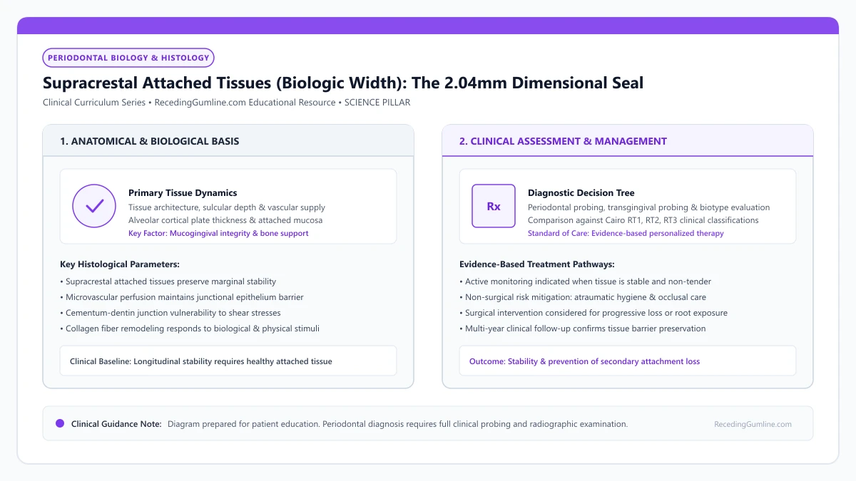
Educational illustration: Supracrestal Attached Tissues (Biologic Width): The 2.04mm Dimensional Seal. Clinical management requires comprehensive periodontal evaluation rather than isolated self-assessment.
Source: RecedingGumline.com Clinical Editorial Team (Proprietary educational diagram for RecedingGumline.com)
Key Clinical Distinctions & Diagnostic Boundaries
- Biologic width does not include the sulcus depth; the total dentogingival unit (sulcus + JE + connective tissue) averages approximately 3.0 mm.
- The connective tissue attachment (1.07 mm) is the most physiologically constant dimension; the junctional epithelium is more variable.
- Placing restorative margins supragingivally (above the gumline) completely eliminates the risk of biologic width violation.
- Thin phenotypes respond to biologic width invasion with rapid recession; thick phenotypes respond with persistent, painful, purplish inflammation.
The Blueprint: Gargiulo's 1961 Histological Measurements
In 1961, Dr. Anthony Gargiulo, Dr. Frank Wentz, and Dr. Balint Orban examined 287 teeth from 30 human autopsy specimens ranging from 19 to 50 years of age. They conducted meticulous microscopic measurements of the tissue layers coronal to the alveolar bone crest.
Their research established that the soft-tissue attachment consists of three specific vertical zones: an average sulcus depth of 0.69 mm, an epithelial attachment (junctional epithelium) averaging 0.97 mm, and a supracrestal connective tissue attachment averaging 1.07 mm. Combining the epithelial and connective tissue attachments yielded the famous 2.04 mm biologic width.
The biologic width, officially redesignated as the supracrestal tissue attachment by the AAP/EFP, consists of the junctional epithelium and the supracrestal connective tissue attachment. In healthy human periodontium, this biological complex measures approximately two millimeters in vertical height coronal to the alveolar bone crest.
Clinical Considerations:
- Junctional epithelium averages 0.97 mm in vertical height
- Connective tissue attachment averages 1.07 mm and is the most dimensionally stable layer
- Total supracrestal attached tissue dimension equals an average of 2.04 mm
The Connective Tissue Seal: Sharpey's Fiber Groups
The 1.07 mm connective tissue attachment is the biological anchor of the complex. Unlike the junctional epithelium (which merely sticks via cellular glue), this zone consists of dense Type I collagen fiber bundles that physically insert into root cementum.
These fibers are organized into distinct architectural groups: dentogingival fibers (radiating upward into the free gingiva), dentoperiosteal fibers (running over the alveolar crest), circular fibers (encircling the tooth like a belt), and transseptal fibers (connecting adjacent teeth over the bone crest). This dense collagen seal physically blocks bacteria from entering the bone marrow.
When dental restorations, crowns, or orthodontics encroach within this two-millimeter zone, the body perceives the foreign restorative material as a biological threat. In response, chronic inflammation develops, triggering osteoclastic bone resorption as the body attempts to recreate its physiological biological width.
Clinical Considerations:
- Dense collagen fiber bundles insert directly into root cementum above the bone crest
- Dentogingival, circular, and transseptal fiber groups form an impermeable biological gasket
- Physically separates the microbial oral environment from the sterile alveolar bone marrow
Consequences of Violation: Unpredictable Bone Resorption and Recession
When a restorative dental crown margin or veneer is placed within 2.0 mm of the alveolar crest, the body perceives the artificial restorative material as a foreign body penetrating its sterile internal connective tissue.
Because soft tissue cannot form hemidesmosomes or anchor collagen into artificial porcelain or metal, chronic inflammation ensues. Osteoclasts resorb the alveolar bone crest until the mandatory 2.04 mm distance is re-established. In thin phenotypes, this bone resorption causes the overlying gum tissue to recede apically, exposing the dark edge of the restoration.
Surgical crown lengthening or orthodontic tooth extrusion is routinely performed to re-establish adequate supracrestal space before final restorative margins are placed. Respecting this fundamental biological dimension is the most critical principle in preventing iatrogenic gingival recession.
Clinical Considerations:
- Encroaching within 2 mm of bone crest triggers foreign body inflammatory reactions
- Osteoclasts resorb the alveolar crest to re-establish nature's required 2.04 mm buffer
- Causes progressive gum recession in thin phenotypes and chronic bleeding in thick phenotypes
Gargiulo's Landmark Dimensions: The 2.04 mm Biological Constant
In 1961, Gargiulo, Wentz, and Orban published the landmark histological study establishing the dimensions of the dentogingival junction in human cadavers. They established that healthy supra-alveolar soft tissue comprises three distinct vertical components.
On average, the histological sulcular depth measures 0.69 mm; the junctional epithelial attachment measures 0.97 mm; and the supra-alveolar connective tissue attachment measures 1.07 mm. The sum of the junctional epithelium and connective tissue attachment (averaging 2.04 mm) was coined "biologic width."
In the 2017 World Workshop, this anatomical complex was formally renamed "supracrestal attached tissues" to reflect that these dimensions are biological variables that differ between individuals and tooth types.
Clinical Considerations:
- Gargiulo et al. (1961) established the classic 2.04 mm biologic width dimension in humans.
- Components comprise ~0.97 mm junctional epithelium and ~1.07 mm supra-alveolar connective tissue.
- The 2017 World Workshop renamed this biological constant "supracrestal attached tissues."
Restorative Infringement & Surgical Crown Lengthening Principles
Violating the supracrestal attached tissues by placing dental crown, veneer, or filling margins closer than 2.0 mm to the alveolar bone crest provokes severe biological consequences. Host tissues cannot tolerate foreign materials within the biological seal, triggering chronic inflammation, persistent bleeding, and unpredictable bone resorption.
In thin phenotypes, the bone resorbs rapidly to re-establish biological space for the soft-tissue attachment, leading to immediate marginal gum recession. In thick phenotypes, deep chronic periodontal pockets develop.
When restorative margins must be placed subgingivally, clinicians perform surgical crown lengthening. Removing crestal bone via ostectomy ensures that at least 3.0 mm of sound tooth structure exists between the future crown margin and the alveolar bone crest.
Clinical Considerations:
- Placing restoration margins within 2 mm of bone crest induces chronic inflammation and bone loss.
- Thin phenotypes respond to margin infringement with bone resorption and soft-tissue recession.
- Surgical crown lengthening ensures a mandatory 3 mm biological clearance from margin to bone crest.
Clinical Reality Check
If your gum has receded around a dental crown and the tissue is dark red, swollen, and bleeds every time you brush, the crown margin was almost certainly placed too close to your jawbone.
Questions to Ask Your Periodontist or Dentist
- Is my crown margin invading my supracrestal attached tissue (biologic width)?
- What is the measured distance between my crown margin and my alveolar bone crest on x-rays?
- Would crown lengthening surgery be needed to move the bone crest down before remaking the crown?
- Can my replacement crown be designed with a margin at or slightly above the gumline to protect the tissue?
Unsure What Your Gum Changes Mean?
Take our free, evidence-based Gum Recession Assessment — approximately 3 minutes. Identify potential risk factors, evaluate symptoms, and receive personalized discussion questions for your dentist or periodontist.
Non-diagnostic educational triage. Private, secure, completed in your browser.
Related Educational Topics
Clinical Evidence & Claim Traceability (2 Mapped Assertions)
Scientific Literature & Clinical Guidelines
3sources · Hide ▲
- Gargiulo AW, Wentz FM, Orban B (1961).
"Dimensions and relations of the dentogingival junction in humans." Journal of Periodontology.Peer-Reviewed Study doi:10.1902/jop.1961.32.3.261
Clinical relevance: Foundational morphometric human autopsy investigation defining average histological dimensions of the dentogingival junction: sulcus depth of 0.69 mm, junctional epithelium of 0.97 mm, and supracrestal connective tissue attachment of 1.07 mm, establishing the biological concept of supracrestal tissue attachment.
- Jepsen S, Caton JG, Albandar JM, Bissada NF, Bouchard P, Cortellini P, et al. (2018).
"Periodontal manifestations of systemic diseases and developmental and acquired conditions: Consensus report of workgroup 3 of the 2017 World Workshop on the Classification of Periodontal and Peri-Implant Diseases and Conditions." Journal of Periodontology.
Clinical relevance: Consensus report defining mucogingival conditions, gingival phenotype (replacing biotype), non-carious cervical lesions, and the multifactorial etiology of gingival recession; emphasizes that recession can occur without periodontitis and classifies recession by interdental clinical attachment loss.
- Pihlstrom BL, Michalowicz BS, Johnson NW (2005).
"Periodontal diseases." The Lancet.
Clinical relevance: Peer-reviewed Lancet seminar review synthesizing global periodontal epidemiology, microbial etiology, host immunopathology, and systemic interactions, emphasizing prevention, biofilm disruption, and early risk factor modification.
Important Medical Notice
The contents of RecedingGumline.com, including text, graphics, self-assessment calculators, and other materials, are intended solely for educational and informational purposes. This content is not intended to replace professional dental examination, diagnosis, or treatment. Always seek the advice of a qualified dentist, periodontist, or other licensed oral healthcare provider with any questions you may have regarding a medical or dental condition. Never disregard professional medical advice or delay seeking it because of something you read on this website.