Dental Restorations & Biologic Width Violations in Gum Recession
Why do gums recede around dental crowns and veneers, and what is biologic width violation?
Dental restorations—such as crowns, veneers, and composite fillings—are designed to restore tooth function and aesthetics. However, when a restoration margin is placed too deeply beneath the gumline, it can invade a delicate anatomical boundary known as the biologic width (supracrestal attached tissues). This iatrogenic violation triggers chronic inflammation and bone resorption, culminating in localized receding gums.
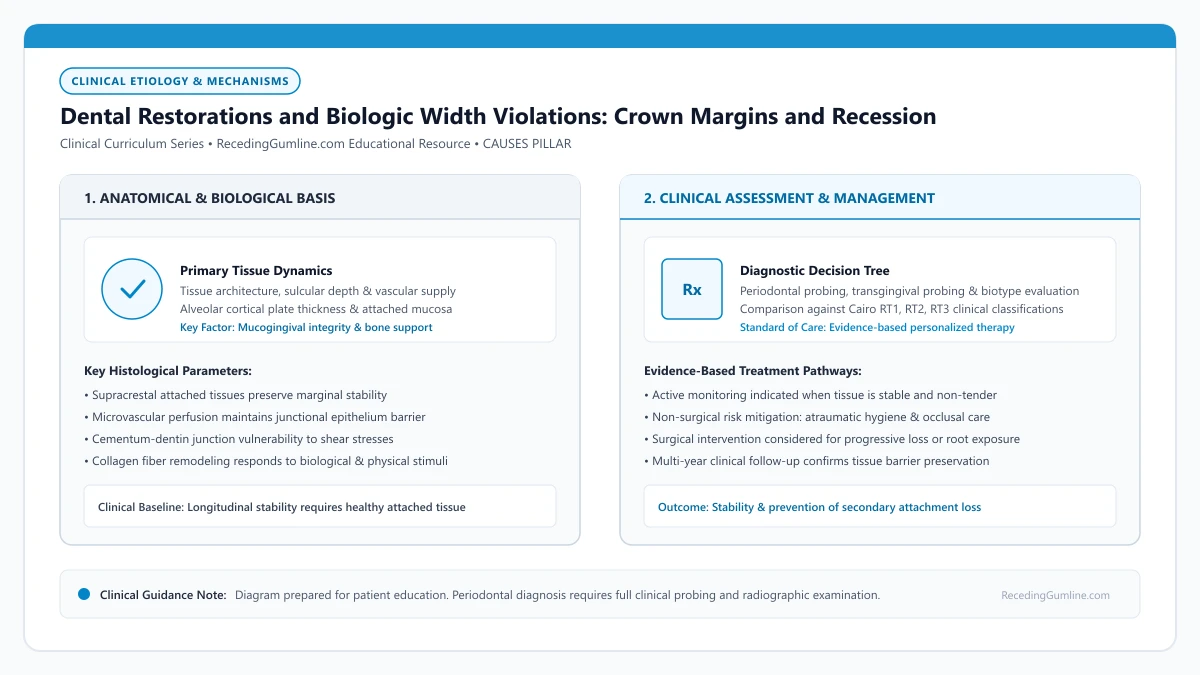
Educational illustration: Dental Restorations and Biologic Width Violations: Crown Margins and Recession. Clinical management requires comprehensive periodontal evaluation rather than isolated self-assessment.
Source: RecedingGumline.com Clinical Editorial Team (Proprietary educational diagram for RecedingGumline.com)
Key Clinical Distinctions & Diagnostic Boundaries
- Subgingival margins placed above the junctional epithelium can remain healthy if well-polished; margins invading the connective tissue attachment will always trigger bone resorption.
- Recession around a crown is not necessarily caused by patient brushing; it is frequently an iatrogenic (dentist-induced) anatomical violation.
- A purplish, puffy, continuously bleeding margin around a crown indicates active biologic width impingement.
- Correcting a biologic width violation often requires crown lengthening surgery to re-contour the bone crest before replacing the crown.
The 2.04 mm Law: Gargiulo's Biologic Width Dimensions
In 1961, Dr. Anthony Gargiulo and colleagues published landmark research measuring the dentogingival junction in human cadavers. They discovered that nature strictly reserves an average of 2.04 millimeters of root space between the base of the sulcus and the crest of the alveolar bone.
This dimension consists of approximately 0.97 mm of junctional epithelium and 1.07 mm of supracrestal connective tissue fibers anchored into root cementum. In the 2017 World Workshop, this space was officially renamed the supracrestal attached tissues. It serves as an impermeable biological gasket sealing the bloodstream from oral bacteria.
Crowns, veneers, and composite fillings that extend subgingivally into the supracrestal attached tissues violate the patient’s biological width. The body’s immune system recognizes this foreign restorative encroachment as an irritant, initiating chronic marginal inflammation.
Clinical Considerations:
- Supracrestal attached tissue requires ~2.04 mm of space above the bone crest
- Consists of ~1 mm junctional epithelium and ~1 mm connective tissue attachment
- Functions as a biological gasket sealing bone from oral microbial pathogens
The Invasion Cascade: Why Gums Recede Around Crowns
When a dentist cuts a crown preparation too deep beneath the gumline to hide the metal margin, the artificial material invades this 2.04 mm space. The human body recognizes the restoration margin as a foreign object wedged into its internal connective tissue.
Because soft tissue cannot anchor to artificial ceramic or metal in the presence of subgingival microleakage, the immune system initiates localized osteoclastic bone resorption. The bone crest melts backward until it re-establishes its required 2.04 mm buffer zone from the crown edge. As the bone recedes, the gumline inevitably follows, exposing the unsightly crown margin.
Overhanging restoration margins and rough restorative seams harbor dense colonies of anaerobic bacteria that cannot be cleaned with standard floss or toothbrushes. Persistent bacterial colonization and cement overhangs drive rapid osteoclastic crestal bone resorption and secondary gum recession.
Clinical Considerations:
- Crown margins placed too close to bone trigger foreign body inflammatory reactions
- Osteoclasts resorb the bone crest to restore the required 2 mm biological distance
- Soft-tissue margin collapses apically, exposing the dark edge of the dental crown
Overhanging Restorations and Crown Lengthening Surgery
A related iatrogenic trigger is an overhanging restoration—where a filling or crown edge juts out beyond the natural contour of the root. Overhangs create microscopic ledges that trap food and anaerobic bacteria where dental floss shreds and cannot reach, inducing rapid localized periodontitis.
Correcting a biologic width violation requires an interdisciplinary approach. The offending crown must be removed and a procedure called crown lengthening surgery is performed. The periodontist gently removes 1 to 2 mm of bone crest around the tooth, recreating adequate biological space so a new crown can be placed with perfectly healthy margins.
Correcting restorative-induced recession requires surgically re-establishing the biological width through crown lengthening or replacing the defective restoration with supragingival margins. Once harmonious anatomical contours are restored, the surrounding periodontal tissues can achieve long-term inflammatory stability.
Clinical Considerations:
- Overhanging margins create permanent plaque traps that shred dental floss
- Crown lengthening surgery removes a small collar of bone to re-establish biologic width
- Allows a new, properly fitted restoration to be placed without triggering recession
Violation of Supracrestal Attached Tissues (Biologic Width)
When crown, veneer, or filling margins are placed too deeply beneath the gingival margin, they frequently violate the zone historically termed "biologic width"—now designated as the supracrestal attached tissues. This anatomical zone comprises approximately 1.0 mm of junctional epithelium and 1.0 mm of supracrestal connective tissue attachment above the alveolar crest.
Restoration margins extending closer than 2.0 millimeters to the alveolar bone create chronic mechanical and microbiological irritation. Host immune cells perceive the restorative material as a foreign body, mounting a persistent inflammatory response that cannot resolve as long as the subgingival overhang or contour exists.
To re-establish biological space for connective tissue attachment, the underlying alveolar bone resorbs apically. As the bone recedes, the overlying gingival margin collapses and recedes alongside it, exposing root surfaces and margin lines.
Clinical Considerations:
- Restoration margins must respect the 2.0 mm supracrestal attached tissue zone above the bone crest.
- Violating this zone induces chronic foreign-body inflammation that does not respond to brushing.
- Alveolar bone undergoes osteoclastic resorption to escape the margin, causing immediate tissue recession.
Clinical Correction: Margin Revision & Crown Lengthening
Resolving restoration-induced gum recession requires correcting the offending restorative margin before attempting any soft-tissue grafting. Simply placing a gum graft over a biologically invasive crown margin guarantees complete graft failure.
Clinicians begin by removing the ill-fitting restoration and placing a meticulously contoured, highly polished provisional restoration with supragingival margins. In cases where the tooth margin cannot be elevated, surgical crown lengthening (ostectomy and osteoplasty) is performed to reposition the alveolar crest 3.0 millimeters apical to the future restoration margin.
Following a 3- to 6-month healing period to allow soft-tissue maturation and biological attachment stabilization, periodontists can perform connective tissue grafting if root coverage is desired, followed by final crown fabrication.
Clinical Considerations:
- Defective restorations must be removed and replaced with provisional restorations before grafting.
- Surgical crown lengthening repositions bone 3.0 mm apical to the planned restorative margin.
- A 3- to 6-month tissue stabilization period ensures predictable final aesthetic and functional outcomes.
Clinical Reality Check
If your gum is red, swollen, and bleeding around a single crown despite flossing, it is likely a biologic width violation caused by crown placement rather than poor home care on your part.
Questions to Ask Your Periodontist or Dentist
- Does my dental x-ray show that my crown margin is invading my biologic width or sitting too close to the bone?
- Is there an overhang on my restoration that is trapping plaque and causing this localized gum recession?
- Would crown lengthening surgery be required to fix the bone spacing before remaking this crown?
- Can we replace this crown with a supragingival margin (placed at or above the gumline) to protect the tissue?
Unsure What Your Gum Changes Mean?
Take our free, evidence-based Gum Recession Assessment — approximately 3 minutes. Identify potential risk factors, evaluate symptoms, and receive personalized discussion questions for your dentist or periodontist.
Non-diagnostic educational triage. Private, secure, completed in your browser.
Related Educational Topics
Clinical Evidence & Claim Traceability (2 Mapped Assertions)
Scientific Literature & Clinical Guidelines
3sources · Hide ▲
- Gargiulo AW, Wentz FM, Orban B (1961).
"Dimensions and relations of the dentogingival junction in humans." Journal of Periodontology.Peer-Reviewed Study doi:10.1902/jop.1961.32.3.261
Clinical relevance: Foundational morphometric human autopsy investigation defining average histological dimensions of the dentogingival junction: sulcus depth of 0.69 mm, junctional epithelium of 0.97 mm, and supracrestal connective tissue attachment of 1.07 mm, establishing the biological concept of supracrestal tissue attachment.
- Jepsen S, Caton JG, Albandar JM, Bissada NF, Bouchard P, Cortellini P, et al. (2018).
"Periodontal manifestations of systemic diseases and developmental and acquired conditions: Consensus report of workgroup 3 of the 2017 World Workshop on the Classification of Periodontal and Peri-Implant Diseases and Conditions." Journal of Periodontology.
Clinical relevance: Consensus report defining mucogingival conditions, gingival phenotype (replacing biotype), non-carious cervical lesions, and the multifactorial etiology of gingival recession; emphasizes that recession can occur without periodontitis and classifies recession by interdental clinical attachment loss.
- Pihlstrom BL, Michalowicz BS, Johnson NW (2005).
"Periodontal diseases." The Lancet.
Clinical relevance: Peer-reviewed Lancet seminar review synthesizing global periodontal epidemiology, microbial etiology, host immunopathology, and systemic interactions, emphasizing prevention, biofilm disruption, and early risk factor modification.
Important Medical Notice
The contents of RecedingGumline.com, including text, graphics, self-assessment calculators, and other materials, are intended solely for educational and informational purposes. This content is not intended to replace professional dental examination, diagnosis, or treatment. Always seek the advice of a qualified dentist, periodontist, or other licensed oral healthcare provider with any questions you may have regarding a medical or dental condition. Never disregard professional medical advice or delay seeking it because of something you read on this website.