Gingival Margin Microanatomy in Gum Recession: Free Gingiva, Sulcus & Junctional Epithelium
What is the microscopic cellular anatomy of the gumline, and how does it attach to the tooth?
In any comprehensive understanding gum recession overview, the gingival margin represents the biological interface between the external microbial environment of the mouth and the sterile internal connective tissues of the human body. Understanding its microscopic architecture—consisting of the keratinized free gingival margin, the shallow gingival sulcus, and the specialized junctional epithelium—explains how health is maintained and how early recession initiates.
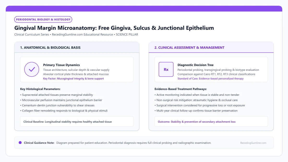
Educational illustration: Gingival Margin Microanatomy: Free Gingiva, Sulcus & Junctional Epithelium. Clinical management requires comprehensive periodontal evaluation rather than isolated self-assessment.
Source: RecedingGumline.com Clinical Editorial Team (Proprietary educational diagram for RecedingGumline.com)
Key Clinical Distinctions & Diagnostic Boundaries
- The outer oral epithelium is heavily keratinized to resist mechanical chew friction; the inner junctional epithelium is non-keratinized and porous.
- The junctional epithelium is the only epithelial tissue in the human body that forms a direct structural attachment to mineralized tooth structure.
- In health, the junctional epithelial attachment terminates exactly at the cementoenamel junction (CEJ).
- Apical migration of the junctional epithelium below the CEJ defines the physical onset of periodontal attachment loss and recession.
Macroscopic and Microscopic Architecture: Free Gingiva and Sulcus
The gingival margin is divided into unattached and attached compartments. The free gingiva (marginal gingiva) is the most coronal ribbon of tissue, approximately 1.0 to 1.5 millimeters wide, that wraps around the neck of the tooth. It forms the soft-tissue wall of the gingival sulcus.
The gingival sulcus is the microscopic space bounded by the tooth surface on one side and the sulcular epithelium on the other. In a state of pristine periodontal health, the sulcular depth measures between 1 and 3 millimeters. It is continually bathed by gingival crevicular fluid (GCF), a serum exudate rich in antibodies and antimicrobial proteins.
The terminal edge of the free gingiva forms a delicate knife-edged scalloped border that contours around the cervical enamel of each tooth. This unattached marginal cuff extends coronally from the free gingival groove to form the external soft-tissue boundary of the gingival sulcus.
Clinical Considerations:
- Free gingiva forms the terminal 1.0 to 1.5 mm unattached border around the tooth
- Sulcular depth of 1 to 3 mm represents normal, non-pathological periodontal anatomy
- Gingival crevicular fluid flows continuously outward, delivering immunoglobulins and neutrophils
The Junctional Epithelium: The Body's Specialized Adhesive Gasket
At the base of the sulcus lies the junctional epithelium (JE)—a specialized non-keratinized epithelial band that is 15 to 30 cells thick coronally, tapering to only 1 to 3 cells thick at its apical termination. The JE attaches directly to the enamel or cementum via an internal basal lamina and cellular anchoring units called hemidesmosomes.
Unlike the protective outer skin, the junctional epithelium is highly porous, possessing wide intercellular spaces. This deliberate biological design allows host immune defenders (neutrophils) to migrate freely from deep blood vessels into the sulcus to patrol for invading bacteria.
Internally, the sulcular epithelium transitions seamlessly into the specialized non-keratinized junctional epithelium. The junctional epithelium forms an organic biological seal along the tooth surface via hemidesmosomes and an internal basal lamina, shielding alveolar structures from microbial invasion.
Clinical Considerations:
- Attaches directly to tooth mineral via hemidesmosomes and internal basal lamina
- Wide intercellular spaces facilitate rapid transit of defensive white blood cells
- Highly permeable nature makes it the primary pathway where bacterial toxins penetrate
The Onset of Recession: Apical Migration of the Epithelial Seal
When bacterial biofilms linger at the margin, persistent leukocytic infiltration and inflammatory cytokines dissolve the coronal hemidesmosomal attachments. The junctional epithelium detaches from the enamel surface, transforming into a pocket epithelium.
To protect the underlying connective tissue, the basal epithelial cells proliferate and migrate apically down the root surface onto the cementum. Once this epithelial seal migrates below the cementoenamel junction, the anatomical tooth crown is permanently lengthened, and clinical gingival recession becomes visible.
Variations in marginal gingival contour reflect the underlying architectural profile of the alveolar bone crest and root prominence. Maintaining continuous microvascular perfusion through the supraperiosteal capillary plexus is essential to preserve marginal tissue height against ischemic atrophy.
Clinical Considerations:
- Enzyme activation dissolves hemidesmosomes, detaching the coronal epithelial seal
- Junctional epithelium proliferates apically down the root to seal off deeper tissues
- Apical migration below the CEJ represents the irreversible onset of attachment loss
Gingival Crevicular Fluid (GCF) Dynamics & Local Host Defense
The gingival crevice is not a static anatomical gutter; it is a dynamically perfused micro-environment flushed by Gingival Crevicular Fluid (GCF). GCF is a serum transudate in health that transforms into an inflammatory exudate during microbial challenge.
Emerging from post-capillary venules in the dentogingival plexus, GCF flows continuously through the junctional epithelium into the oral cavity. It delivers circulating immunoglobulins (predominantly IgG), complement cascade factors, and migrating polymorphonuclear neutrophils (PMNs) to neutralize subgingival bacteria.
In active periodontitis or localized recession, GCF flow rates increase dramatically, transporting host-derived collagenases (MMP-8) and pro-inflammatory cytokines that can be sampled as biochemical biomarkers of active tissue destruction.
Clinical Considerations:
- GCF transitions from a physiological serum transudate to an inflammatory exudate under microbial challenge.
- Delivers host immunoglobulins (IgG), complement proteins, and neutrophils to defend the sulcular margin.
- Elevated GCF flow transports destructive host collagenases (MMP-8) during active attachment loss.
The Free Gingival Groove & Marginal Epithelial Histology
The boundary separating the free marginal gingiva from the attached gingiva is marked clinically in about 50% of individuals by the free gingival groove—a shallow, scalloped depression running parallel to the gingival margin.
Histologically, this groove corresponds to the coronal termination of dense collagen fibers inserting into the periosteum. The marginal tissue is surfaced by parakeratinized stratified squamous epithelium characterized by prominent rete pegs that interlock with connective tissue papillae, providing mechanical resistance against masticatory shear.
Coronal to the attached tissue, the marginal collar tapers to a knife-edge tip. Loss of this parakeratinized barrier through toothbrush abrasion or chronic inflammation leaves the underlying vascular plexus unprotected, facilitating rapid marginal retraction.
Clinical Considerations:
- The free gingival groove demarcates free marginal tissue from firmly anchored attached gingiva.
- Parakeratinized epithelium with dense rete pegs resists mechanical shear forces from mastication.
- Loss of the keratinized epithelial barrier leaves the underlying capillary bed vulnerable to mechanical recession.
Clinical Reality Check
Gentle probing with light force (0.25 N) rests the probe tip at the coronal border of the junctional epithelium; excessive force punctures the delicate cells, giving a falsely deep pocket reading.
Questions to Ask Your Periodontist or Dentist
- Are my sulcular probing depths within the healthy 1 to 3 millimeter physiological range?
- Has my junctional epithelial attachment migrated below the cementoenamel junction on my receded teeth?
- Is my gingival crevicular fluid showing signs of active inflammatory cell bleeding?
- How does gentle home hygiene help maintain the delicate hemidesmosomal seal?
Unsure What Your Gum Changes Mean?
Take our free, evidence-based Gum Recession Assessment — approximately 3 minutes. Identify potential risk factors, evaluate symptoms, and receive personalized discussion questions for your dentist or periodontist.
Non-diagnostic educational triage. Private, secure, completed in your browser.
Related Educational Topics
Clinical Evidence & Claim Traceability (2 Mapped Assertions)
Scientific Literature & Clinical Guidelines
3sources · Hide ▲
- Gargiulo AW, Wentz FM, Orban B (1961).
"Dimensions and relations of the dentogingival junction in humans." Journal of Periodontology.Peer-Reviewed Study doi:10.1902/jop.1961.32.3.261
Clinical relevance: Foundational morphometric human autopsy investigation defining average histological dimensions of the dentogingival junction: sulcus depth of 0.69 mm, junctional epithelium of 0.97 mm, and supracrestal connective tissue attachment of 1.07 mm, establishing the biological concept of supracrestal tissue attachment.
- Pihlstrom BL, Michalowicz BS, Johnson NW (2005).
"Periodontal diseases." The Lancet.
Clinical relevance: Peer-reviewed Lancet seminar review synthesizing global periodontal epidemiology, microbial etiology, host immunopathology, and systemic interactions, emphasizing prevention, biofilm disruption, and early risk factor modification.
- Chapple ILC, Mealey BL, Van Dyke TE, Bartold PM, Dommisch H, Eickholz P, et al. (2018).
"Periodontal health and gingival diseases and conditions on an intact and a reduced periodontium: Consensus report of workgroup 1 of the 2017 World Workshop on the Classification of Periodontal and Peri-Implant Diseases and Conditions." Journal of Clinical Periodontology.
Clinical relevance: Consensus report establishing diagnostic criteria for periodontal health and gingivitis across intact and reduced periodontia, defining clinical gingival health as <10% bleeding on probing without attachment loss and strictly differentiating gingivitis from periodontitis.
Important Medical Notice
The contents of RecedingGumline.com, including text, graphics, self-assessment calculators, and other materials, are intended solely for educational and informational purposes. This content is not intended to replace professional dental examination, diagnosis, or treatment. Always seek the advice of a qualified dentist, periodontist, or other licensed oral healthcare provider with any questions you may have regarding a medical or dental condition. Never disregard professional medical advice or delay seeking it because of something you read on this website.