Biology of Periodontal Wound Healing: Clot Formation, Epithelial Migration & Maturation
What happens at the cellular level during periodontal wound healing after gum surgery?
Following gum recession plastic surgery, scaling, or soft-tissue grafting, the body initiates a complex, highly coordinated biological repair sequence. Unlike wound healing in skin, oral tissues must heal in an open, warm, saliva-filled environment populated by hundreds of bacterial species while constantly exposed to mechanical movement from speech and chewing. Understanding this wound healing cascade is vital for safeguarding recovery.
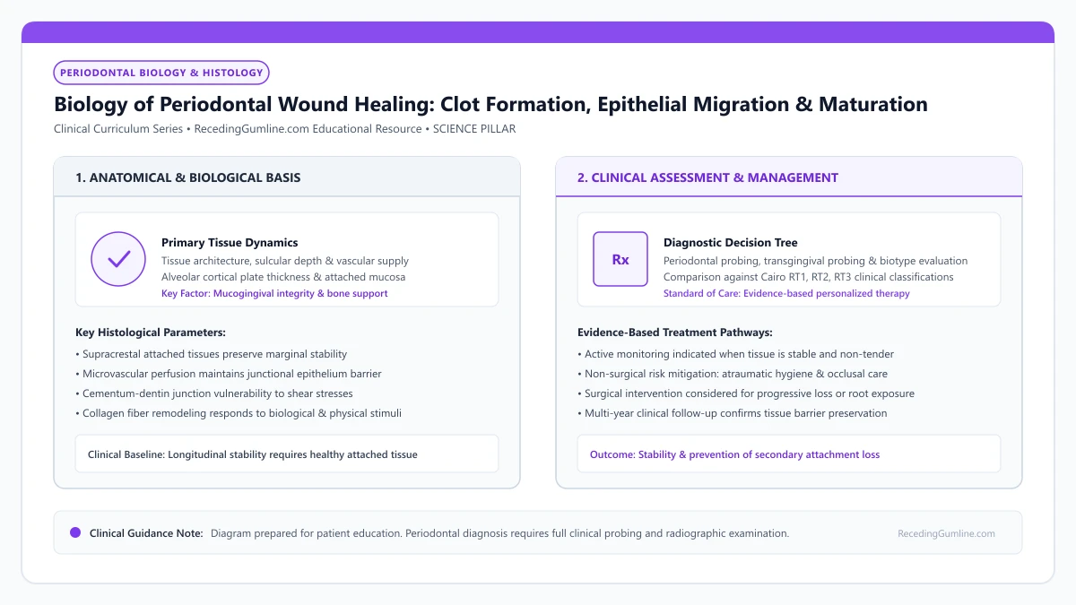
Educational illustration: Biology of Periodontal Wound Healing: Clot Formation, Epithelial Migration & Maturation. Clinical management requires comprehensive periodontal evaluation rather than isolated self-assessment.
Source: RecedingGumline.com Clinical Editorial Team (Proprietary educational diagram for RecedingGumline.com)
Key Clinical Distinctions & Diagnostic Boundaries
- Oral mucosal wounds heal significantly faster and with less scarring than cutaneous skin wounds due to salivary growth factors.
- The race between fast-moving epithelial cells and slow-moving connective tissue cells dictates whether a wound heals by repair or regeneration.
- Micro-movement of only 20 to 50 micrometers during early healing can rupture anchoring fibrin strands, causing flap failure.
- Complete collagen remodeling and orientation of mature gingival fibers requires 6 to 12 months.
Phase 1 & 2: Hemostasis, Fibrin Clot Scaffolding & Inflammation
Within seconds of a surgical incision, platelets aggregate at severed capillaries, releasing clotting factors that convert soluble fibrinogen into an insoluble mesh of fibrin strands. This creates a stable blood clot between the tooth root and the overlying gum flap.
This fibrin clot is not just a temporary plug; it is a vital biological highway. Platelets trapped in the clot degranulate, releasing growth factors such as Platelet-Derived Growth Factor (PDGF) and Transforming Growth Factor-beta (TGF-β). Neutrophils and macrophages infiltrate the clot within 24 to 48 hours to clean necrotic tissue and kill bacteria.
Periodontal wound healing following surgical intervention is a tightly orchestrated biological cascade progressing through four distinct phases: hemostasis, inflammation, proliferation, and tissue remodeling. Within seconds of surgical incision, a fibrin clot forms, providing a temporary provisional matrix for migrating cells.
Clinical Considerations:
- Platelet aggregation creates an insoluble fibrin mesh that anchors the surgical flap
- Release of PDGF and TGF-β growth factors recruits healing repair cells
- Neutrophils and macrophages debride the wound space within 24 to 48 hours
Phase 3: Angiogenesis, Granulation Tissue & Rapid Epithelial Migration
Between days 3 and 7, capillary endothelial cells sprout from adjacent vascular beds, forming new capillary loops (angiogenesis) that invade the fibrin mesh. Concurrently, fibroblasts proliferate and secrete a provisional extracellular matrix rich in Type III collagen and hyaluronic acid, transforming the clot into vascular granulation tissue.
Simultaneously, basal epithelial cells at the wound margins begin migrating across the surface at a rapid rate of 0.5 to 1.0 millimeter per day. This rapid epithelial migration seals the external barrier within 7 to 10 days, shielding the delicate granulation tissue beneath.
Epithelial cells exhibit the fastest migration velocity of all periodontal tissues, migrating across wound margins at approximately 0.5 mm per day to re-establish a surface barrier. If the epithelial front reaches the root surface first, it forms a long junctional epithelium rather than new fibrous connective tissue attachment.
Clinical Considerations:
- Capillary sprouting (angiogenesis) establishes active blood flow by days 4 to 7
- Fibroblasts synthesize provisional Type III collagen, forming granulation tissue
- Epithelial cells migrate rapidly across the surface, closing the external wound within 10 days
Phase 4: Collagen Maturation and Tissue Remodeling
From week 2 through month 6, the wound enters the prolonged remodeling phase. Fibroblasts replace weak provisional Type III collagen with dense, highly organized bundles of mature Type I collagen. Blood vessel density gradually diminishes, transitioning red granulation tissue into firm, pale pink, healthy gingiva.
Over weeks 8 to 12, the new tissue achieves functional tensile strength, and the junctional epithelium forms a permanent hemidesmosomal seal against the root surface. Creeping attachment—the gradual coronal migration of the margin—may continue for up to a year.
Periodontal regenerative therapies utilize barrier membranes to physically exclude rapidly migrating epithelial cells from the root surface. This exclusion creates protected space that allows slower-migrating pluripotential cells from the periodontal ligament and alveolar bone to regenerate authentic attachment tissues.
Clinical Considerations:
- Weak Type III collagen is gradually replaced by dense, mature Type I collagen bundles
- Vascular density normalizes, transitioning pink tissue from red granulation tissue
- Complete collagen reorganization and functional tissue maturation requires 6 to 12 months
Melcher's Four-Compartment Theory: The Cellular Race to the Root
In 1976, A.H. Melcher published a foundational biological concept governing periodontal wound healing: the four-compartment theory. Melcher demonstrated that the nature of periodontal repair depends entirely on which of four distinct cellular compartments repopulates the root surface first following surgery.
The four cellular compartments are: (1) oral epithelium, (2) gingival connective tissue, (3) alveolar bone, and (4) periodontal ligament (PDL). Epithelial cells possess the fastest migration velocity, advancing across a denuded root at approximately 0.5 to 1.0 mm per day.
If epithelial cells win the race, they form a "long junctional epithelium"—a weak, adhesion-based seal without true fibrous reattachment. True periodontal regeneration requires excluding epithelial cells to allow slower-moving PDL and bone cells to repopulate the root.
Clinical Considerations:
- Melcher's theory identifies four cellular compartments: epithelium, gingival CT, bone, and PDL.
- Epithelial cells migrate fastest (0.5-1.0 mm/day), forming a non-regenerative long junctional epithelium.
- True regeneration requires blocking epithelial downgrowth to allow PDL and bone cells to colonize the root.
Barrier Mechanics: Guiding Cellular Repopulation and Stability
The clinical application of Melcher's biological theory led directly to the development of Guided Tissue Regeneration (GTR) and contemporary periodontal plastic surgical flaps. By placing a physical biocompatible barrier membrane over the alveolar bone and PDL, epithelial down-growth is mechanically blocked.
This barrier isolation creates a protected, secluded space over the root, allowing undifferentiated mesenchymal stem cells from the remaining periodontal ligament space and adjacent bone marrow to migrate, proliferate, and differentiate.
These cells differentiate into functional cementoblasts, osteoblasts, and fibroblasts, synthesizing new acellular extrinsic fiber cementum, inserting new Sharpey's fibers, and regenerating genuine alveolar bone.
Clinical Considerations:
- Barrier membranes physically exclude rapidly migrating epithelial cells from the root surface.
- Protected secluded spaces allow slower-migrating PDL and bone mesenchymal cells to colonize.
- Differentiating progenitor cells regenerate true acellular cementum, Sharpey's fibers, and alveolar bone.
Clinical Reality Check
Do not touch, brush, or stretch your surgical site for the first two weeks; the entire success of your surgery depends on keeping the micro-thin fibrin clot completely immobile.
Questions to Ask Your Periodontist or Dentist
- What healing phase is my surgical site currently in at my one-week checkup?
- Has the surface epithelium completely sealed over the underlying graft tissue?
- When is the fibrin clot secure enough for me to safely resume brushing with an ultra-soft brush?
- How long will full collagen maturation take before my gums reach their permanent final position?
Unsure What Your Gum Changes Mean?
Take our free, evidence-based Gum Recession Assessment — approximately 3 minutes. Identify potential risk factors, evaluate symptoms, and receive personalized discussion questions for your dentist or periodontist.
Non-diagnostic educational triage. Private, secure, completed in your browser.
Related Educational Topics
Clinical Evidence & Claim Traceability (3 Mapped Assertions)
Scientific Literature & Clinical Guidelines
3sources · Hide ▲
- Sculean A, Nikolidakis D, Schwarz F (2008).
"Regeneration of periodontal tissues: combinations of barrier membranes and grafting materials - biological foundation and preclinical evidence: a systematic review." Journal of Clinical Periodontology.
Clinical relevance: Comprehensive systematic review analyzing clinical and histological outcomes of biomaterials in periodontal regeneration; confirmed that enamel matrix derivative (EMD) and barrier membranes for guided tissue regeneration achieve histological evidence of true regeneration (new cementum, periodontal ligament, and bone).
- Melcher AH (1976).
"On the repair potential of periodontal tissues." Journal of Periodontology.
Clinical relevance: Foundational biological treatise describing the four distinct cellular compartments during periodontal wound healing (lamina propria, periodontal ligament, bone, and cementum); established the biological premise of guided tissue regeneration by excluding rapidly migrating gingival epithelium to permit PDL cell repopulation.
- Zucchelli G, Mounssif I (2015).
"Periodontal plastic surgery." Periodontology 2000.
Clinical relevance: Peer-reviewed review of periodontal plastic surgery modalities, detailing flap design, coronally advanced flaps, autogenous connective tissue grafting, tunneling techniques, and anatomical factors governing aesthetic and functional root coverage.
Important Medical Notice
The contents of RecedingGumline.com, including text, graphics, self-assessment calculators, and other materials, are intended solely for educational and informational purposes. This content is not intended to replace professional dental examination, diagnosis, or treatment. Always seek the advice of a qualified dentist, periodontist, or other licensed oral healthcare provider with any questions you may have regarding a medical or dental condition. Never disregard professional medical advice or delay seeking it because of something you read on this website.