Biological Realities of Periodontal Regeneration in Gum Recession
As detailed in our central receding gums overview guide, one of the most widespread questions asked by individuals experiencing gum recession is whether receded gums can spontaneously regenerate. In modern periodontal biology, the definitive scientific answer is that once periodontal attachment—comprising alveolar bone, periodontal ligament, and root cementum—has been destroyed, it does not spontaneously regrow. Understanding the biological mechanisms governing wound healing, cellular competition, and the strict biological limits separating tissue repair from true multi-tissue regeneration is essential for separating clinical reality from unscientific claims.
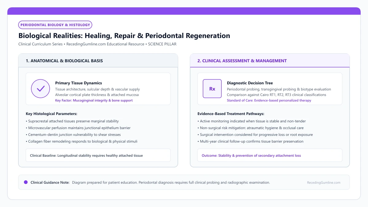
Educational illustration: Biological Realities: Healing, Repair & Periodontal Regeneration. Clinical management requires comprehensive periodontal evaluation rather than isolated self-assessment.
Source: RecedingGumline.com Clinical Editorial Team (Proprietary educational diagram for RecedingGumline.com)
The Myth of Spontaneous Natural Regrowth
A pervasive misconception promoted in commercial advertising and home-remedy forums is that receding gums can "spontaneously regrow" through the use of topical herbal oils, specialty toothpastes, or nutritional supplements. From a biological and histological standpoint, spontaneous soft-tissue regrowth over an exposed root is physically impossible.
Gingival recession is not merely a superficial skin problem; it reflects the underlying loss of the entire attachment apparatus. The marginal gingiva is anchored to the tooth by collagenous Sharpey's fibers inserting into mineralized root cementum, which in turn is anchored into alveolar bone crests.
Once bacterial enzymes or mechanical forces sever these inserting fibers and resorb the alveolar bone plate, the denuded root surface becomes contaminated with bacterial endotoxins and loses its blood supply. Without a vascular scaffold, undifferentiated stem cells, and space provision, soft tissue cannot crawl coronally across an avascular root surface.
Key Scientific Insights
- Spontaneous regrowth of receded gum tissue does not occur under any natural conditions.
- Recession reflects loss of underlying alveolar bone and inserting Sharpey's fibers, not just surface tissue.
- Exposed root surfaces are avascular and contaminated, preventing unassisted soft-tissue coronal migration.
Melcher's Principle: Cellular Competition in Periodontal Wounds
The biological foundation of periodontal wound healing was established in 1976 by A.H. Melcher in a landmark theoretical paper. Melcher identified that periodontal wounds are populated by cells originating from four distinct anatomical compartments: gingival epithelium, gingival connective tissue, alveolar bone, and the periodontal ligament (PDL).
Each cell type possesses vastly different rates of migration and biological potential. Epithelial cells from the oral mucosa are the fastest-moving cells in the body, migrating across an instrumented root surface at rates up to 0.5 mm per day. In contrast, osteoblasts from bone and mesenchymal stem cells from the PDL migrate substantially more slowly.
In an untreated healing wound, rapid epithelial down-growth wins the race, covering the instrumented root surface before slower-moving bone and PDL cells can arrive. This down-growth forms a Long Junctional Epithelium (LJE). While an LJE provides a stable, healthy biological seal, it physically blocks bone and ligament cells from contacting the root, preventing true regeneration.
Key Scientific Insights
- Melcher (1976) demonstrated that healing outcome is dictated by which cell type repopulates the root first.
- Oral epithelial cells migrate up to ten times faster than regenerative osteoblasts and PDL stem cells.
- Rapid epithelial down-growth forms a long junctional epithelium, permanently blocking true regeneration.
Repair vs. True Periodontal Regeneration: Histological Standards
In periodontal science, a rigorous distinction exists between "repair" and "regeneration." Repair is defined as the healing of a wound by tissue that does not fully restore the architecture or function of the lost parts. Formation of a long junctional epithelium or dense scar-like connective tissue adhesion without cementum insertion represents repair.
True Periodontal Regeneration is defined histologically as the complete reproduction or reconstitution of lost tissues: the simultaneous, coordinated regeneration of new alveolar bone, a newly oriented periodontal ligament with functional inserting Sharpey's fibers, and new root cementum coronal to the pre-existing baseline.
Demonstrating true regeneration requires decalcified histological sectioning of human biopsy specimens; clinical probing depth reduction and radiographic bone fill alone cannot prove true regeneration, as they frequently represent dense connective tissue repair.
Key Scientific Insights
- Periodontal repair restores clinical health via a long junctional epithelium without new bone or cementum.
- True regeneration requires simultaneous histological reformation of bone, PDL fibers, and cementum.
- Histological biopsy is the only definitive scientific proof separating true regeneration from repair.
Surgical Regenerative Modalities: GTR, Biomaterials & Enamel Matrix Proteins
Overcoming Melcher's cell-competition barrier requires advanced surgical interventions designed to selectively guide tissue repopulation. Guided Tissue Regeneration (GTR) achieves this by placing a physical barrier membrane (resorbable collagen or non-resorbable PTFE) over the debrided defect, beneath the surgical flap.
The membrane acts as a physical shield, excluding fast-migrating epithelial cells and gingival fibroblasts while tenting open a protected space over the root. This sheltered space allows slower-migrating pluripotential stem cells from the periodontal ligament and bone marrow to populate the root surface, synthesizing new cementum, ligament, and bone.
Biomaterials enhance this regenerative cascade: Enamel Matrix Derivative (Emdogain, a porcine-derived amelogenin protein formulation) mimics embryological cementogenesis, stimulating cementoblasts and PDL fibroblasts to achieve true histological regeneration. Autologous Platelet-Rich Fibrin (PRF) provides sustained release of growth factors (PDGF, VEGF, TGF-beta) that accelerate capillary angiogenesis and tissue maturation.
Key Scientific Insights
- Guided Tissue Regeneration (GTR) membranes physically block epithelial cells, allowing PDL stem cells to regenerate.
- Enamel Matrix Derivative (Emdogain) chemically stimulates cementoblasts to replicate embryological development.
- Platelet-Rich Fibrin (PRF) delivers autologous growth factors that accelerate microvascular revascularization.
Anatomical Limits: Defect Geometry & Surgical Predictability
While surgical regenerative therapies offer remarkable biological potential, their success is strictly governed by local defect geometry. Periodontal regeneration requires bony walls to contain the graft biomaterial, provide blood supply, and stabilize the blood clot.
Three-wall intrabony defects (surrounded by three intact walls of bone) and narrow vertical bone craters offer the highest regenerative predictability. In contrast, flat horizontal bone loss or two-wall defects offer significantly lower predictability because blood supply is diminished and space cannot be maintained.
For isolated buccal recession defects where the facial bone plate has completely resorbed (Cairo RT1), root coverage is achieved through soft-tissue connective tissue grafting. This reconstructs a thick, protective soft-tissue barrier and covers denuded root dentin, successfully resolving aesthetic deficits and sensitivity even where the facial bone plate cannot be restored.
Key Scientific Insights
- Regenerative predictability depends heavily on local defect geometry and remaining bony walls.
- Deep, three-wall intrabony pockets offer the highest predictability for true bone and PDL regeneration.
- Buccal recession defects are successfully covered with soft-tissue grafts that rebuild durable attached gingiva.
Tissue Engineering in Periodontics: Enamel Matrix Derivative (EMD), PRF & Growth Factors
In modern periodontal surgery, clinicians no longer rely solely on passive barrier membranes and inert bone grafts. The cutting edge of periodontology utilizes biomimetic tissue engineering—employing biological signaling proteins, autologous blood concentrates, and recombinant growth factors to instruct host stem cells to regenerate lost periodontal tissues. Understanding these advanced biomaterials reveals what is realistically possible in modern tissue regeneration.
• Tissue engineering relies on the triad of stem cells, conductive scaffolds, and biological signaling molecules.
• Enamel Matrix Derivative (EMD, Emdogain) mimics tooth developmental biology, stimulating cementoblasts to form new acellular cementum.
• Platelet-Rich Fibrin (PRF) provides a concentrated autologous matrix of platelets, leukocytes, and sustained growth factor release.
• Recombinant human Platelet-Derived Growth Factor (rhPDGF-BB) accelerates angiogenesis and cellular proliferation in bone defects.
The Tissue Engineering Triad: Cells, Scaffolds & Signals
Modern periodontal tissue engineering is founded on a classic biomedical triad: progenitor cells, biomaterial scaffolds, and signaling molecules (morphogens). For regeneration to occur, undifferentiated mesenchymal stem cells from the periodontal ligament must be present.
These cells require an extracellular matrix scaffold (such as a collagen sponge, allograft matrix, or bone particulate) to guide their three-dimensional migration. Finally, biological signaling molecules bind to cell-surface receptors, delivering biochemical instructions that command cells to divide, synthesize collagen, and differentiate into cementoblasts and osteoblasts.
Periodontal tissue engineering combines three essential components—biological signaling molecules, biocompatible physical scaffolds, and responsive progenitor cells—to regenerate lost attachment structures. Recombinant human platelet-derived growth factor (rhPDGF-BB) and enamel matrix derivative (EMD) stimulate osteoblastic and cementoblastic proliferation.
Key Scientific Insights
- Progenitor stem cells from the periodontal ligament provide the cellular machinery
- Biomaterial scaffolds provide structural three-dimensional scaffolding for cell growth
- Biological growth factors deliver the molecular commands triggering cellular differentiation
Enamel Matrix Derivative (Emdogain): Biomimetic Amelogenins
Enamel Matrix Derivative (EMD, commercially known as Emdogain) is one of the most extensively researched biologics in dentistry. Developed in Sweden, EMD consists of a purified protein extract derived from developing porcine tooth buds, dominated by amelogenins.
During natural embryological tooth development, amelogenins are secreted onto the newly formed dentin root, triggering mesenchymal cells to differentiate into cementoblasts that deposit root cementum. By applying EMD to a cleaned root surface during surgery, the periodontist mimics embryological tooth development, stimulating host cells to deposit new acellular cementum and form new inserting Sharpey's fibers.
Engineered biomaterial scaffolds provide three-dimensional structural stability, maintaining space against soft-tissue flap collapse while facilitating cellular infiltration and angiogenesis. As new cellular matrices are deposited, the temporary biodegradable scaffold resorbs harmlessly without leaving foreign debris.
Key Scientific Insights
- Composed of natural amelogenin proteins that mimic embryological tooth development
- Precipitates onto the root surface, stimulating stem cells to differentiate into cementoblasts
- Proven in human histology to regenerate true acellular cementum, PDL, and alveolar bone
Autologous Biologics: Platelet-Rich Fibrin (PRF) and Growth Factors
Another major advancement is Platelet-Rich Fibrin (PRF). Before surgery, a small vial of the patient's blood is drawn and spun in a specialized centrifuge at low speeds. This separates red blood cells, concentrating platelets and white blood cells into a dense, autologous fibrin matrix.
This PRF clot is pressed into a biological membrane and placed over the receded root. PRF slowly releases a steady physiological cascade of growth factors—including Vascular Endothelial Growth Factor (VEGF) and PDGF—over 10 to 14 days, accelerating capillary ingrowth, reducing postoperative pain, and enhancing soft-tissue healing.
Advanced research into autologous stem cells derived from periodontal ligament tissues offers tremendous potential for personalized regenerative therapies. Incorporating molecular tissue engineering into clinical periodontal surgery transforms unpredictable complex defects into highly predictable reconstructive outcomes.
Key Scientific Insights
- PRF is 100% natural and autologous, prepared chairside from a simple blood draw
- Concentrates platelets, leukocytes, and sustained-release growth factors (VEGF, PDGF)
- Dramatically accelerates capillary revascularization and reduces surgical recovery discomfort
The Periodontal Tissue Engineering Triad: Scaffolds, Cells & Signals
Tissue engineering represents the cutting edge of regenerative periodontics, shifting therapy from passive tissue replacement to active, de novo biological tissue construction. The paradigm relies on the "tissue engineering triad": cells, biomimetic scaffolds, and signaling molecules.
Biomimetic scaffolds (such as 3D-printed polycaprolactone, collagen matrices, and bioceramics) provide a physical three-dimensional extracellular template with calibrated pore sizes (100 to 300 micrometers) that permit cellular ingrowth and vascular angiogenesis.
Autologous mesenchymal stem cells (harvested from periodontal ligament, dental pulp, or adipose tissue) are seeded into these scaffolds and stimulated by recombinant growth factors (rhPDGF, BMP-2, FGF-2) to generate fully functional attachment complexes.
Key Scientific Insights
- The tissue engineering triad integrates stem cells, 3D biomimetic scaffolds, and signaling molecules.
- Scaffolds with interconnected 100-300 micrometer pores facilitate cell ingrowth and capillary angiogenesis.
- Periodontal stem cells combined with growth factors enable de novo reconstruction of lost tissues.
Clinical Translators: Acellular Dermal Matrices & 3D Printed Constructs
In contemporary daily periodontal practice, tissue engineering principles are translated through advanced biomaterials that eliminate the morbidity of palatal autograft harvesting. Acellular Dermal Matrix (ADM) allografts—processed human donor skin stripped of cellular components—provide a pure collagen-elastin scaffold.
When placed over denuded root surfaces, the ADM scaffold is repopulated by recipient host endothelial cells and fibroblasts, guiding orderly tissue regeneration while providing outcomes comparable to autogenous tissue grafts in wide Cairo RT1 defects.
The next generation of tissue engineering utilizes patient-specific 3D-printed bioresorbable micro-channeled scaffolds custom-fabricated from CBCT data. These constructs guide fiber orientation perpendicular to the root surface, promising true structural regeneration for complex periodontal defects.
Key Scientific Insights
- Acellular Dermal Matrix (ADM) provides a ready-to-use collagen scaffold, avoiding palatal surgery.
- Host blood vessels and fibroblasts infiltrate the ADM scaffold, regenerating a thick tissue barrier.
- Patient-specific 3D-printed micro-channeled scaffolds guide functional fiber orientation toward the root.
Clinical Reality Check
No commercial mouthwash, supplement, or toothpaste can regrow lost periodontal bone or receded gums. Patients must be protected from fraudulent marketing claims that promise "natural gum regrowth," which delay essential clinical periodontal evaluation.
Questions to Ask Your Dentist or Periodontist
- Can any of the bone or gum tissue I have lost be surgically regenerated using GTR or Emdogain?
- What type of bone defect geometry (intrabony vs horizontal) do I have around these teeth?
- Does my soft-tissue recession require a connective tissue graft to restore root coverage?
- What is the difference between the clinical repair achieved by deep cleaning and true regeneration?
- What is the realistic timeline for healing and tissue maturation following regenerative surgery?
- Do you recommend incorporating enamel matrix derivative (Emdogain) or Platelet-Rich Fibrin (PRF) into my gum graft procedure?
- Does my specific recession defect have the anatomical shape that benefits from biological growth factors?
- Will using PRF from my own blood help reduce my postoperative swelling and palatal discomfort?
- What additional costs are associated with adding tissue engineering biologics to my surgical plan?
Unsure What Your Gum Changes Mean?
Take our free, evidence-based Gum Recession Assessment — approximately 3 minutes. Identify potential risk factors, evaluate symptoms, and receive personalized discussion questions for your dentist or periodontist.
Non-diagnostic educational triage. Private, secure, completed in your browser.
Related Educational Topics
Scientific Literature & Clinical Guidelines
6sources · Hide ▲
- Melcher AH (1976).
"On the repair potential of periodontal tissues." Journal of Periodontology.
Clinical relevance: Foundational biological treatise describing the four distinct cellular compartments during periodontal wound healing (lamina propria, periodontal ligament, bone, and cementum); established the biological premise of guided tissue regeneration by excluding rapidly migrating gingival epithelium to permit PDL cell repopulation.
- Sculean A, Nikolidakis D, Schwarz F (2008).
"Regeneration of periodontal tissues: combinations of barrier membranes and grafting materials - biological foundation and preclinical evidence: a systematic review." Journal of Clinical Periodontology.
Clinical relevance: Comprehensive systematic review analyzing clinical and histological outcomes of biomaterials in periodontal regeneration; confirmed that enamel matrix derivative (EMD) and barrier membranes for guided tissue regeneration achieve histological evidence of true regeneration (new cementum, periodontal ligament, and bone).
- Tonetti MS, Greenwell H, Kornman KS (2018).
"Staging and grading of periodontitis: Framework and proposal of a new classification and case definition." Journal of Clinical Periodontology.
Clinical relevance: Consensus framework establishing the multidimensional staging (severity and extent of periodontal tissue breakdown) and grading (biological rate of disease progression, incorporating smoking and diabetes as grade modifiers) for periodontitis. It addresses periodontitis diagnosis and staging, not the classification of localized gingival recession defects.
- Cairo F, Nieri M, Cincinelli S, Mervelt J, Pagliaro U (2011).
"The interproximal clinical attachment level to classify gingival recessions and predict root coverage outcomes: an explorative and reliability study." Journal of Clinical Periodontology.
Clinical relevance: Exploratory and reliability study establishing the Cairo classification based on interdental clinical attachment level (CAL): RT1 (no interproximal attachment loss; complete root coverage is clinically predictable), RT2 (interproximal attachment loss <= buccal loss; partial coverage predictable), and RT3 (interproximal loss exceeds buccal recession; complete coverage not predictable).
- Chambrone L, Salinas Ortega MA, Sukekava F, Rotundo R, Kalemaj Z, Buti J, Pini Prato GP (2018).
"Root coverage procedures for treating localised and multiple recession-type defects." Cochrane Database of Systematic Reviews.
Clinical relevance: Cochrane systematic review evaluating root-coverage procedures for localized and multiple recession-type defects. While subepithelial connective tissue grafts (SCTG) combined with coronally advanced flaps demonstrated higher rates of complete root coverage and keratinized tissue gain compared to flap advancement alone, evidence quality varied across outcomes and clinical decisions must balance donor site morbidity and patient-reported outcomes.
- Cortellini P, Bissada NF (2018).
"Mucogingival conditions in the natural dentition: Narrative review, case definitions, and diagnostic considerations." Journal of Periodontology.
Clinical relevance: World Workshop 2017 comprehensive review defining mucogingival conditions, establishing diagnostic criteria for thin vs thick periodontal phenotypes, and detailing surgical indications including progressive recession, hypersensitivity, aesthetic dissatisfaction, and root caries vulnerability.
Important Medical Notice
The contents of RecedingGumline.com, including text, graphics, self-assessment calculators, and other materials, are intended solely for educational and informational purposes. This content is not intended to replace professional dental examination, diagnosis, or treatment. Always seek the advice of a qualified dentist, periodontist, or other licensed oral healthcare provider with any questions you may have regarding a medical or dental condition. Never disregard professional medical advice or delay seeking it because of something you read on this website.