Long Junctional Epithelium vs. True Periodontal Regeneration: The Melcher Hypothesis
What is the biological difference between periodontal repair and true periodontal regeneration?
In our comprehensive guide to gingival recession conditions, a vital scientific distinction separates periodontal repair from true periodontal regeneration. When gums heal after deep cleaning or traditional surgery, the body almost universally heals by repair—forming an elongated, scar-like junctional epithelium along the root. Understanding Dr. Antony Melcher's landmark 1976 hypothesis reveals the cellular competition that governs whether tissues merely heal or truly regenerate.
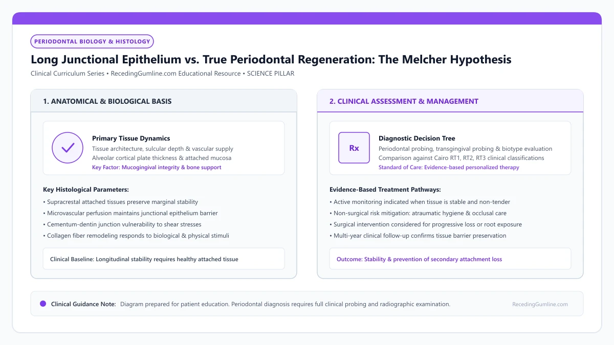
Educational illustration: Long Junctional Epithelium vs. True Periodontal Regeneration: The Melcher Hypothesis. Clinical management requires comprehensive periodontal evaluation rather than isolated self-assessment.
Source: RecedingGumline.com Clinical Editorial Team (Proprietary educational diagram for RecedingGumline.com)
Key Clinical Distinctions & Diagnostic Boundaries
- A long junctional epithelium provides an effective biological seal against bacteria, but lacks Sharpey's fibers and bone support.
- Guided Tissue Regeneration (GTR) uses barrier membranes to physically block fast-moving epithelial cells, allowing slow PDL cells to populate the root.
- Enamel matrix derivatives (Emdogain) chemically signal mesenchymal cells to differentiate into new cementoblasts.
- Repair stops disease progression and stabilizes teeth; regeneration structurally restores what was lost.
The Melcher Hypothesis: Four Competing Cell Compartments
In 1976, Dr. Antony Melcher published a revolutionary paper in the Journal of Periodontology. He proposed that the periodontium contains four distinct cellular compartments participating in wound healing: (1) oral epithelium, (2) gingival connective tissue, (3) alveolar bone, and (4) periodontal ligament (PDL).
Melcher hypothesized that the ultimate nature of periodontal healing is determined exclusively by which of these four cell types migrates first onto the instrumented root surface. If epithelial cells populate the root, an epithelial seal forms. If gingival connective tissue cells populate the root, root resorption occurs. Only if cells originating from the PDL and perivascular bone populate the root can true regeneration occur.
In periodontal biology, clinical healing occurs via two fundamentally distinct pathways: tissue repair or true biological regeneration. Periodontal repair describes the healing of a wound by tissue that does not fully restore the original architecture or function, typically manifesting as a long junctional epithelium.
Clinical Considerations:
- Four competing cellular compartments: epithelium, gingival connective tissue, bone, and PDL
- The phenotype of the cell that first colonizes the root surface dictates the healing outcome
- Only periodontal ligament-derived cells possess the genetic capacity to form new cementum and PDL
The Cellular Race: Why Long Junctional Epithelium Is the Default
In natural healing, there is an unfair biological race. Epithelial cells are rapid sprinters, migrating across wound surfaces at speeds of 0.5 to 1.0 millimeter per day. In contrast, osteoblasts and periodontal ligament progenitor cells are slow crawlers.
Consequently, after conventional scaling or flap surgery, epithelial cells easily win the race. They migrate down the instrumented root surface, interposing themselves between the tooth and the healing bone. This creates a Long Junctional Epithelium (LJE)—a thin, elongated epithelial sheet attached via hemidesmosomes. This is periodontal repair: it seals the pocket, but regenerates zero bone or inserting fibers.
True periodontal regeneration represents the complete reconstitution of lost structural tissues, including new alveolar bone, new cementum, and functionally oriented periodontal ligament fibers inserting into both. While soft-tissue grafting repairs the mucosal margin, it rarely achieves histological regeneration on denuded root surfaces.
Clinical Considerations:
- Fast-moving epithelial cells (0.5–1.0 mm/day) consistently out-race slow bone and PDL cells
- Epithelium sheets down the root surface, forming a Long Junctional Epithelium (LJE)
- LJE provides a healthy, functional biological seal, but is a repair mechanism, not regeneration
Achieving True Regeneration: Guided Tissue Regeneration (GTR)
To overcome this epithelial takeover, periodontal researchers developed Guided Tissue Regeneration (GTR). The surgeon places a biocompatible barrier membrane (collagen or PTFE) over the bone defect beneath the gum flap.
The membrane acts as a physical shield, mechanically blocking fast-moving epithelial cells and gingival connective tissue from touching the root. This creates a protected, secluded space where slow-moving PDL cells and osteoprogenitor cells can colonize the root, successfully regenerating new cementum, new Sharpey's fibers, and true alveolar bone.
Achieving authentic regeneration requires combining osteoconductive bone scaffolds, barrier membranes for cell exclusion, and biological bioactive growth factors like enamel matrix derivatives. Meticulous surgical immobility and tension-free wound closure are mandatory to protect fragile regenerating microvascular networks.
Clinical Considerations:
- GTR barrier membranes physically exclude fast-migrating epithelial cells from the root
- Creates a secluded biological chamber where slow-moving PDL cells can thrive
- Enables true de novo regeneration of cementum, inserting Sharpey's fibers, and bone
Histologic Endpoints: Long Junctional Epithelium vs. True Regeneration
In periodontal literature, a fundamental biological distinction exists between "periodontal repair" and "true periodontal regeneration." Periodontal repair describes the healing of a defect by tissue that does not fully restore the original architecture or function.
Following conventional scaling and root planing or flap repositioning, healing almost universally occurs by repair via a Long Junctional Epithelium (LJE). Hemidesmosomes attach the epithelial cells to root cementum or dentin without forming collagen fiber insertions into bone.
True periodontal regeneration, by contrast, is defined histologically as the complete de novo reformation of all three lost attachment tissues: new cementum with inserting Sharpey's fibers, a functionally oriented periodontal ligament, and new alveolar bone crest height.
Clinical Considerations:
- Periodontal repair restores tissue continuity but fails to recreate original functional architecture.
- Long Junctional Epithelium (LJE) attaches via hemidesmosomes without true fibrous bone anchorage.
- True regeneration histologically demands new cementum, inserting Sharpey's fibers, and alveolar bone.
Biologic Mediators: Enamel Matrix Derivatives & Recombinant Growth Factors
Achieving true periodontal regeneration on exposed root surfaces requires biologic mediators that stimulate embryological developmental cascades. Enamel Matrix Derivative (EMD, derived from porcine tooth buds) contains amelogenins that mimic the biochemical signals of Hertwig's Epithelial Root Sheath.
When applied to an acid-etched, detoxified root, EMD precipitates into an insoluble protein matrix that promotes cementoblast differentiation and suppresses epithelial cell down-growth, stimulating true cementogenesis and periodontal ligament regeneration.
Recombinant human Platelet-Derived Growth Factor-BB (rhPDGF-BB) combined with an osteoconductive beta-tricalcium phosphate (β-TCP) scaffold acts as a potent mitogen and chemotactic agent, accelerating angiogenesis and recruiting osteoprogenitor cells to rebuild osseous defects.
Clinical Considerations:
- Enamel Matrix Derivatives (EMD) mimic root embryogenesis to stimulate new cementum formation.
- EMD suppresses epithelial proliferation while promoting cementoblast and fibroblast migration.
- Recombinant PDGF-BB stimulates powerful angiogenesis and recruits bone-forming osteoprogenitor cells.
Clinical Reality Check
A long junctional epithelium is not a "failed" result; it is nature's robust scar tissue that seals off bacteria and allows teeth to remain stable for a lifetime.
Questions to Ask Your Periodontist or Dentist
- Will my proposed treatment result in periodontal repair (long junctional epithelium) or true regeneration?
- Am I a candidate for Guided Tissue Regeneration (GTR) using a barrier membrane?
- Does my bone loss have vertical defect walls that can support bone grafting materials?
- How does an epithelial seal protect my tooth compared to a regenerated ligament?
Unsure What Your Gum Changes Mean?
Take our free, evidence-based Gum Recession Assessment — approximately 3 minutes. Identify potential risk factors, evaluate symptoms, and receive personalized discussion questions for your dentist or periodontist.
Non-diagnostic educational triage. Private, secure, completed in your browser.
Related Educational Topics
Clinical Evidence & Claim Traceability (2 Mapped Assertions)
Scientific Literature & Clinical Guidelines
3sources · Hide ▲
- Melcher AH (1976).
"On the repair potential of periodontal tissues." Journal of Periodontology.
Clinical relevance: Foundational biological treatise describing the four distinct cellular compartments during periodontal wound healing (lamina propria, periodontal ligament, bone, and cementum); established the biological premise of guided tissue regeneration by excluding rapidly migrating gingival epithelium to permit PDL cell repopulation.
- Sculean A, Nikolidakis D, Schwarz F (2008).
"Regeneration of periodontal tissues: combinations of barrier membranes and grafting materials - biological foundation and preclinical evidence: a systematic review." Journal of Clinical Periodontology.
Clinical relevance: Comprehensive systematic review analyzing clinical and histological outcomes of biomaterials in periodontal regeneration; confirmed that enamel matrix derivative (EMD) and barrier membranes for guided tissue regeneration achieve histological evidence of true regeneration (new cementum, periodontal ligament, and bone).
- Pihlstrom BL, Michalowicz BS, Johnson NW (2005).
"Periodontal diseases." The Lancet.
Clinical relevance: Peer-reviewed Lancet seminar review synthesizing global periodontal epidemiology, microbial etiology, host immunopathology, and systemic interactions, emphasizing prevention, biofilm disruption, and early risk factor modification.
Important Medical Notice
The contents of RecedingGumline.com, including text, graphics, self-assessment calculators, and other materials, are intended solely for educational and informational purposes. This content is not intended to replace professional dental examination, diagnosis, or treatment. Always seek the advice of a qualified dentist, periodontist, or other licensed oral healthcare provider with any questions you may have regarding a medical or dental condition. Never disregard professional medical advice or delay seeking it because of something you read on this website.