Stillman Clefts and McCall Festoons in Gum Recession: Slits and Rolled Margins in Tissue
What are Stillman clefts and McCall festoons, and what causes slits or rolled rims in gum tissue?
When examining receding gumline guide, clinicians occasionally identify distinctive tissue deformities that deviate from typical smooth, curved recession. First described in early 20th-century dental literature, Stillman's clefts (narrow, slit-like vertical fissures in the gum margin) and McCall's festoons (thickened, rolled, doughnut-shaped margin collars) represent classic anatomical reactions to traumatic forces and localized inflammation.
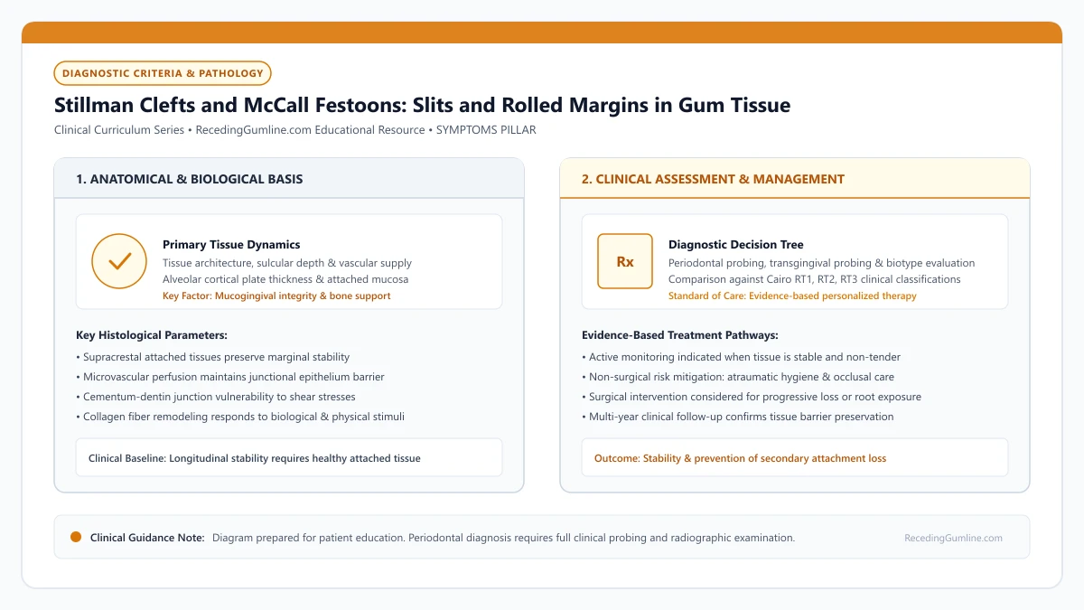
Educational illustration: Stillman Clefts and McCall Festoons: Slits and Rolled Margins in Gum Tissue. Clinical management requires comprehensive periodontal evaluation rather than isolated self-assessment.
Source: RecedingGumline.com Clinical Editorial Team (Proprietary educational diagram for RecedingGumline.com)
Key Clinical Distinctions & Diagnostic Boundaries
- A Stillman cleft looks like a vertical paper-cut in the gum; it is not an accidental laceration, but a chronic destructive mucogingival defect.
- McCall festoons look like swollen, doughnut-like rims around the tooth neck, frequently affecting lower premolars and canines.
- Clefts are classified as simple (single vertical split) or compound (cleaved margin with lateral tissue loss).
- Treatment requires eliminating the offending mechanical habit or occlusal interference before surgical repair.
Stillman Clefts: The "Paper Cut" Fissure in Gum Tissue
Described by Dr. Paul Stillman in 1921, a Stillman cleft is a narrow, vertical, slit-like indentation extending from the marginal gingiva down toward the root apex. It often looks as if the gum has been sliced cleanly with a razor blade or paper cut.
Clefts can be simple (a single straight vertical split) or compound (where the fissure branches or exhibits ragged edges). They most frequently occur on the facial surfaces of canines, premolars, and lower incisors. If left unmanaged, the cleft deepens until it penetrates through the mucogingival junction, exposing the root completely.
Stillman’s clefts present as narrow, vertical slit-like fissures extending apically from the gingival margin along the root surface. McCall’s festoons manifest as exaggerated, life-preserver-like rolled enlargements of the marginal gingiva circling the cervical neck of the tooth.
Clinical Considerations:
- Presents as a sharp, vertical, slit-like fissure extending apically from the margin
- Commonly triggered by aggressive horizontal brushing, fingernail picking, or occlusal stress
- Can rapidly progress into severe, full-thickness root exposure if left untreated
McCall Festoons: Enlarged, Rolled Marginal Collars
Described by periodontist J.O. McCall, a McCall festoon is an exaggerated, rounded, lifesaver-shaped enlargement of the marginal gingiva. Instead of tapering to a fine, knife-edged margin against the enamel, the gum tissue rolls into a distinct, swollen rim.
Festoons most commonly affect the facial surfaces of mandibular premolars and canines. While they may appear inflamed and red, festoons in health can be pale pink and fibrous. They represent the body's attempt to reinforce a gingival collar subjected to chronic mechanical friction or heavy biting forces.
Both clefting and festooning represent localized soft-tissue reactions to chronic mechanical trauma, such as aggressive fingernail picking or severe occlusal trauma. The localized micro-tears disrupt normal microcirculation, creating characteristic indented clefts or thickened fibrous collars.
Clinical Considerations:
- Presents as an exaggerated, rolled, doughnut-shaped thickening of the marginal tissue
- Most prevalent on mandibular canines and premolars
- Represents compensatory fibrous tissue proliferation in response to mechanical strain
Etiological Triad and Treatment Strategies
Historical periodontics blamed clefts and festoons almost exclusively on "occlusal trauma" (bite interference). Modern periodontal science recognizes a multifactorial triad: biomechanical bite forces create micro-strain at the cervical margin, which is then exacerbated by aggressive toothbrush scrubbing and bacterial plaque accumulation.
Treating Stillman clefts requires removing the causative trauma: adjusting high bite interferences, breaking fingernail picking habits, and switching to ultra-soft brushes. For deep, open clefts, periodontal plastic surgery—such as a connective tissue graft or coronally advanced flap—is performed to close the fissure and restore smooth marginal continuity.
Resolution requires completely eliminating the underlying traumatic stimulus through occlusal adjustment or habit cessation therapy. Once mechanical trauma is removed, shallow clefts often resolve spontaneously, while deep clefts may be surgically repaired using soft-tissue grafting.
Clinical Considerations:
- Multifactorial cause involving bite forces, toothbrush trauma, and plaque inflammation
- Bite adjustment (equilibration) relieves heavy traumatic forces on the affected tooth
- Periodontal plastic surgery (gum grafting) reconstructs smooth, continuous tissue margins
Diagnostic Features: Stillman's Clefts vs. McCall's Festoons
Stillman's clefts and McCall's festoons are classic clinical morphological manifestations of localized mucogingival pathology. A Stillman's cleft is a narrow, slit-like vertical indentation or fissure extending from the gingival margin apically along the root surface.
In contrast, McCall's festoons appear as distinct, doughnut-shaped or rolled, exaggerated enlargements of the marginal gingiva, most commonly observed around the labial surfaces of canines and premolars. Both lesions are often triggered by heavy occlusal trauma combined with aggressive horizontal toothbrushing.
Under excessive mechanical force, the marginal vascular plexus undergoes localized thrombosis. In thin phenotypes, the ischemic tissue collapses into a sharp cleft, whereas in thicker phenotypes, the tissue responds with hyperplastic fibrous enlargement (festooning).
Clinical Considerations:
- Stillman's clefts are narrow, slit-like vertical fissures running apically along the root.
- McCall's festoons are rolled, doughnut-shaped fibrous enlargements of the marginal gumline.
- Ischemic vascular thrombosis from trauma causes clefting in thin tissue and festoons in thick tissue.
Clinical Protocols: Occlusal Equilibration, Debridement & Grafting
Resolving Stillman's clefts and McCall's festoons requires identifying and eliminating the underlying mechanical trauma before attempting soft-tissue surgery. When occlusal interferences or clenching habits are detected, precision occlusal adjustment and custom nightguard therapy are implemented immediately.
Simultaneously, the patient's brushing technique is adjusted to gentle sulcular cleansing with extra-soft filaments. In early or mild cases, eliminating trauma allows the cleft to spontaneously re-epithelialize and shallow over several months.
However, if the cleft persists, extends to the mucogingival junction, or causes plaque stagnation, surgical connective tissue grafting is performed to re-establish a continuous, harmonious marginal architecture and seal the exposed root.
Clinical Considerations:
- Eliminating occlusal trauma and aggressive brushing must precede any surgical repair.
- Early Stillman's clefts frequently re-epithelialize and resolve spontaneously once trauma ceases.
- Persistent clefts extending past the mucogingival junction require connective tissue grafting.
Clinical Reality Check
A Stillman cleft is not a cut that will close with simple ointment; it is an anatomical loss of underlying tissue support that requires modifying brushing habits and often surgical repair.
Questions to Ask Your Periodontist or Dentist
- Is the slit in my gum tissue a classic Stillman cleft, and how deep does it extend?
- Does my bite show excessive pressure or interference on this specific tooth?
- Do you see signs of a rolled McCall festoon around my premolars or canines?
- Will this cleft heal on its own if I stop brushing it hard, or does it require a gum graft?
Unsure What Your Gum Changes Mean?
Take our free, evidence-based Gum Recession Assessment — approximately 3 minutes. Identify potential risk factors, evaluate symptoms, and receive personalized discussion questions for your dentist or periodontist.
Non-diagnostic educational triage. Private, secure, completed in your browser.
Related Educational Topics
Clinical Evidence & Claim Traceability (2 Mapped Assertions)
Scientific Literature & Clinical Guidelines
3sources · Hide ▲
- Jepsen S, Caton JG, Albandar JM, Bissada NF, Bouchard P, Cortellini P, et al. (2018).
"Periodontal manifestations of systemic diseases and developmental and acquired conditions: Consensus report of workgroup 3 of the 2017 World Workshop on the Classification of Periodontal and Peri-Implant Diseases and Conditions." Journal of Periodontology.
Clinical relevance: Consensus report defining mucogingival conditions, gingival phenotype (replacing biotype), non-carious cervical lesions, and the multifactorial etiology of gingival recession; emphasizes that recession can occur without periodontitis and classifies recession by interdental clinical attachment loss.
- Kassab MM, Cohen RE (2003).
"The etiology and prevalence of gingival recession." The Journal of the American Dental Association.
Clinical relevance: Comprehensive epidemiological and etiological review establishing that gingival recession affects more than 50% of adults aged 18 to 64 and over 88% of individuals aged 65 and older, driven by interactions between predisposing anatomical factors and precipitating mechanical or inflammatory stimuli.
- Grippo JO, Simring M, Coleman TA (2012).
"Abfraction, abrasion, biocorrosion, and the enigma of noncarious cervical lesions: a 20-year perspective." Journal of Esthetic and Restorative Dentistry.
Clinical relevance: Authoritative review defining and clarifying cervical hard tissue lesions: stress-induced abfraction, frictional abrasion from dentifrices/brushes, and chemical biocorrosion (acidic erosion), explaining how multi-factorial mechanisms accelerate cervical notching once gingival recession exposes root dentin.
Important Medical Notice
The contents of RecedingGumline.com, including text, graphics, self-assessment calculators, and other materials, are intended solely for educational and informational purposes. This content is not intended to replace professional dental examination, diagnosis, or treatment. Always seek the advice of a qualified dentist, periodontist, or other licensed oral healthcare provider with any questions you may have regarding a medical or dental condition. Never disregard professional medical advice or delay seeking it because of something you read on this website.