Gum Recession Treatment Comparisons & Clinical Decision Matrix
When treating a receding gums management, selecting the optimal therapy requires navigating a complex clinical decision framework. Treatment options range from conservative oral hygiene modifications and ongoing monitoring to minimally invasive tunneling, autogenous connective tissue grafting, and biomaterial-assisted regeneration. No single procedure is universally optimal for every patient; the ideal approach depends on anatomical biotype, interproximal bone levels, aesthetic expectations, medical risk factors, and financial considerations.
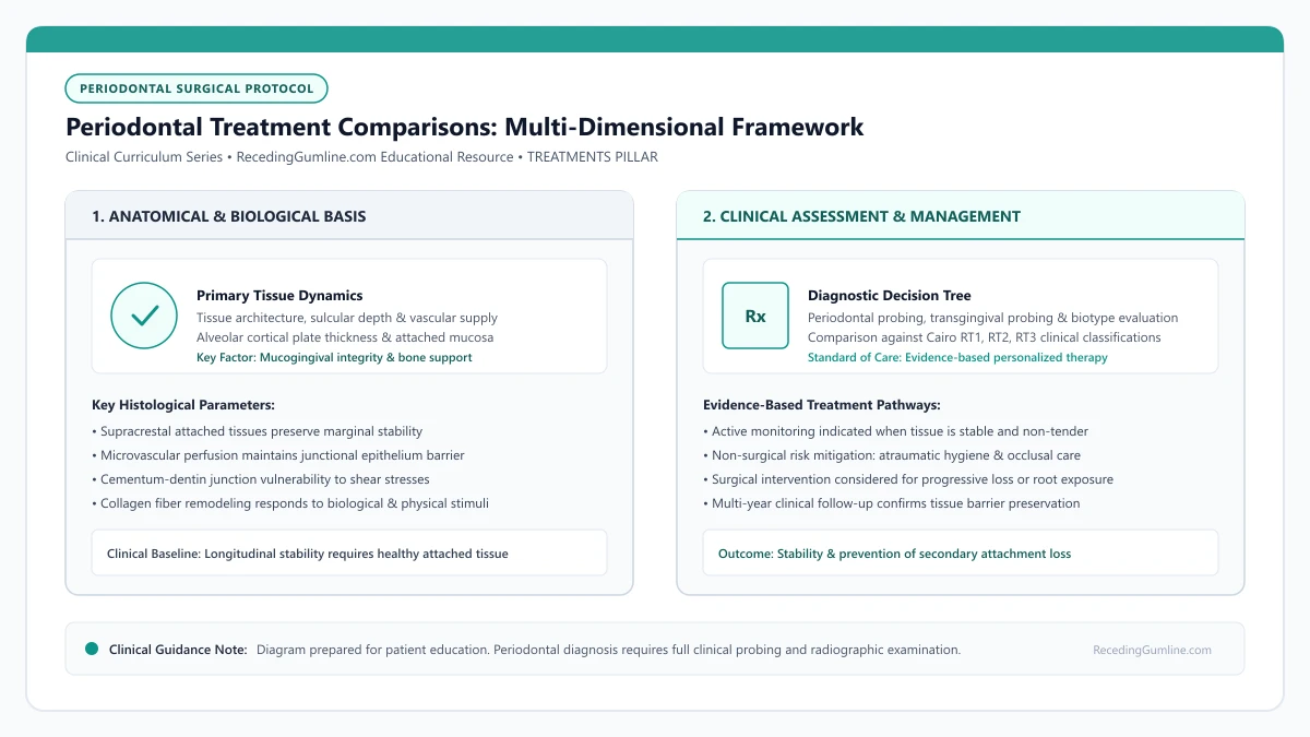
Educational illustration: Periodontal Treatment Comparisons: Multi-Dimensional Framework. Clinical management requires comprehensive periodontal evaluation rather than isolated self-assessment.
Source: RecedingGumline.com Clinical Editorial Team (Proprietary educational diagram for RecedingGumline.com)
Active Monitoring vs. Surgical Intervention
The first fundamental clinical decision is determining whether a receded site warrants surgical intervention or active conservative monitoring. Not all receded gingiva requires surgery. If a site exhibits shallow recession (1 to 2 mm), an adequate zone of attached keratinized tissue (>= 2 mm), zero bleeding on probing, and zero progression on sequential 6-month periodontal charts, surgical grafting offers minimal therapeutic advantage over vigilant monitoring.
Conversely, surgical consultation is strongly indicated when sequential measurements demonstrate progressive attachment loss, when attached gingiva is completely absent (< 1 mm), when root hypersensitivity fails to respond to desensitizing agents, or when the patient experiences severe aesthetic concern in the smile zone.
Intervening proactively in progressive cases preserves bone support and allows the surgeon to operate while the defect remains a highly predictable Cairo RT1 lesion, before interdental bone loss transforms it into a less predictable RT2 or RT3 defect.
Key Clinical Insights
- Stable, asymptomatic recession with adequate attached tissue is appropriately managed via monitoring.
- Progressive tissue retreat, absent keratinized tissue, or severe sensitivity prompts surgical consultation.
- Treating defects early (Cairo RT1) ensures the highest surgical predictability for complete root coverage.
Graft Material Selection: Autogenous CTG vs. Allograft Biomaterials
When soft-tissue grafting is indicated, clinicians must choose between autogenous donor tissue harvested from the patient's palate and processed biomaterials. The Subepithelial Connective Tissue Graft (CTG) harvested from the palate remains an established clinical benchmark in terms of complete root coverage percentage and long-term tissue thickness.
However, harvesting palatal tissue introduces a second surgical site, increasing postoperative morbidity and discomfort during the first 7 to 10 days. Furthermore, the volume of harvestable palatal tissue is anatomically limited, making it challenging to treat full-arch, multiple-tooth recession in a single operation.
Acellular Dermal Matrix (ADM) allografts and xenogeneic collagen matrices completely eliminate the palatal donor wound, significantly reducing post-surgical pain and eliminating donor anatomical constraints. While allografts achieve high root coverage (typically 80% to 90% in RT1 sites), extensive systematic reviews show that autogenous CTG provides greater long-term keratinized tissue width and marginal stability over 5- to 10-year follow-up intervals.
Key Clinical Insights
- Autogenous CTG provides the highest rate of complete root coverage and durable tissue thickening.
- Palatal harvesting introduces a secondary surgical wound and is limited by available donor anatomy.
- Allografts avoid donor site pain and treat extensive multi-tooth defects, with slightly lower complete coverage.
Flap Design Comparison: Coronally Advanced Flap vs. Tunneling
The surgical approach utilized to position the graft over denuded roots dramatically impacts both aesthetic integration and postoperative recovery. The Coronally Advanced Flap (CAF) with vertical releasing incisions provides exceptional surgical access, allowing the clinician to visualize root anatomy, thoroughly debride root surfaces, and mobilize tissues with maximum tension-free coronal advancement.
However, vertical releasing incisions sever the collateral supraperiosteal vascular supply and can leave permanent, visible mucosal scar lines. In contrast, the Coronally Advanced Tunnel (CAT) and vestibular access techniques preserve the interdental papillae and eliminate vertical cuts entirely, maximizing blood supply and achieving seamless aesthetic blending.
While tunneling offers refined aesthetic results and reduced swelling, it is technically demanding and requires sufficient baseline flap thickness. For deep, isolated defects with prominent root bulges, an open coronally advanced flap remains a highly predictable, versatile modality.
Key Clinical Insights
- Coronally Advanced Flaps provide unmatched surgical access but may produce minor mucosal scarring.
- Tunneling techniques preserve interdental papillae and lateral blood supply, avoiding all vertical scars.
- Surgeon experience and anatomical defect geometry dictate whether a tunnel or flap approach is optimal.
Multi-Dimensional Clinical Decision Framework
Synthesizing these therapeutic variables into a cohesive decision framework ensures that clinical treatment plans align with both biological reality and patient priorities. The decision matrix evaluates four key dimensions: Clinical Indication, Anatomical Predictability (Cairo classification), Patient Morbidity Profile, and Long-Term Value.
For a Cairo RT1 defect on a single anterior tooth with high aesthetic demands, a tunnel procedure paired with an autogenous CTG represents the premium treatment choice. For a non-aesthetic mandibular premolar with zero attached tissue, a free gingival graft offers the most durable, cost-effective structural solution.
Patients must participate actively in this decision-making process, weighing trade-offs between secondary donor site discomfort, material costs, procedural predictability, and aesthetic outcomes in consultation with their periodontist.
Key Clinical Insights
- Treatment planning must balance clinical predictability, aesthetic goals, and surgical morbidity.
- Aesthetic anterior defects favor microsurgical tunneling with subepithelial connective tissue grafts.
- Non-aesthetic functional deficits are efficiently stabilized with durable free gingival grafts.
Long-Term 5- to 10-Year Clinical Stability & Creeping Attachment
Evaluating periodontal treatment options requires examining long-term outcomes beyond immediate 6-month postoperative metrics. Longitudinal clinical studies tracking patients over 5 to 10 years reveal distinct differences in stability among grafting modalities. Subepithelial connective tissue grafts combined with coronally advanced flaps or tunnels demonstrate the highest rates of long-term stability, with over 80% of complete root coverage sites maintaining full coverage a decade later.
In contrast, non-surgical monitoring without tissue augmentation frequently exhibits gradual, slow apical migration of the margin over time, particularly in individuals who maintain high-pressure brushing habits or possess less than 1 mm of attached keratinized gingiva.
An intriguing biological phenomenon observed following autogenous grafting is creeping attachment—the slow, post-surgical coronal migration of the gingival margin over months and years. Documented primarily on narrow recession defects on anterior teeth, creeping attachment can provide an additional 0.5 to 1.5 mm of spontaneous root coverage as mature collagen fibers reorganize and stabilize.
Key Clinical Insights
- Autogenous connective tissue grafts exhibit greater 10-year stability compared to non-augmented sites.
- Sites with inadequate attached tissue (< 1 mm) show ongoing vulnerability to progressive recession.
- Creeping attachment can spontaneously enhance root coverage by 0.5 to 1.5 mm over multi-year healing.
Clinical Reality Check
There is no universal "best" treatment for gum recession. A successful outcome requires matching the specific anatomical defect, biotype, and Cairo classification with the appropriate surgical or non-surgical modality.
Questions to Ask Your Dentist or Periodontist
- Which treatment modality (monitoring, CTG, tunneling, or allograft) is best suited for my specific teeth?
- What are the trade-offs between using my own palatal tissue versus a donor allograft material?
- What is your personal clinical success rate with complete root coverage for defects like mine?
- How does the expected recovery time and pain level differ between these surgical approaches?
- What are the total cost differences between the various treatment options available for my condition?
Unsure What Your Gum Changes Mean?
Take our free, evidence-based Gum Recession Assessment — approximately 3 minutes. Identify potential risk factors, evaluate symptoms, and receive personalized discussion questions for your dentist or periodontist.
Non-diagnostic educational triage. Private, secure, completed in your browser.
Related Educational Topics
Scientific Literature & Clinical Guidelines
4sources · Hide ▲
- Chambrone L, Salinas Ortega MA, Sukekava F, Rotundo R, Kalemaj Z, Buti J, Pini Prato GP (2018).
"Root coverage procedures for treating localised and multiple recession-type defects." Cochrane Database of Systematic Reviews.
Clinical relevance: Cochrane systematic review evaluating root-coverage procedures for localized and multiple recession-type defects. While subepithelial connective tissue grafts (SCTG) combined with coronally advanced flaps demonstrated higher rates of complete root coverage and keratinized tissue gain compared to flap advancement alone, evidence quality varied across outcomes and clinical decisions must balance donor site morbidity and patient-reported outcomes.
- Cairo F, Nieri M, Cincinelli S, Mervelt J, Pagliaro U (2011).
"The interproximal clinical attachment level to classify gingival recessions and predict root coverage outcomes: an explorative and reliability study." Journal of Clinical Periodontology.
Clinical relevance: Exploratory and reliability study establishing the Cairo classification based on interdental clinical attachment level (CAL): RT1 (no interproximal attachment loss; complete root coverage is clinically predictable), RT2 (interproximal attachment loss <= buccal loss; partial coverage predictable), and RT3 (interproximal loss exceeds buccal recession; complete coverage not predictable).
- Zucchelli G, Mounssif I (2015).
"Periodontal plastic surgery." Periodontology 2000.
Clinical relevance: Peer-reviewed review of periodontal plastic surgery modalities, detailing flap design, coronally advanced flaps, autogenous connective tissue grafting, tunneling techniques, and anatomical factors governing aesthetic and functional root coverage.
- Tonetti MS, Greenwell H, Kornman KS (2018).
"Staging and grading of periodontitis: Framework and proposal of a new classification and case definition." Journal of Clinical Periodontology.
Clinical relevance: Consensus framework establishing the multidimensional staging (severity and extent of periodontal tissue breakdown) and grading (biological rate of disease progression, incorporating smoking and diabetes as grade modifiers) for periodontitis. It addresses periodontitis diagnosis and staging, not the classification of localized gingival recession defects.
Important Medical Notice
The contents of RecedingGumline.com, including text, graphics, self-assessment calculators, and other materials, are intended solely for educational and informational purposes. This content is not intended to replace professional dental examination, diagnosis, or treatment. Always seek the advice of a qualified dentist, periodontist, or other licensed oral healthcare provider with any questions you may have regarding a medical or dental condition. Never disregard professional medical advice or delay seeking it because of something you read on this website.