Conservative & Non-Invasive Gum Recession Care
Conservative care represents the foundational layer of gum recession management. When gingival margins have receded without active microbial periodontal destruction or severe deficiency of attached keratinized tissue, non-surgical approaches aim to eliminate mechanical friction, manage marginal inflammation, desensitize exposed root surfaces, and stabilize existing attachment levels. While conservative protocols cannot reposition a receded margin back to the cementoenamel junction, they are essential for halting progressive tissue loss.
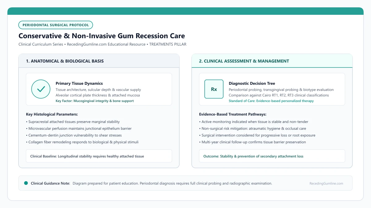
Educational illustration: Conservative & Non-Invasive Gum Recession Care. Clinical management requires comprehensive periodontal evaluation rather than isolated self-assessment.
Source: RecedingGumline.com Clinical Editorial Team (Proprietary educational diagram for RecedingGumline.com)
Oral Hygiene Mechanics & Bristle Selection
Vigorous horizontal scrubbing with medium or hard toothbrush bristles is a well-documented mechanical contributor to gingival recession and wedge-shaped cervical defects. When patients scrub back and forth with high pressure, stiff nylon filaments exert concentrated shear forces across the free gingival margin. In areas where overlying tissue is naturally thin, this repetitive physical microtrauma strips away the fragile epithelial attachment.
Periodontal clinical consensus strongly recommends transitioning to the Modified Bass Technique. In this atraumatic approach, the brush head is angled at 45 degrees toward the gingival sulcus. Gentle vibratory micro-movements are applied without dislodging the bristle tips, disrupting subgingival biofilm within the sulcular crevice before the brush is rolled coronally away from the gumline.
Selecting the proper brush is paramount. Patients should utilize only extra-soft or ultra-soft tapered bristles with microscopically polished, end-rounded tips. Electric toothbrushes equipped with smart pressure sensors offer valuable real-time feedback, automatically slowing oscillation or pulsing warning lights when applied force exceeds safe physiological thresholds (typically 1.5 to 2.0 Newtons).
Key Clinical Insights
- Use only extra-soft, multi-tufted bristles with polished, end-rounded filament tips.
- The Modified Bass Technique cleans the sulcus without abrading the delicate marginal gingival collar.
- Smart pressure-sensing electric toothbrushes provide biofeedback to eliminate traumatic scrubbing.
Low-Abrasivity Dentifrices & Chemical Plaque Control
Toothpastes contain varying concentrations of abrasive mineral particles formulated to remove extrinsic surface stains. However, exposed root cementum has a mineral hardness less than half that of enamel, rendering it highly susceptible to rapid gouging from abrasive toothpastes.
The Relative Dentin Abrasivity (RDA) scale provides a standardized measurement of dentifrice abrasiveness. For patients with exposed roots or thin marginal tissue, periodontists recommend selecting formulations with an RDA score below 70 to 80. Whitening, tartar-control, and charcoal formulations frequently exceed 100 to 150 RDA, accelerating root wear and cervical notch formation.
When mechanical plaque removal must be minimized—such as around hypersensitive or inflamed margins—adjunctive chemical plaque control can be introduced. Therapeutic mouth rinses containing essential oils, cetylpyridinium chloride (CPC), or short-term chlorhexidine digluconate help suppress bacterial biofilm formation without inflicting physical friction.
Key Clinical Insights
- Exposed root cementum wears rapidly under high-abrasivity toothpastes; select RDA < 70.
- Avoid commercial whitening and charcoal pastes that accelerate cervical gouging.
- Non-abrasive therapeutic antimicrobial rinses assist in biofilm control around tender margins.
Managing Exposed Root Sensitivity: Hydrodynamic Occlusion
When gingival recession exposes root cementum, the protective mineral layer can easily wear away, uncapping thousands of microscopic dentinal tubules. According to Brännström's hydrodynamic theory, external thermal, osmotic, or tactile stimuli cause rapid fluid displacement within these open tubules, mechanically deforming A-delta and A-beta nerve fibers in the dental pulp and triggering sharp, transient pain.
Conservative desensitization therapies operate via two distinct biological pathways: nerve depolarization and mechanical tubule occlusion. Nerve-depolarizing agents—predominantly potassium nitrate (5%)—increase extracellular potassium ion concentrations around pulpal sensory nerves, blocking axonal action potential repolarization and calming nerve excitability over 2 to 4 weeks of consistent use.
Tubule-occluding agents act immediately by physically sealing the patent tubule orifices. In-office professional varnishes (such as 5% sodium fluoride or glutaraldehyde-HEMA formulations) and at-home toothpastes containing stannous fluoride, calcium sodium phosphosilicate (NovaMin), or arginine-calcium carbonate precipitate insoluble mineral plugs that block fluid movement and provide durable relief.
Key Clinical Insights
- Brännström's hydrodynamic theory explains that fluid movement in exposed tubules triggers nerve pain.
- Potassium nitrate calms pulpal sensory nerves via prolonged membrane depolarization.
- Stannous fluoride and mineralizing agents physically plug tubule orifices, stopping fluid shift.
Occlusal Splints for Bruxism and Abfraction Management
Nocturnal bruxism (teeth grinding) and chronic daytime clenching generate tremendous non-axial forces along the dentition. When teeth absorb heavy lateral loading, the crown undergoes microscopic bending flexure at the cervical fulcrum near the cementoenamel junction. Over time, this flexural strain can cause fatigue microfractures in the crystalline cervical enamel and dentin, contributing to Non-Carious Cervical Lesions (abfractions).
While occlusal trauma alone does not induce periodontal pockets in a plaque-free environment, severe tooth flexure disrupts the marginal soft-tissue seal and accelerates attachment loss in individuals with thin underlying alveolar bone. Fabricating a custom-fitted hard acrylic occlusal splint (nightguard) provides a vital protective shield.
The occlusal guard establishes mutually protected occlusion, ensuring that canine guidance discludes posterior teeth during lateral excursions and distributing biting forces evenly across the entire arch. This eliminates localized hyperfunction and stabilizes fragile cervical margins.
Key Clinical Insights
- Lateral clenching forces cause tooth flexure, concentrating mechanical strain at the cervical gumline.
- Abfraction microfractures weaken cervical tooth structure and accelerate soft-tissue retreat.
- Custom hard acrylic nightguards redistribute biting forces and eliminate destructive lateral stresses.
Clinical Monitoring Protocols: Assessing Longitudinal Stability
Conservative therapy is not a static endpoint; it requires structured clinical surveillance to confirm that recession has truly halted. During regular periodontal recall appointments (every 3 to 6 months), the dental professional performs meticulous measurements using a calibrated periodontal probe (such as the UNC-15 probe).
Key clinical parameters recorded include probing depth (PD), clinical attachment level (CAL), recession depth from the CEJ, width of keratinized tissue, and presence of bleeding on probing (BOP). High-resolution intraoral clinical photography or digital 3D surface scanning provides objective baseline documentation to detect sub-millimeter tissue changes over time.
If sequential measurements demonstrate progressive attachment loss, recurrent inflammation, or a complete absence of attached keratinized tissue, conservative care must be re-evaluated, and a referral to a periodontist for surgical root coverage or tissue augmentation should be considered.
Key Clinical Insights
- Regular 3- to 6-month clinical exams measure probing depth, recession depth, and keratinized width.
- Intraoral photographs and digital scans provide objective comparisons to detect minor tissue changes.
- Documented progression or lack of attached tissue prompts surgical periodontal consultation.
Coronally Advanced Flap (CAF) Surgery for Gum Recession
For patients seeking root coverage for a receding gumline, the Coronally Advanced Flap (CAF) is a foundational periodontal plastic surgery technique designed to cover denuded root surfaces and restore the marginal gingiva. By mobilizing adjacent keratinized soft tissue and advancing it coronally over the exposed root, surgeons can achieve esthetic root coverage without harvesting a large external graft, or combine CAF with an underlying connective tissue graft for maximum predictability.
• CAF mobilizes existing keratinized tissue apically and advances it over the receded root surface.
• Sufficient pre-existing keratinized tissue width (ideally ≥ 2 mm) is an essential anatomical requirement when CAF is performed alone.
• Combining CAF with a subepithelial connective tissue graft (CAF + SCTG) represents the established benchmark surgical protocol for complete root coverage.
• Tension-free flap mobilization achieved through periosteal releasing incisions is the single most critical factor for surgical flap stability.
Surgical Mechanics: Incision Design and Tension-Free Flap Release
The success of a coronally advanced flap hinges on micro-surgical precision. The surgeon creates horizontal sulcular and beveled papilla-sparing incisions to mobilize the full-thickness mucoperiosteal flap up to the mucogingival junction, transitioning into a split-thickness dissection in the alveolar mucosa.
The decisive step is the periosteal releasing incision: micro-incisions made through the deep elastic fibers of the periosteum. This relieves all muscle and tissue tension, allowing the flap to passively drape 1 to 2 millimeters coronal to the cementoenamel junction without any retraction force.
Micro-surgical instruments and high-magnification loupes ensure atraumatic handling of thin gingival tissues during incision and split-thickness dissection. Preserving the interdental papillae intact ensures that the lateral vascular supply remains uninterrupted throughout the procedure.
Key Clinical Insights
- Split-thickness dissection in mucosal tissue preserves deep vascular supply
- Periosteal release eliminates muscle tension pulling the flap downward
- The flap must sit passively coronal to the CEJ without requiring suture tension
Anatomical Prerequisites: Tissue Thickness and Keratinized Width
Not every receded tooth is suitable for CAF alone. For a coronally advanced flap to succeed as a monotherapy, the patient must possess a baseline band of keratinized gingiva apical to the recession (ideally at least 1.5 to 2.0 mm) and a flap thickness exceeding 0.8 to 1.0 mm.
If the pre-existing tissue is paper-thin (< 0.8 mm) or lacks keratinization entirely, moving that tissue upward will result in gradual re-recession over time. In such cases, surgeons place a connective tissue graft beneath the flap (CAF + SCTG) to transform the biotype from thin to thick.
Complete release of the underlying periosteum at the base of the flap eliminates all muscular tension from the surrounding facial expressions. When tension is eliminated, the flap passively rests at or coronal to the cementoenamel junction without relying on suture tension.
Key Clinical Insights
- Flap thickness ≥ 0.8 mm is required for predictable root coverage with CAF alone
- At least 2 mm of keratinized tissue must be present apical to the recession defect
- Thin tissues require an underlying connective tissue graft to prevent relapse
Microsurgical Suturing and Vascular Bed Integration
Once mobilized, the flap is secured using ultra-fine microsurgical sutures (6-0 or 7-0 gauge). Sling sutures or suspended mattress sutures anchor the flap securely around the neck of the tooth, compressing the margins against the de-epithelialized interdental papillae.
Because the flap remains pedicled (attached at its base), it maintains its original apical blood supply while simultaneously receiving new collateral circulation from the de-epithelialized papillae, ensuring rapid revascularization within the first 5 to 7 days.
Long-term periodontal trials demonstrate favorable aesthetic color blending and contour harmony with coronally advanced flaps compared to traditional free gingival grafts. Patient satisfaction is consistently high due to the avoidance of open palatal wound harvesting.
Key Clinical Insights
- 6-0 and 7-0 microsurgical sutures minimize tissue trauma and scarring
- De-epithelialized anatomical papillae act as vital vascular recipient beds
- Pedicle base maintains uninterrupted blood supply during initial healing
Surgical Biomechanics: Split-Full-Split Thickness Flap Elevation
The coronally advanced flap (CAF) is a cornerstone of periodontal plastic surgery for single and multiple recession defects. Achieving predictable, tension-free coronal advancement requires the meticulous "split-full-split" flap elevation technique.
Flap elevation commences with a split-thickness incision at the interdental surgical papillae, preserving periosteum over the bone. Over the denuded root and marginal bone crest, a full-thickness mucoperiosteal flap is elevated to include the periosteum, protecting the marginal vascular plexus.
Apical to the mucogingival junction, the dissection converts back to split-thickness, severing muscle insertions and elastic fibers within the alveolar mucosa. This releases all basal tension, allowing the flap to passively drape coronally past the cementoenamel junction with zero elastic recoil.
Key Clinical Insights
- Split-thickness papilla dissection preserves periosteal vascular beds on interdental bone.
- Full-thickness dissection over marginal bone crest preserves the periosteal microvascular plexus.
- Apical muscle release allows the flap to passively drape coronally with zero tissue tension.
Suture Materials & Papillary Anchorage Dynamics
Securing the coronally advanced flap in a stable, coronal position during the critical 14-day healing window depends on suture selection and microsurgical anchoring techniques. Surgeons utilize ultra-fine 6-0 or 7-0 monofilament sutures (polypropylene or polyvinylidene fluoride) with precision micro-needles.
Monofilament sutures eliminate the "wicking effect" characteristic of braided silk or vicryl sutures, preventing the migration of oral bacteria through the suture tract into the healing graft bed. The sutures are placed using sling or suspended mattress configurations that anchor around tooth contact points.
Tension-free coronal stabilization positioned 1.0 to 1.5 millimeters coronal to the cementoenamel junction compensates for minor postoperative tissue contraction, ensuring stable long-term root coverage.
Key Clinical Insights
- Ultra-fine 6-0 or 7-0 monofilament sutures eliminate capillary bacterial wicking.
- Sling and suspended mattress sutures anchor securely around interproximal tooth contact points.
- Positioning the flap 1.0-1.5 mm coronal to the CEJ anticipates physiological contraction during healing.
Free Gingival Graft (FGG) Surgery for Gum Recession
The Free Gingival Graft (FGG) is one of the earliest and most extensively documented surgical procedures in periodontics. First described in the 1960s, its primary clinical purpose in the management of receding gums is not cosmetic root coverage, but the predictable creation of a wide, dense band of attached, keratinized gingiva to arrest progressive recession and establish periodontal stability.
• The primary indication for an FGG is augmenting the band of attached keratinized tissue rather than achieving cosmetic root coverage.
• FGG harvests both surface epithelium and underlying lamina propria from the palate as a full-thickness soft-tissue unit.
• It is frequently indicated on mandibular incisors and premolars where muscle pull and lack of attached tissue threaten tooth stability.
• FGG heals with a distinct, paler appearance and firmer texture that resembles palatal mucosa rather than native gingiva.
When Is a Free Gingival Graft Indicated? Halting Progressive Recession
A free gingival graft is recommended when a tooth has zero or minimal remaining attached keratinized tissue—a condition where the fragile, movable alveolar mucosa extends directly to the gingival margin. Without an attached collar, daily toothbrushing and lip movements pull the margin away from the tooth, facilitating bacterial penetration and progressive attachment loss.
FGG is most commonly performed on the facial surfaces of lower incisors and premolars. In these areas, aesthetic appearance is rarely a primary concern, but structural preservation against strong mentalis muscle attachments or high frenal pulls is paramount for keeping the teeth.
The primary objective of a free gingival graft is to establish a dense, resilient collar of non-movable keratinized tissue that resists mechanical brushing abrasion. While root coverage is often minimal, halting progressive apical migration of the mucosal margin protects underlying alveolar bone.
Key Clinical Insights
- Indicated when movable alveolar mucosa extends directly to the tooth margin
- Commonly performed on lower incisors where strong muscle pull accelerates recession
- Arrests progressive recession even if exposed roots are not fully recovered
Surgical Protocol: Recipient Bed Preparation and Palatal Harvest
The procedure begins at the recipient site, where the clinician performs a partial-thickness dissection to separate movable mucosa and muscle fibers from the underlying periosteum, creating a stable, vascular recipient bed firmly bound to the alveolar bone.
At the palate, the surgeon harvests a graft consisting of both the keratinized oral epithelium and the dense underlying lamina propria (approximately 1.0 to 1.5 mm thick). The graft is trimmed and meticulously sutured onto the periosteal bed, immobilizing it completely to allow revascularization.
Patients with severe high frenum pull and minimal attached gingiva benefit substantially from the deepening of the vestibular fornix provided by the graft. The transplanted fibrous tissue halts the transmission of lip tension directly to the fragile marginal tissue.
Key Clinical Insights
- Recipient bed is anchored to fixed periosteum to eliminate muscle movement
- Full-thickness graft includes epithelium and dense lamina propria from the palate
- Immobilization with compressive sutures is vital to establish plasmatic diffusion
Tissue Maturation: Why FGG Leaves a Distinct Cosmetic "Patch"
Because the free gingival graft retains its palatal epithelial genetics, it heals with the histology of the roof of the mouth. This means the grafted area becomes thick, dense, and heavily keratinized, but appears noticeably paler and whiter than the adjacent pink gingiva.
This "tire-patch" appearance is completely functional and healthy, providing an impenetrable barrier against recession. However, because of this stark aesthetic difference, periodontists rarely place free gingival grafts on upper front teeth where they would be visible when smiling.
Palatal donor harvesting for free gingival grafts involves taking both surface epithelium and underlying dense lamina propria. The resulting palatal donor site heals via secondary intention over three to four weeks, necessitating protective stents and gentle oral hygiene.
Key Clinical Insights
- Palatal genetic memory results in a paler, firmer, heavily keratinized band
- Provides unmatched structural resilience against brushing and plaque trauma
- Rarely used in the maxillary aesthetic smile zone due to visible color contrast
Clinical Indications: Eliminating High Muscle Pull in Mandibular Sites
The free gingival graft (FGG) is primarily indicated when the clinical objective is widening the zone of keratinized, attached gingiva rather than achieving primary cosmetic root coverage. It is most frequently deployed in the anterior mandible, where shallow vestibular depth and aberrant frenal attachments create dynamic mechanical pull on the gingival margin.
When teeth exhibit thin, movable alveolar mucosa extending directly to the margin, even minor lip movements pull the sulcus open, facilitating plaque accumulation and rapid recession. An FGG surgically arrests this breakdown by creating a wide, dense, immovable band of keratinized tissue anchored to the periosteum.
By establishing a static, collagenous mucogingival barrier and deepening the vestibular fornix, the FGG halts further apical attachment loss and provides a maintainable foundation for lifelong hygiene.
Key Clinical Insights
- FGG is the established benchmark procedure for increasing the width of keratinized, attached tissue.
- Highly effective in the anterior mandible where shallow vestibules and muscle pull drive recession.
- Creates an immovable, dense collagenous barrier that halts further clinical attachment loss.
Donor Palatal Harvest & Secondary Intention Epithelialization
Unlike a subepithelial connective tissue graft, a free gingival graft includes both the lamina propria connective tissue and the overlying stratum corneum epithelium. The donor tissue is harvested from the premolar region of the hard palate at a calibrated uniform thickness of 1.0 to 1.5 millimeters.
Because the surface epithelium is excised, the palatal donor site is left as an open wound that must heal by secondary intention. Epithelial cells must migrate across the bare connective tissue bed from the wound margins, a process requiring 14 to 21 days for complete re-epithelialization.
At the recipient site, the grafted epithelium undergoes complete desquamation during the first four days as the graft survives on plasmatic circulation, followed by re-epithelialization from the graft margins to form durable, pale keratinized mucosa.
Key Clinical Insights
- FGG harvests include both surface epithelium and underlying dense connective tissue (1.0-1.5 mm thick).
- The donor palatal wound heals by secondary intention, requiring 2-3 weeks for full re-epithelialization.
- Grafted tissue undergoes initial surface desquamation before establishing a durable keratinized zone.
Scaling & Root Planing vs. Routine Cleaning for Gum Recession
In understanding gum recession and periodontal infection, many patients are confused when told that a standard six-month cleaning is no longer sufficient and that they require scaling and root planing. Understanding the clear anatomical, diagnostic, and procedural distinctions between preventive hygiene and therapeutic deep cleaning clarifies why this non-surgical therapy is vital for arresting tissue breakdown.
• Routine prophylaxis (CDT D1110) is a preventive procedure confined to supragingival tooth surfaces in patients with healthy periodontium.
• Scaling and root planing (CDT D4341/D4342) is a therapeutic intervention treating subgingival root surfaces affected by active attachment loss.
• Deep cleaning removes calcified calculus and bacterial endotoxins embedded within the cementum to promote soft-tissue re-adaptation.
• Prophylaxis cannot stop active periodontitis or resolve subgingival microbial biofilms residing in deep pockets.
Routine Prophylaxis (D1110): Scope and Clinical Indications
A standard dental cleaning, known clinically as dental prophylaxis (ADA code D1110), is purely preventive. It is indicated for patients who exhibit clinically healthy gingival tissues or mild gingivitis without any radiographic bone loss or clinical attachment loss.
During prophylaxis, the hygienist uses ultrasonic scalers and hand instruments to polish the crowns of the teeth and remove supragingival plaque and tartar. The instrumentation stops at or just slightly below the healthy marginal gingival collar (within normal 1 to 3 mm sulci).
Routine dental prophylaxis focuses on supra-gingival polishing and superficial calculus removal for patients with intact periodontal attachment and minimal inflammation. In contrast, scaling and root planing treats diseased cementum within pathological subgingival pockets.
Modern ultrasonic scaling tips operate with specialized linear micro-vibrations, flushing subgingival crevices with cooled antimicrobial irrigants while gently removing deep calculus deposits without damaging the underlying sound dentin matrix.
Key Clinical Insights
- Intended exclusively for patients with intact periodontium or mild gingivitis
- Removes plaque and tartar from coronal enamel surfaces above the gumline
- Does not address subgingival pathogens or debride root surfaces in pockets ≥ 4 mm
Scaling and Root Planing (D4341/D4342): Therapeutic Debridement
Scaling and root planing (SRP), colloquially termed "deep cleaning," is a specialized therapeutic procedure. It is prescribed when diagnostic charting reveals periodontal pocket depths of 4 mm or deeper accompanied by bleeding and radiographic bone loss.
SRP involves two clinical actions: scaling removes tenacious hard calculus deposits from the root surface, and root planing meticulously shaves away microscopic layers of necrotic cementum impregnated with bacterial lipopolysaccharide endotoxins, creating a biocompatible surface for tissue re-adaptation.
Thorough ultrasonic instrumentation combined with sharp curettes disrupts complex anaerobic bacterial biofilms residing within deep pocket niches. Removing endotoxin-impregnated necrotic cementum provides a biocompatible biological surface for junctional epithelial reattachment.
Key Clinical Insights
- Prescribed for active periodontitis with pocketing ≥ 4 mm and bone loss
- Removes subgingival calculus and bacterial endotoxins embedded in root cementum
- Performed under local anesthesia to ensure thorough debridement without discomfort
Healing Responses and the Transition to Periodontal Maintenance
Following scaling and root planing, inflamed, swollen pocket tissues undergo dramatic biological resolution. Over 4 to 8 weeks, edema subsides, collagen fibers reorganize, and a long junctional epithelium forms against the cleaned root surface, reducing pocket depths by 1 to 2 millimeters.
Because periodontal pathogens repopulate subgingival pockets within 90 to 120 days, patients who complete SRP cannot return to standard twice-yearly prophylaxis. They transition into ongoing Periodontal Maintenance (CDT D4910) at 3- to 4-month intervals to maintain clinical stability.
A formal periodontal re-evaluation examination is conducted four to six weeks following scaling and root planing. This appointment measures pocket reduction, resolves residual bleeding on probing, and determines whether localized periodontal surgery or ongoing maintenance is required.
Key Clinical Insights
- Tissues shrink as inflammation subsides, which may increase visual root exposure
- Long junctional epithelium seals against the planed root to reduce pocket depths
- Requires 3- to 4-month periodontal maintenance recalls to prevent bacterial repopulation
Therapeutic Endpoints: Biofilm Detoxification vs. Over-Instrumentation
Scaling and Root Planing (SRP) differs fundamentally from a routine prophylaxis in its therapeutic intent and anatomical scope. While a standard cleaning removes supragingival plaque and calculus from crown enamel, SRP is a therapeutic intervention performed within deep periodontal pockets along diseased root cementum.
Historical periodontal concepts emphasized aggressive root planing until the root felt glass-smooth, inadvertently stripping away healthy cementum and exposing sensitive dentinal tubules. Modern clinical protocols prioritize biofilm detoxification and selective calculus removal using ultrasonic micro-inserts with light overlapping strokes.
The primary endpoint is biological compatibility: removing endotoxins embedded in superficial cementum while preserving as much healthy cementum and native collagen fibers as possible to facilitate cellular reattachment.
Key Clinical Insights
- Prophylaxis is preventive and supragingival; SRP is therapeutic and subgingival.
- Modern SRP emphasizes biofilm detoxification rather than aggressive cementum removal.
- Preserving sound root cementum minimizes post-treatment root sensitivity and pulp irritation.
The Six-Week Periodontal Re-Evaluation: Measuring Clinical Endpoints
The success of Scaling and Root Planing is formally assessed at a mandatory four- to six-week re-evaluation visit. This interval allows adequate biological time for junctional epithelial re-attachment, connective tissue fiber remodeling, and resolution of inflammatory edema.
During this examination, the clinician repeats complete full-mouth periodontal probing to evaluate primary clinical endpoints: reduction in pocket probing depths, elimination of bleeding on probing (BOP), and gain of clinical attachment.
Pocket depth reductions of 1 to 2 millimeters are typical as swollen marginal tissues shrink and a long junctional epithelium forms. Persistent deep pockets (≥ 5 mm) with active bleeding indicate refractory disease or complex root anatomy, warranting referral for advanced periodontal surgery.
Key Clinical Insights
- Mandatory re-evaluation occurs 4-6 weeks post-SRP to assess biological tissue response.
- Primary success metrics include pocket depth reduction and elimination of bleeding on probing.
- Residual bleeding pockets ≥ 5 mm require advanced surgical periodontal therapy.
Periodontal Maintenance Frequency for Gum Recession Patients
Following active treatment for periodontal disease—whether scaling and root planing or surgical grafting—patients are universally placed on a 3- to 4-month periodontal maintenance schedule. This strict recall interval is not an arbitrary dental scheduling habit; it is grounded in landmark microbiological and clinical trials tracking the exact repopulation timeline of pathogenic subgingival biofilms.
• Subgingival bacterial biofilms repopulate treated pockets and re-establish pathogenic virulence within 9 to 12 weeks.
• Landmark longitudinal trials demonstrate that 3-month recalls preserve bone and attachment levels, whereas 6-month recalls allow recurrent disease.
• Periodontal maintenance (CDT D4910) includes continuous site-specific debridement and full-mouth pocket depth monitoring.
• Lifelong maintenance is required because periodontitis is a manageable chronic disease with high relapse potential if left unmonitored.
The Bacterial Succession Timeline: Why Biofilms Recover at 90 Days
When a skilled clinician performs scaling and root planing, subgingival bacterial loads are reduced by over 99%. However, bacteria are microscopic organisms inhabiting deep dentinal tubules and tissue micro-crevices, meaning the subgingival pocket is never completely sterilized.
In the weeks following debridement, beneficial pioneer streptococcal species colonize the pocket first. However, between 9 and 12 weeks, virulent anaerobic pathogens—including Porphyromonas gingivalis, Treponema denticola, and Tannerella forsythia—regain predominance. Disrupting this pathogenic shift at the 12-week mark stops tissue destruction before attachment loss resumes.
Clinical microbiology demonstrates that pathogenic anaerobic bacterial complexes recolonize subgingival periodontal pockets within 9 to 12 weeks following thorough mechanical debridement. A 3-month recall interval disrupts this repopulation cycle before mature pathogenic biofilms can trigger bone destruction.
Key Clinical Insights
- Scaling reduces subgingival bacteria dramatically but cannot sterilize pockets
- Beneficial pioneer bacteria are gradually replaced by virulent anaerobes over 9 to 12 weeks
- Quarterly professional debridement resets the biofilm clock before bone destruction triggers
Clinical Proof: The Landmark Michigan and Scandinavian Studies
The clinical necessity of the 3-month recall was proven in historic longitudinal studies led by Dr. Sigurd Ramfjord at the University of Michigan and researchers in Gothenburg, Sweden. Across 10- to 15-year trials, patients who received thorough periodontal maintenance every 3 months maintained their bone levels and suffered almost zero tooth loss.
Conversely, patients who received excellent initial treatment but were returned to unmonitored standard 6-month cleanings experienced recurrent pocket deepening, accelerated bone resorption, and high rates of tooth extraction. The maintenance schedule, not the initial surgery alone, proved to be the ultimate protector of teeth.
During maintenance visits, selective subgingival scaling is performed at persistent deep sites alongside targeted topical antimicrobial irrigation. Evaluating bleeding on probing and gingival margin stability at each recall allows rapid intervention before systemic attachment loss advances.
Key Clinical Insights
- Patients on 3-month recalls maintained bone levels over 15+ years of tracking
- Patients returning to 6-month intervals suffered recurrent pocketing and tooth loss
- Supportive maintenance is the critical determinant of long-term tooth preservation
What Happens During a Periodontal Maintenance Visit (D4910)
A periodontal maintenance appointment is vastly more comprehensive than a standard polish. The clinician performs full-mouth probing to compare millimeter numbers against previous visits, detecting any localized disease reactivation immediately.
They provide localized subgingival debridement with ultrasonic tips and fine curettes in all sites exhibiting 4 mm depths or bleeding, evaluate tooth mobility, review home hygiene effectiveness, and apply topical fluoride or desensitizing agents to exposed root surfaces.
Patients diagnosed with generalized severe periodontitis or medical comorbidities like diabetes may require 8-week maintenance intervals during active stabilization phases. Maintaining strict adherence to prescribed recall schedules is the single most reliable predictor of long-term tooth retention.
Key Clinical Insights
- Comprehensive probing comparison against baseline records at every visit
- Targeted subgingival instrumentation in residual or recurrent pockets
- Application of remineralizing varnishes to protect receded root surfaces from decay
Microbial Kinetics: The 90-Day Subgingival Biofilm Recolonization Cycle
The scientific justification for scheduling periodontal maintenance at 3-month (90-day) intervals is rooted in bacterial recolonization microbiology. Following thorough professional subgingival debridement, pathogenic subgingival microflora are drastically suppressed, replaced by benign, aerobic gram-positive cocci.
However, in patients with a history of periodontitis, red-complex pathogens (including Porphyromonas gingivalis, Treponema denticola, and Tannerella forsythia) gradually re-emerge and repopulate deep periodontal pockets over a 9 to 12-week period.
If undisturbed beyond 90 days, these pathogens reach critical virulent densities capable of re-initiating inflammatory attachment loss and bone destruction. A 3-month maintenance interval physically disrupts the biofilm before it can re-establish its pathogenic potential.
Key Clinical Insights
- Subgingival debridement resets the pocket microbiome to a healthy, non-pathogenic state.
- Red-complex pathogens recolonize deep pockets and regain virulence within 9 to 12 weeks.
- A 3-month recall interval disrupts maturing biofilms before tissue destruction can restart.
Risk-Stratified Recall Scheduling: Modifying Maintenance Frequency
While a 3-month interval is the standard clinical baseline for treated periodontal patients, maintenance schedules must be customized according to individual patient risk profiles. Clinicians utilize the Periodontal Risk Assessment (PRA) tool to stratify patients into low-, moderate-, or high-risk categories.
High-risk patients—such as active smokers, individuals with poorly controlled diabetes (HbA1c > 7.5%), or those with persistent bleeding on probing in > 16% of sites—frequently require more frequent 2-month maintenance intervals to prevent disease recurrence.
Conversely, patients who demonstrate exemplary home plaque control, zero bleeding sites, and stable attachment levels over two consecutive years may be evaluated for lengthening the recall interval to 4 or 6 months.
Key Clinical Insights
- Maintenance intervals are customized using standardized Periodontal Risk Assessment metrics.
- High-risk patients (smokers, diabetics, high bleeding scores) benefit from 2-month recall visits.
- Stable patients with exemplary hygiene may transition to 4-month maintenance after 2 years.
Orthodontic Movement and Gum Grafting: Sequencing Interventions for Optimal Stability
The relationship between orthodontic tooth movement and receding gums requires careful interdisciplinary coordination. Moving tooth roots through thin alveolar bone plates can cause dehiscence and rapid recession. Clinicians must strategically determine whether soft-tissue grafting should occur before orthodontic treatment to reinforce the phenotype, or after alignment when roots are properly centered.
• Moving prominent tooth roots labially through a thin cortical bone plate is a primary trigger of orthodontic-induced recession.
• Pre-orthodontic grafting is indicated when keratinized tissue is thin (< 1 mm) to prevent soft-tissue breakdown during expansion.
• Post-orthodontic grafting is indicated when tooth roots can first be moved lingually back into the alveolar bone housing.
• Interdisciplinary collaboration between periodontist and orthodontist prior to bonding brackets prevents irreversible attachment loss.
The Bone Envelope: Biological Boundaries of Tooth Movement
Every tooth root is enclosed by the alveolar bone process. When an orthodontist expands dental arches or proinclines lower front teeth to relieve crowding, the tooth root is pushed closer to the paper-thin outer cortical bone plate.
If root movement pushes the root completely outside the cortical plate, an alveolar bone dehiscence (cleft) forms. If the overlying gingiva is thin and delicate, the soft tissue collapses into the cleft, manifesting as severe, progressive gingival recession.
Pre-orthodontic periodontal assessment evaluates whether tooth roots are being moved outside the protective cortical bone plates of the alveolar ridge. Expanding dental arches beyond the natural skeletal envelope dramatically elevates the risk of progressive buccal bone dehiscence.
Key Clinical Insights
- Orthodontic arch expansion pushes roots toward the outer cortical bone plate
- Moving roots beyond the bone plate creates structural bone dehiscences
- Thin gingival tissue over a dehiscence rapidly breaks down into recession
When to Graft Before Orthodontics: Prophylactic Phenotype Augmentation
A pre-orthodontic gum graft is recommended when a patient presents with a very thin gingival biotype (translucent tissue where the probe shines through) and less than 1 to 2 mm of attached keratinized tissue on teeth scheduled for labial expansion or proinclination.
In this scenario, a free gingival graft or connective tissue graft is performed prior to placing brackets or aligners. The primary goal is not root coverage, but converting the delicate biotype into a thick, resilient fibrotic cuff capable of withstanding orthodontic stress without tearing.
When thin phenotypes are identified prior to clear aligner or fixed appliance therapy, prophylactic soft-tissue grafting thickens the gingival biotype. This preemptive surgical reinforcement shields delicate marginal tissue from subsequent orthodontic forces and rotational stress.
Key Clinical Insights
- Indicated when attached tissue is < 1 mm and labial movement is planned
- Converts thin tissue into a dense, protective phenotype
- Typically requires 6 to 8 weeks of healing before starting tooth movement
When to Graft After Orthodontics: Centering Roots for Complete Coverage
In other situations, delaying the gum graft until after orthodontic treatment is completed offers distinct advantages. If a tooth is currently tilted forward (proclined) and already has recession, attempting root coverage surgery while the root is protruding from the arch has a low success rate.
By having the orthodontist first upright the tooth and move the root lingually back inside the alveolar housing, the periodontist can later perform a coronally advanced flap and connective tissue graft with substantially higher predictability for complete root coverage.
Close interdisciplinary collaboration between orthodontists and periodontists ensures forces are kept light, continuous, and directed along optimal physiological vectors. Regular three-month periodontal check-ups during active tooth movement catch emerging mucogingival complications early.
Key Clinical Insights
- Lingual root movement reduces root prominence and narrow defect width
- Centering the root within bone creates a stable vascular recipient bed
- Surgical root coverage achieves higher success rates after orthodontic alignment
Orthodontic Decompensation: Re-Centering Roots in the Bone Housing
When past orthodontic treatment has tipped or expanded tooth roots outward through the facial cortical plate, attempting soft-tissue grafting alone often produces disappointing results. Without an underlying bony vascular bed, the grafted tissue lacks the biological support needed for survival.
In these complex cases, interdisciplinary orthodontic retreatment is essential to "decompensate" the teeth. Using customized bracket positioning or clear aligner torque staging, the orthodontist applies light, continuous biomechanical forces to torque the displaced root lingually back into the center of the cancellous bone envelope.
Once the root is centered and surrounded by supportive bone, the recipient bed anatomy is normalized, transforming an unpredictable surgical site into a receptive environment for soft-tissue coverage.
Key Clinical Insights
- Roots positioned outside the cortical plate lack the vascular bed required for graft survival.
- Orthodontic torque mechanics move roots lingually back into the cancellous alveolar envelope.
- Centering the root within the bone housing dramatically increases subsequent graft predictability.
Surgical Decision Framework: Grafting Before vs. After Tooth Movement
Deciding whether to perform soft-tissue grafting before initiating orthodontic retreatment or after completing tooth movement is a critical clinical determination. The decision depends primarily on baseline keratinized tissue width and gingival thickness.
If a tooth has less than 1.0 mm of keratinized tissue or exhibits a paper-thin phenotype (< 0.5 mm), pre-orthodontic grafting is mandatory. Moving a tooth through bone with deficient soft-tissue coverage inevitably triggers rapid, severe recession. In this scenario, prophylactic grafting thickens the phenotype to withstand orthodontic forces.
Conversely, if the tissue biotype is moderate and the primary defect is a prominent root contour, moving the root into the bone first often results in spontaneous tissue rebound, allowing a smaller, more conservative graft procedure after tooth movement is completed.
Key Clinical Insights
- Thin phenotypes (< 1.0 mm KTW) mandate prophylactic grafting before active orthodontic retreatment.
- Prophylactic grafting prevents severe soft-tissue breakdown during tooth repositioning.
- Moving prominent roots into bone first may facilitate spontaneous tissue rebound.
Frenectomy and Gum Recession: When Muscle Attachment Release Is Indicated
A frenum is a fold of mucous membrane containing connective tissue and muscle fibers that connects the lips or cheeks to the alveolar mucosa and periosteum. When a frenum inserts too close to the marginal gingiva—a condition known as a high frenal attachment—normal lip movements generate mechanical tension that pulls the gumline away from the tooth, accelerating recession.
• A high or aberrant frenum attachment exerts continuous mechanical tension on the marginal gingiva during speaking and chewing.
• A positive blanching test—where pulling the lip causes the marginal tissue to turn pale—confirms dynamic tension.
• A frenectomy surgically releases the fibrous attachment but does not replace already lost gum tissue on exposed roots.
• Combining a frenectomy with a soft-tissue graft simultaneously eliminates muscle pull and restores receded tissue.
Biomechanics of Frenal Tension: The Blanching Test
In a healthy mouth, the labial frenum attaches comfortably within the alveolar mucosa several millimeters apical to the attached gingiva. However, when the frenum attaches directly into the marginal gingiva or interdental papilla, facial muscle actions—such as talking, smiling, or chewing—transmit physical tension directly to the tooth margin.
Clinicians evaluate this mechanical tension using the "blanching test." The clinician gently retracts the lip outward and upward. If the marginal tissue around the tooth exhibits ischemic blanching or separates from the root surface, pathological frenal pull is identified.
A high labial frenum attachment exerts continuous tensile traction on the marginal gingiva every time the patient talks, smiles, or chews. This persistent dynamic pulling separates the junctional epithelium from the root surface, creating an anatomical pathway for rapid recession.
Key Clinical Insights
- Healthy frenal attachments sit well below the zone of attached keratinized tissue
- High frenal insertions transmit direct tensile pull to the delicate marginal collar
- Blanching test demonstrates dynamic tissue ischemia and margin retraction
Surgical Techniques: Scalpel, Electrosurgery & Laser Excision
A frenectomy is a minor outpatient procedure performed under local anesthesia. The clinician can choose from three primary surgical modalities: traditional scalpel excision, electrosurgery, or soft-tissue dental laser (diode, Nd:YAG, or CO2).
Traditional scalpel excision allows precise removal of deep fibrous attachments down to the periosteum followed by fine sutures. Laser frenectomies offer the advantage of instantaneous coagulation, minimal bleeding, and often eliminate the need for sutures, resulting in faster post-operative recovery.
Soft-tissue diode or carbon dioxide lasers provide efficient frenum excision with minimal intraoperative bleeding and reduced post-operative edema compared to conventional cold scalpel techniques. Laser coagulation eliminates the need for extensive sutures, improving patient comfort.
Key Clinical Insights
- Scalpel excision removes deep fibrous and muscle attachments with suture closure
- Soft-tissue lasers vaporize tissue with instant hemostasis and minimal swelling
- The entire fibrous band must be released down to the bone to prevent re-attachment
Why a Frenectomy Alone Does Not Restore Receded Gums: Combined Protocols
Patients sometimes assume that having a frenectomy will cause receded gum tissue to regenerate spontaneously. A frenectomy merely eliminates the offending pulling force; it cannot regenerate lost bone or reconstruct attached tissue over an already exposed root.
For this reason, periodontists frequently perform a frenectomy simultaneously with a soft-tissue graft (such as a free gingival graft or coronally advanced flap). By releasing the muscle pull and placing a graft in the same surgical session, the clinician both removes the root cause and repairs the anatomical defect.
A frenectomy alone cannot restore lost gingival tissue or cover exposed cementum; it merely halts ongoing mechanical traction. In cases where significant recession has already occurred, the frenectomy is staged simultaneously or sequentially with a connective tissue graft.
Key Clinical Insights
- Frenectomy removes the pulling force but cannot replace lost attached tissue
- Combining frenectomy with a gum graft provides comprehensive structural repair
- Healing requires 2 to 3 weeks of soft food and gentle oral hygiene
Surgical Modalities: Conventional Scalpel vs. Diode & CO2 Lasers
Surgically releasing an aberrant labial frenum can be executed via conventional scalpel excision or soft-tissue laser ablation (diode, Nd:YAG, or CO2 lasers). The conventional scalpel technique involves diamond-shaped excision of the fibrous band down to the periosteum, followed by muscle fiber undermining and suturing.
While scalpel frenectomy is highly effective, soft-tissue lasers offer substantial clinical advantages. The laser energy simultaneously vaporizes the collagenous band and seals micro-capillaries and nerve endings, providing a bloodless operative field, zero requirement for sutures, and dramatically reduced postoperative swelling.
Regardless of the instrument used, the critical surgical objective is complete disinsertion of the muscular fibers (transverse fibers of the orbicularis oris) anchored into the periosteum above the marginal gingiva.
Key Clinical Insights
- Conventional scalpel excision requires surgical undermining and suture placement.
- Soft-tissue lasers vaporize fibrous bands while coagulating vessels, eliminating sutures and bleeding.
- Complete release of deep muscular insertions into the periosteum is essential to prevent recurrence.
Preventing Relapse: Muscle Retraining & Cicatrization Management
A significant complication following frenectomy is fibrous cicatrization and muscle re-attachment. As secondary intention healing occurs, scar tissue contracture can pull the mucosal margins back toward their original cervical position, re-establishing pathogenic tension on the gumline.
To prevent relapse, periodontists prescribe structured lip mobilization exercises beginning 48 hours postoperatively. Patients are instructed to pull the lip outward and upward several times daily for four weeks to stretch healing connective tissue fibers and encourage healing in an extended, apical orientation.
When frenectomy is combined with connective tissue grafting, the apical relocation of the frenal attachment must be secured with deep non-resorbable periosteal sutures to ensure the muscular attachment remains permanently positioned within the vestibular fornix.
Key Clinical Insights
- Scar contracture during healing can pull muscle fibers back to their original position.
- Postoperative lip mobilization exercises stretch healing tissues and prevent muscle re-attachment.
- Combined grafting protocols anchor released muscle fibers apically with deep periosteal sutures.
Clinical Reality Check
Conservative therapy stabilizes tissues and prevents further loss. It cannot reposition the receded gingival margin back to the cementoenamel junction once attachment has been lost. Patients must understand that stabilization is the clinical objective, and surgical therapy is required if root coverage is desired.
Questions to Ask Your Dentist or Periodontist
- Is my current recession stable, or is there evidence of ongoing active progression?
- Would my cervical sensitivity benefit from an in-office desensitizing varnish or resin glaze?
- Do I exhibit signs of bruxism or wear facets that warrant a custom night guard?
- Do I have an adequate width of attached keratinized tissue to maintain long-term stability without surgery?
- What specific toothpaste and mouthwash regimen is safest for my exposed root surfaces?
- Is my receded tooth a candidate for a coronally advanced flap alone, or does it require an underlying graft?
- How thick is my tissue flap, and do I have enough keratinized tissue to prevent recession from returning?
- What size and type of sutures will be used to secure the flap, and when will they be removed?
- Will this technique provide a natural color and texture match with my surrounding gums?
- Is the primary objective of this procedure to cover my exposed root or to create protective attached tissue?
- Why did you choose a free gingival graft over a connective tissue graft for this specific tooth?
- What measures will be taken to manage pain and protect the open donor site on my palate?
- How many millimeters of attached keratinized tissue do you expect this graft to produce?
- What pocket depths and bone loss measurements specifically indicate that I need scaling and root planing?
- Will my treatment be completed in a single visit or divided into two quadrant appointments with local anesthesia?
- How long after deep cleaning will you perform a re-evaluation probing examination?
- What should I expect regarding root sensitivity and gum appearance as my tissues heal?
- What specific pocket depths or bleeding scores determine my current 3- or 4-month recall interval?
- If my pockets stabilize at 2 to 3 millimeters, will I ever be able to transition back to 6-month recalls?
- How does your team document my measurements from visit to visit to track longitudinal stability?
- What home hygiene tools (interdental brushes, water flossers) will best support my maintenance visits?
- Does my orthodontic treatment plan involve expanding my dental arches or moving my lower incisors forward?
- Do I have a thin gum phenotype that puts me at risk of developing recession during braces or aligners?
- Would it be safer for me to have a soft-tissue graft before starting tooth movement or wait until treatment is completed?
- How frequently should my periodontist evaluate my gum tissue during my orthodontic treatment?
- Did my blanching test indicate that my frenum is actively pulling on my receded gums?
- Do you recommend performing the frenectomy with a surgical scalpel or a soft-tissue laser?
- Should we combine the frenectomy with a gum graft during the same appointment?
- What exercises or precautions should I take after surgery to keep the frenum from re-attaching?
Unsure What Your Gum Changes Mean?
Take our free, evidence-based Gum Recession Assessment — approximately 3 minutes. Identify potential risk factors, evaluate symptoms, and receive personalized discussion questions for your dentist or periodontist.
Non-diagnostic educational triage. Private, secure, completed in your browser.
Related Educational Topics
Scientific Literature & Clinical Guidelines
15sources · Hide ▲
- Slot DE, Wiggelinkhuizen L, Rosema NA, Van der Weijden GA (2012).
"The efficacy of manual toothbrushes following a brushing exercise: a systematic review." International Journal of Dental Hygiene.
Clinical relevance: Systematic review evaluating plaque removal efficacy of manual toothbrushes following a single brushing exercise; observed that bristle design variations produce modest differences in plaque scores, while aggressive force or stiff bristles do not improve plaque removal and clinical evidence linking mechanical brushing technique directly to gingival recession remains contradictory.
- West NX, Seong J, Davies M (2015).
"Management of dentine hypersensitivity: efficacy of professionally and self-administered agents." Journal of Clinical Periodontology.
Clinical relevance: Systematic review evaluating professionally and self-administered desensitizing agents; found evidence supporting tubule-occluding dentifrices and potassium-based nerve desensitizers in providing transient to moderate symptom relief, with substantial heterogeneity across clinical trials.
- Jepsen S, Caton JG, Albandar JM, Bissada NF, Bouchard P, Cortellini P, et al. (2018).
"Periodontal manifestations of systemic diseases and developmental and acquired conditions: Consensus report of workgroup 3 of the 2017 World Workshop on the Classification of Periodontal and Peri-Implant Diseases and Conditions." Journal of Periodontology.
Clinical relevance: Consensus report defining mucogingival conditions, gingival phenotype (replacing biotype), non-carious cervical lesions, and the multifactorial etiology of gingival recession; emphasizes that recession can occur without periodontitis and classifies recession by interdental clinical attachment loss.
- Tonetti MS, Greenwell H, Kornman KS (2018).
"Staging and grading of periodontitis: Framework and proposal of a new classification and case definition." Journal of Clinical Periodontology.
Clinical relevance: Consensus framework establishing the multidimensional staging (severity and extent of periodontal tissue breakdown) and grading (biological rate of disease progression, incorporating smoking and diabetes as grade modifiers) for periodontitis. It addresses periodontitis diagnosis and staging, not the classification of localized gingival recession defects.
- Chambrone L, Salinas Ortega MA, Sukekava F, Rotundo R, Kalemaj Z, Buti J, Pini Prato GP (2018).
"Root coverage procedures for treating localised and multiple recession-type defects." Cochrane Database of Systematic Reviews.
Clinical relevance: Cochrane systematic review evaluating root-coverage procedures for localized and multiple recession-type defects. While subepithelial connective tissue grafts (SCTG) combined with coronally advanced flaps demonstrated higher rates of complete root coverage and keratinized tissue gain compared to flap advancement alone, evidence quality varied across outcomes and clinical decisions must balance donor site morbidity and patient-reported outcomes.
- Cortellini P, Bissada NF (2018).
"Mucogingival conditions in the natural dentition: Narrative review, case definitions, and diagnostic considerations." Journal of Periodontology.
Clinical relevance: World Workshop 2017 comprehensive review defining mucogingival conditions, establishing diagnostic criteria for thin vs thick periodontal phenotypes, and detailing surgical indications including progressive recession, hypersensitivity, aesthetic dissatisfaction, and root caries vulnerability.
- Zucchelli G, Mounssif I (2015).
"Periodontal plastic surgery." Periodontology 2000.
Clinical relevance: Peer-reviewed review of periodontal plastic surgery modalities, detailing flap design, coronally advanced flaps, autogenous connective tissue grafting, tunneling techniques, and anatomical factors governing aesthetic and functional root coverage.
- Lang NP, Löe H (1972).
"The relationship between the width of keratinized gingiva and gingival health." Journal of Periodontology.
Clinical relevance: Landmark clinical investigation observing that gingival sites with less than 2 mm of keratinized gingiva (corresponding to less than 1 mm of attached gingiva) frequently exhibited clinical signs of persistent marginal inflammation despite plaque control, historically establishing the 2 mm keratinized tissue reference point.
- Kassab MM, Cohen RE (2003).
"The etiology and prevalence of gingival recession." The Journal of the American Dental Association.
Clinical relevance: Comprehensive epidemiological and etiological review establishing that gingival recession affects more than 50% of adults aged 18 to 64 and over 88% of individuals aged 65 and older, driven by interactions between predisposing anatomical factors and precipitating mechanical or inflammatory stimuli.
- Heitz-Mayfield LJ, Trombelli L, Heitz F, Needleman I, Moles D (2002).
"A systematic review of the effect of surgical debridement vs non-surgical debridement for the treatment of chronic periodontitis." Journal of Clinical Periodontology.
Clinical relevance: Systematic review comparing surgical versus non-surgical debridement; concluded that both modalities effectively reduce probing pocket depths and bleeding on probing, with non-surgical scaling and root planing providing the foundation of periodontal therapy and surgical debridement offering greater probing depth reduction in deep pockets (>6 mm).
- Sanz M, Herrera D, Kebschull M, Chapple ILC, Jepsen S, Beglundh T, et al. (2020).
"Treatment of stage I-III periodontitis—The EFP S3 level clinical practice guideline." Journal of Clinical Periodontology.
Clinical relevance: Evidence-based clinical practice guideline establishing the 4-step therapeutic pathway for periodontal management: Step 1 (behavioral and supragingival hygiene), Step 2 (subgingival instrumentation), Step 3 (surgical therapy where indicated), and Step 4 (supportive periodontal care at 3- to 4-month intervals).
- American Dental Association (2026).
"Code on Dental Procedures and Nomenclature (CDT)." American Dental Association.Coding Standard Official Publication
Clinical relevance: Standardized dental procedure nomenclature maintained and updated annually by the American Dental Association (ADA, currently referencing CDT 2026/2027) for administrative classification and billing (including codes D4273, D4275, D4277, D4341, D4910). The site references CDT procedure numbers for high-level educational and administrative context and does not reproduce proprietary CDT descriptors or substantial CDT text. Commercial use/licensing requirements should be reviewed separately with the ADA or qualified counsel. Procedure codes facilitate administrative reporting and do not dictate clinical necessity or insurance benefit coverage.
- Ramfjord SP, Caffesse RG, Morrison EC, Burgett FG, Nissle RR, Shick RA (1987).
"4 modalities of periodontal treatment compared over 5 years." Journal of Clinical Periodontology.
Clinical relevance: Longitudinal clinical trial comparing four periodontal treatment modalities over 5 years; established that rigorous professional maintenance at 3-month intervals successfully maintains clinical attachment levels and prevents further periodontal tissue breakdown regardless of the initial surgical or non-surgical modality used.
- Pihlstrom BL, Michalowicz BS, Johnson NW (2005).
"Periodontal diseases." The Lancet.
Clinical relevance: Peer-reviewed Lancet seminar review synthesizing global periodontal epidemiology, microbial etiology, host immunopathology, and systemic interactions, emphasizing prevention, biofilm disruption, and early risk factor modification.
- Wennström JL (1987).
"Lack of association between width of attached gingiva and development of soft tissue recession. A 5-year longitudinal study." Journal of Clinical Periodontology.
Clinical relevance: Clinical study demonstrating that in the presence of meticulous plaque control, an extremely narrow zone or absence of attached keratinized gingiva does not inevitably lead to soft-tissue breakdown or recession progression, qualifying historical mandatory width dogmas.
Important Medical Notice
The contents of RecedingGumline.com, including text, graphics, self-assessment calculators, and other materials, are intended solely for educational and informational purposes. This content is not intended to replace professional dental examination, diagnosis, or treatment. Always seek the advice of a qualified dentist, periodontist, or other licensed oral healthcare provider with any questions you may have regarding a medical or dental condition. Never disregard professional medical advice or delay seeking it because of something you read on this website.