Toothbrush Abrasion & Brushing Mechanics in Gum Recession
Toothbrush abrasion is a primary non-inflammatory physical etiology associated with a localized receding gumline guide. While diligent mechanical plaque removal is the cornerstone of periodontal health, improper oral hygiene practices—characterized by excessive brush force, stiff nylon filaments, and abrasive dentifrices—can inflict cumulative microtrauma on thin marginal gingiva. Over time, this mechanical friction strips away the delicate keratinized tissue collar and erodes softer root cementum in susceptible anatomical sites.
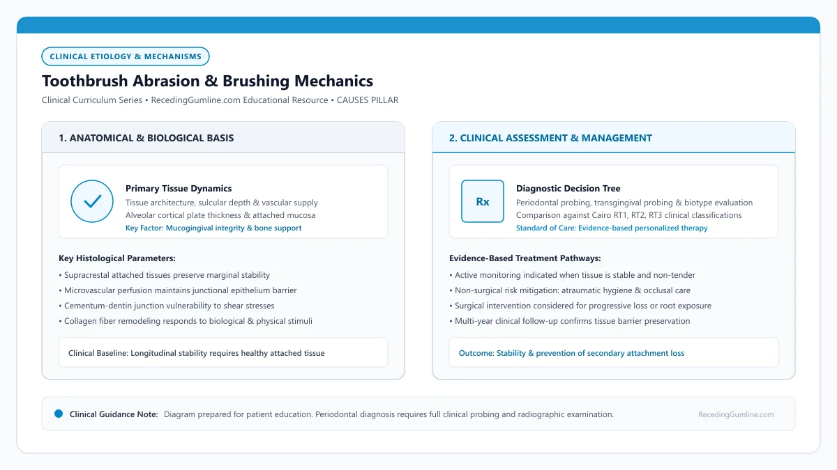
Educational illustration: Toothbrush Abrasion & Brushing Mechanics. Clinical management requires comprehensive periodontal evaluation rather than isolated self-assessment.
Source: RecedingGumline.com Clinical Editorial Team (Proprietary educational diagram for RecedingGumline.com)
Mechanical Shear Forces & Marginal Tissue Microtrauma
The facial gingival margin protecting anterior and premolar teeth is remarkably delicate, frequently measuring less than 1.0 millimeter in thickness in individuals with a thin periodontal phenotype. Unlike enamel, which is a highly calcified crystalline structure, the gingival margin consists of stratified squamous epithelium anchored to underlying alveolar bone and root cementum by collagenous connective tissue fibers. When subjected to repetitive, high-frequency horizontal friction, this thin epithelial barrier sustains chronic cellular micro-ulcerations.
Repetitive horizontal scrub strokes exert substantial lateral shear stress across the marginal tissue. When patients brush with excessive force—often exceeding 2.5 to 3.0 Newtons compared to the physiologically recommended 1.5 Newtons—the mechanical pressure compresses and shears the supracrestal soft tissue attachment. Over months and years of twice-daily abrasion, the tissue's biological reparative capacity is overwhelmed, leading to permanent, apical retreat of the gingival margin.
Crucially, toothbrush abrasion typically occurs in individuals with exemplary plaque control and low oral biofilm scores. Unlike inflammatory recession driven by bacterial toxins, toothbrush-induced recession typically presents with pale, firm, knife-edged margins that show zero bleeding upon gentle periodontal probing, masking the severity of structural attachment loss from casual visual inspection.
Key Etiological Insights
- Facial gingival margins measuring under 1.0 mm thickness exhibit extreme vulnerability to physical friction.
- Brushing force exceeding 2.0 Newtons accelerates tissue trauma without conferring any additional plaque removal benefit.
- Abrasion-induced recession characteristically occurs in mouths with clean surfaces and zero inflammatory bleeding.
Relative Dentin Abrasivity (RDA) & Root Surface Erosion
Commercial toothpastes incorporate abrasive mineral particles—including hydrated silica, calcium carbonate, dicalcium phosphate, and alumina—engineered to dislodge extrinsic pellicle and surface stains. While coronal enamel possesses a Mohs hardness of 5 and resists moderate abrasives, exposed root cementum has a Mohs hardness of merely 2 to 2.5, and underlying dentin measures approximately 3. Once the gumline recedes, abrasive toothpastes rapidly gouge the denuded root.
The International Organization for Standardization (ISO 11609) and the American Dental Association utilize the Relative Dentin Abrasivity (RDA) laboratory standard to quantify toothpaste abrasiveness. Although regulatory standards permit RDA values up to 250, clinical evidence indicates that exposed dentin undergoes accelerated dimensional loss at RDA values exceeding 70 to 80, particularly when paired with stiff manual bristles.
Patients presenting with cervical sensitivity or early gingival recession should be guided toward therapeutic, low-abrasivity dentifrices with RDA ratings below 70. Whitening, stain-defense, and smoker-targeted formulations routinely record RDA scores between 100 and 150+, exacerbating wedge-shaped cervical defects (non-carious cervical lesions) and driving further soft-tissue retreat.
Key Etiological Insights
- Root cementum and dentin are substantially softer than enamel, abrading rapidly under abrasive toothpastes.
- Toothpastes with RDA scores exceeding 80 inflict significant physical wear on denuded cervical root surfaces.
- Selecting low-abrasivity dentifrices (RDA under 70) preserves exposed root architecture and protects healing margins.
Bristle Geometry, Filament Stiffness & Grip Dynamics
Toothbrush filaments are manufactured from synthetic nylon polymers whose bending modulus is governed by diameter and length. Medium and hard bristles feature thick filaments that resist deflection, focusing applied force into sharp pinpoint vectors that slice across the free gingival margin. Soft and ultra-soft brushes employ slender filaments (typically 0.12 to 0.15 mm in diameter) that flex easily under physiological pressure, distributing energy across broader contact areas.
Filament end-rounding is an essential manufacturing quality metric. Low-cost or worn brushes often feature jagged, chisel-shaped cut ends that create microscopic abrasions and lacerations on the junctional epithelium. High-quality therapeutic brushes utilize multi-stage polishing processes to achieve smooth, domed hemispherical tips that gently sweep the sulcus without abrading adjacent soft tissue.
Patient grip mechanics directly dictate the magnitude of delivered brushing force. A palm or fist grasp recruits powerful forearm and bicep musculature, routinely generating destructive pressures of 3.0 to 5.0 Newtons. Conversely, holding the toothbrush with a modified pen grasp (using only the thumb, index, and middle fingers) mechanically restricts applied pressure to safe physiological thresholds under 1.5 Newtons.
Key Etiological Insights
- Soft bristles flex under light pressure, shielding the marginal tissue collar from high-impact shear stresses.
- Microscopically polished, end-rounded filaments prevent repetitive micro-lacerations to sulcular epithelium.
- A modified pen grasp physically prevents excessive muscle force recruitment during daily brushing routines.
Anatomical Predilection: Arch Prominence & Hand Dominance
Toothbrush abrasion does not occur randomly throughout the mouth; it demonstrates a pronounced predilection for anatomically prominent teeth positioned at the anterior corners of the dental arch. Maxillary and mandibular canines, along with first premolars, project labially beyond the curvature of adjacent incisors and molars, absorbing the greatest mechanical impact during horizontal scrub strokes.
In addition, patient hand dominance creates a distinct unilateral asymmetry in recession distribution. Right-handed individuals typically exert substantially greater force and spend longer cleaning times on the left maxillary and mandibular quadrants. Left-handed brushers exhibit corresponding contralateral tissue loss on the right dental arch. This asymmetric presentation is a classic clinical indicator of mechanical etiology.
When prominent root positions coincide with congenital alveolar bone dehiscences or thin cortical plates, mechanical toothbrush friction can rapidly strip away the thin overlying soft-tissue cover, creating deep, isolated recession defects while adjacent, lingually positioned teeth remain completely unaffected.
Key Etiological Insights
- Canines and premolars absorb maximal mechanical friction due to their prominent positions at arch transitions.
- Hand dominance frequently produces an asymmetric recession pattern that mirrors the patient's brushing vector.
- Prominent root contours combined with thin alveolar plates form prime anatomical sites for localized tissue loss.
Atraumatic Oral Hygiene Protocols: The Modified Bass Technique
Halting the progression of toothbrush abrasion requires transitioning away from aggressive scrub habits toward atraumatic, biologically sound plaque control methods. The Modified Bass Technique represents an established clinical benchmark protocol recommended by periodontists worldwide: bristle filaments are placed at a 45-degree angle pointing toward the gingival sulcus, vibrated with gentle, short back-and-forth micro-movements to disrupt subgingival biofilm, and then rolled coronally toward the incisal edge.
Electric toothbrushes equipped with dynamic pressure sensors provide an evidence-based intervention for patients with ingrained heavy-scrub habits. When applied pressure exceeds safe clinical thresholds (typically 2.0 to 2.5 Newtons), these smart sensors illuminate visual warning indicators, emit haptic pulses, or automatically throttle oscillation speed, compelling the patient to maintain atraumatic pressure.
Clinical research indicates that plaque removal efficiency plateaus at approximately 1.5 Newtons of pressure. Brushing with greater force does not enhance bacterial biofilm clearance; it purely accelerates mechanical hard- and soft-tissue destruction without therapeutic benefit.
Key Etiological Insights
- The Modified Bass Technique directs cleaning energy into the sulcus while eliminating abrasive scrub vectors.
- Pressure-sensing electric toothbrushes provide objective real-time biofeedback to extinguish traumatic brushing.
- Plaque removal efficiency plateaus at gentle pressure; applying higher force solely increases tissue wear.
Clinical Distinctions: Mechanical Wear vs. Biofilm Periodontitis
Distinguishing mechanical toothbrush abrasion from microbial-induced periodontitis is fundamental to selecting appropriate clinical therapy. While both conditions result in loss of clinical attachment, their underlying pathophysiology, prognosis, and treatment pathways diverge completely. Periodontitis is an infectious disease characterized by deep periodontal pockets (probing depths >= 4 mm), subgingival calculus, bleeding on probing, and interproximal alveolar bone loss.
Conversely, pure toothbrush abrasion presents with shallow, healthy sulcular depths (1 to 2 mm), firm, stippled marginal gingiva, zero bleeding on probing, and intact interdental papillae with normal interproximal bone levels on bitewing radiographs (consistent with Cairo RT1 recession classification).
A comprehensive periodontal evaluation—including a 6-point probing depth chart, clinical attachment level mapping, transgingival biotype assessment, and periapical radiographs—is required to ensure that mechanical modifications are matched to the true clinical etiology.
Key Etiological Insights
- Abrasion recession exhibits shallow probing depths (1-2 mm) with preserved interproximal bone support.
- Periodontal disease exhibits inflammatory pocketing, purulent exudate, bleeding on probing, and bone loss.
- Full-mouth 6-point periodontal charting is essential to differentiate mechanical wear from active infection.
Thin Gingival Phenotype: Anatomical Vulnerability to Recession
In periodontics, tissue phenotype describes the thickness of the soft-tissue margin and underlying alveolar cortical bone. Individuals possessing a thin gingival phenotype have a delicate, translucent mucosal barrier that offers minimal structural resistance against mechanical abrasion, inflammation, and tooth movement, making it the premier anatomical predisposing factor for a receding gumline.
• Gingival phenotype combines gingival thickness (GT), keratinized tissue width (KTW), and alveolar bone morphotype.
• Thin phenotypes exhibit a tissue thickness under 1.0 mm, clinically verified when a periodontal probe shines through the sulcular margin.
• Thin tissue is supported by equally thin underlying labial bone plates that easily develop dehiscences.
• Prophylactic soft-tissue augmentation can convert a thin, vulnerable biotype into a thick, protective phenotype.
Diagnostic Criteria: The Periodontal Probe Transparency Test
Historically referred to as "biotype," the 2017 World Workshop updated clinical nomenclature to periodontal phenotype. Phenotype is characterized by three interdependent anatomical features: gingival thickness (GT), keratinized tissue width (KTW), and bone morphotype.
Clinicians determine phenotype chairside using the simple and reliable probe transparency test. A standard metallic periodontal probe is inserted gently into the facial sulcus. If the silver outline of the probe is clearly visible shining through the overlying gingiva, the tissue is categorized as a thin phenotype (thickness < 1.0 mm). If the probe is completely masked, the phenotype is thick.
Clinical assessment using periodontal probe transparency remains the simplest diagnostic method to identify thin phenotypes in routine practice. When the underlying metal probe shines through the sulcular margin, clinicians recognize an elevated vulnerability to mechanical trauma and surgical margin retraction.
Key Etiological Insights
- Probe transparency test provides a reliable clinical threshold for thin tissue (< 1 mm)
- Thin phenotype features delicate, translucent margins with scalloped architectural contours
- Thick phenotype exhibits dense, fibrous tissue (≥ 1 mm) with flatter architectural contours
The Bone-Tissue Connection: Deficient Cortical Plates
Soft tissue contours faithfully reflect the skeletal architecture beneath them. In individuals with a thin phenotype, the facial cortical bone plate covering the tooth roots is exceptionally thin, often measuring less than 0.5 millimeters in thickness.
In many sites, this cortical plate is naturally absent near the cervical margin, forming developmental defects known as alveolar dehiscences. Without a solid bone foundation to anchor the periosteum and blood supply, the fragile overlying gingiva easily undergoes ischemic breakdown when challenged by bacterial plaque or mechanical friction.
Thin tissue architecture is strongly correlated with delicate, scallop-shaped underlying bone morphology that features naturally thinner labial cortical plates. Any localized inflammatory episode in a thin phenotype can lead to rapid full-thickness bone fenestration and immediate marginal gum recession.
Key Etiological Insights
- Thin soft tissue directly mirrors paper-thin or absent underlying facial bone
- Cortical bone plates under 0.5 mm cannot adequately buffer inflammatory insults
- Alveolar bone dehiscences leave the root covered only by fragile soft tissue
Clinical Management: Converting Vulnerable Phenotypes
Having a thin phenotype does not guarantee tooth loss, but it requires specialized clinical management. Patients must be educated to abandon medium- or hard-bristled toothbrushes and use ultra-soft micro-filaments with zero scrubbing pressure.
When patients with thin phenotypes plan orthodontic therapy or exhibit progressive recession, periodontists can perform phenotype modification therapy. Placing a subepithelial connective tissue graft augments tissue thickness beyond the 1.5 mm threshold, converting a fragile phenotype into a robust, protective barrier.
Clinicians frequently recommend prophylactic connective tissue grafting for patients with thin phenotypes who are undergoing comprehensive orthodontic alignment. Thickening the mucosal biotype creates a durable collagenous barrier that resists future mechanical toothbrush abrasion and marginal recession.
Key Etiological Insights
- Strict avoidance of hard brushes and horizontal scrubbing is non-negotiable
- Phenotype modification surgery thickens tissue to prevent future recession
- Thickened tissue provides greater long-term resistance against bacterial toxins
Microsurgical Phenotype Modification: Converting Biotype Architecture
Modern periodontal therapy approaches the thin gingival phenotype not merely as an unalterable genetic liability, but as an anatomically modifiable tissue structure. Phenotype modification therapy (PMT) utilizes microsurgical subepithelial connective tissue grafting to biologically increase both mucosal thickness and the apicocoronal band of keratinized tissue.
Clinical studies demonstrate that increasing gingival thickness beyond a 1.5-millimeter threshold creates a structural buffer that effectively prevents future recession progression, even in the presence of minor toothbrush friction or bacterial inflammation. The augmented connective tissue matrix establishes a resilient collagen barrier that alters the tissue's underlying biological response to trauma.
During surgical conversion, minimally invasive tunneling techniques preserve the integrity of interdental papillae while positioning the donor tissue beneath delicate marginal tissue. This approach avoids releasing incisions and optimizes microvascular perfusion to ensure predictable graft integration.
Key Etiological Insights
- Phenotype modification converts fragile tissue into a durable biotype exceeding 1.5 mm thickness.
- Augmented tissue provides long-term resistance against mechanical abrasion and inflammatory breakdown.
- Minimally invasive tunneling preserves interdental papillae and maximizes local vascular perfusion.
Long-Term Periodontal Stability & Monitoring Guidelines
Patients identified with a thin gingival phenotype require personalized long-term maintenance protocols to prevent subtle, cumulative attachment loss. Standard periodontal charting must be augmented with high-resolution digital photographic tracking and calibrated periodontal probe depth recording at six-month intervals.
When monitoring thin phenotypes, clinicians evaluate the tension exerted by adjacent muscles and the depth of the vestibular fornix. A shallow vestibule combined with a narrow keratinized tissue band magnifies mechanical stress on the marginal gingiva during mastication and facial animation, accelerating apical margin migration.
Preventive oral hygiene guidance must strictly eliminate aggressive brushing techniques. Recommending extra-soft, rounded filament brushes and modified Bass vibratory strokes ensures effective biofilm disruption without inducing mechanical epithelial abrasion.
Key Etiological Insights
- Standard periodontal charting should be paired with digital photographic tracking every six months.
- Shallow vestibules and muscular tension exacerbate recession risk in thin phenotypes.
- Non-abrasive plaque control protocols protect vulnerable margins from micro-trauma.
Prominent Tooth Roots & Labial Bone Dehiscence in Gum Recession
The three-dimensional position of a tooth root within the dental arch plays a decisive role in soft-tissue stability. When a tooth erupts too far facially (buccally) or tilts outward, its prominent root stretches the overlying cortical bone and gingival tissue, creating an anatomical hot spot for isolated, localized gum recession.
• Roots positioned labially outside the center of the alveolar process have minimal or absent facial bone coverage.
• Canine teeth and maxillary first premolars are the most anatomically prominent teeth in the human dentition.
• Root prominence creates a bulbous contour that receives excessive physical friction during normal toothbrushing.
• Repositioning prominent roots through orthodontics can restore proper bony housing and facilitate graft coverage.
The Arch Trajectory: How Eruption Creates Prominent Contours
The dental arch has a curved, parabolic shape. Certain teeth—most notably maxillary canines, upper premolars, and lower central incisors—frequently erupt at the corners or outer contours of this curve. When dental crowding occurs, these teeth are often squeezed facially outside the main line of the alveolar bone.
As a consequence of this outward position, the facial cortical bone plate fails to form over the prominent root convexities, creating a developmental alveolar dehiscence. Instead of being encased in solid bone, the outer root surface is covered only by a paper-thin layer of periosteum and mucosa.
Teeth that are positioned buccally outside the primary curvature of the dental arch exhibit prominent root profiles that stretch the overlying soft tissue. This anatomical displacement leaves little to no vascularized cancellous bone covering the facial root convexities.
Key Etiological Insights
- Crowding pushes canines and incisors facially outside the alveolar bone envelope
- Cortical bone fails to develop over prominent root convexities
- Leaves the root vulnerable with only soft tissue coverage and no skeletal support
The "Speed Bump" Effect: Concentrated Brushing Abrasion
When an individual brushes their teeth using horizontal scrubbing strokes, prominent roots act like speed bumps in the road. The toothbrush bristles hit the protruding root contour with significantly greater force and friction than the adjacent, receded, or normally positioned teeth.
Over months and years, this concentrated mechanical wear strips away the thin gingival margin. Because there is no underlying bone to resist the force, the tissue rapidly recedes apically until it reaches a level where bone is present, exposing the yellow root surface.
During daily oral hygiene, standard toothbrush bristles exert disproportionately higher shearing forces against protruding root surfaces compared to well-aligned adjacent teeth. Over years of vigorous brushing, the thin tissue band stretched over these convexities recedes rapidly.
Key Etiological Insights
- Protruding roots receive disproportionate mechanical impact during toothbrushing
- Horizontal scrubbing selectively wears down the most prominent teeth in the arch
- Recession continues apically until it encounters a stable bone foundation
Clinical Sequencing: Orthodontics vs. Periodontal Grafting
Treating recession on a prominent tooth requires careful diagnostic planning. If a periodontist simply places a gum graft over a root that juts out 2 millimeters beyond the dental arch, the graft has no lateral vascular bed and is highly likely to fail or relapse.
The ideal interdisciplinary strategy often involves orthodontic alignment first: using braces or clear aligners to upright the tooth and torque the root lingually back inside the alveolar bone envelope. Once the root is centered in the bone, a connective tissue graft can achieve predictable, permanent root coverage.
Periodontists often coordinate with orthodontic specialists to evaluate whether minor lingual root torque can reposition protruding roots back into the center of the alveolar ridge. Bringing the root inside the cortical bone plate significantly enhances the predictability of surgical soft-tissue coverage.
Key Etiological Insights
- Grafting over severely protruding roots has reduced biological predictability
- Orthodontic root torquing moves the root back into the protective bone housing
- Subsequent grafting achieves predictable coverage and long-term attachment stability
Cone-Beam Computed Tomography (CBCT) in Prominent Root Evaluation
Evaluating teeth with prominent root contours requires imaging beyond standard two-dimensional bitewing or periapical radiographs, which cannot resolve the thin labial cortical bone plate. High-resolution Cone-Beam Computed Tomography (CBCT) provides cross-sectional volumetric visualization of the alveolar housing, revealing the exact spatial relationship between the tooth root and the buccal plate.
CBCT analyses consistently show that prominent tooth roots—frequently canines and premolars positioned buccally in the arch—often possess labial bone plates thinner than 0.5 millimeters or exhibit complete congenital dehiscences. In such sites, the root surface is covered exclusively by soft tissue and periosteum, rendering it vulnerable to rapid marginal retraction if exposed to friction or chronic plaque.
This radiographic insight is pivotal before undergoing restorative dentistry, periodontal surgery, or orthodontic tooth movement, enabling clinicians to identify non-visible osseous deficiencies and tailor treatment parameters accordingly.
By analyzing cross-sectional bone dimensions prior to intervention, clinicians can identify whether root prominence requires orthodontic movement or phenotype modification before attempting soft tissue coverage.
Key Etiological Insights
- CBCT cross-sectional imaging reveals cortical bone plates thinner than 0.5 mm that conventional x-rays miss.
- Prominent roots often lack buccal bone coverage, existing with congenital dehiscences under the soft tissue.
- Preoperative 3D imaging prevents surgical surprises and guides protective restorative planning.
Orthodontic Decompensation & Root Centering Strategies
When prominent root contours contribute to localized recession, interdisciplinary orthodontic and periodontal planning is essential. Moving a prominent tooth root back into the center of the alveolar envelope—a process known as orthodontic decompensation—can re-establish protective osseous architecture around the root prominence.
Orthodontists utilize controlled continuous light forces with precise torque mechanics to guide the root lingually without compromising the apical neurovascular bundle. Clinical evidence indicates that centering the root within the cancellous bone creates a more favorable anatomical bed, significantly increasing the success rate of subsequent soft-tissue coverage procedures.
Attempting soft-tissue grafting over a severely displaced or prominent root without first addressing its three-dimensional position frequently yields incomplete root coverage, as the avascular root surface remains outside the biological protective envelope.
Key Etiological Insights
- Torque-controlled orthodontic movement centers prominent roots within the cancellous alveolar housing.
- Centering the root creates an optimal vascular bed for subsequent soft-tissue grafting procedures.
- Grafting over prominent, uncentered roots carries higher rates of partial failure due to lack of osseous support.
Alveolar Bone Dehiscence & Fenestration in Gum Recession
The stability of the gingival margin is directly governed by the skeletal architecture of the alveolar process. In many individuals, the outer cortical bone plate covering tooth roots contains natural anatomical defects known as dehiscences (cleft-like gaps in the crestal bone) and fenestrations (isolated bone windows). Understanding these skeletal variations explains why receding gums can develop even in the absence of active periodontal infection.
• An alveolar bone dehiscence is a V-shaped absence of cortical bone extending from the alveolar crest apically along the root.
• An alveolar bone fenestration is an isolated "window" defect where the root surface is denuded of bone while the crestal margin remains intact.
• Dehiscences provide zero skeletal support for overlying gingival margins, leading to rapid soft-tissue collapse when challenged.
• Standard dental x-rays cannot detect facial dehiscences; three-dimensional CBCT scans are required for definitive visualization.
Anatomical Anatomy: Dehiscence vs. Fenestration
The alveolar bone forms the sockets (alveoli) that support the teeth. Under ideal anatomical conditions, the facial cortical plate extends to within 1.5 to 2.0 mm of the cementoenamel junction, providing continuous circumferential bone support.
However, developmental variations frequently occur. A dehiscence is an isolated cleft or dip where the marginal alveolar crest is completely missing, exposing the root surface in a continuous V-shape. In contrast, a fenestration is an isolated "window" of missing bone where the root penetrates the cortex, but an intact bridge of crestal bone remains above it.
An alveolar bone dehiscence represents an isolated V-shaped or U-shaped absence of cortical bone along the coronal facial aspect of a root. Without underlying cortical bone support, the overlying gingival margin depends entirely on suprabony soft-tissue attachment to maintain its vertical position.
Key Etiological Insights
- Dehiscence: Continuous V-shaped loss of marginal crestal bone exposing the cervical root
- Fenestration: Isolated window defect with an intact bridge of bone at the marginal collar
- Dehiscences directly cause the overlying gingival margin to collapse into recession
Etiology: Developmental Occurrence vs. Acquired Breakdown
Dehiscences can be developmental or acquired. Developmental dehiscences occur during tooth eruption when large tooth roots erupt through narrow alveolar ridges, common in patients with thin facial profiles, prominent canines, or crowded lower incisors.
Acquired dehiscences develop later in life. They are triggered when orthodontic arch expansion pushes roots facially through the cortical plate, or when chronic biofilm-induced inflammation resorbs the razor-thin facial bone. Once the cortical plate is gone, the overlying attached gingiva lacks blood supply from the periosteum, accelerating soft-tissue recession.
Direct trauma from aggressive horizontal scrubbing or accidental hard food impaction can easily shear through this unsupported soft-tissue sleeve. Once the thin junctional epithelium breaks down, marginal tissue rapidly collapses to the level of the existing bone crest.
Key Etiological Insights
- Developmental dehiscences arise when roots erupt through narrow alveolar ridges
- Acquired dehiscences result from orthodontic expansion or inflammatory bone loss
- Loss of underlying periosteal blood supply leaves the gingival margin fragile
Diagnostic Imaging: Why Standard Dental X-Rays Miss Dehiscences
A major source of patient confusion is why their regular dental x-rays look "completely normal" despite significant gum recession. Standard intraoral periapical and bitewing radiographs are two-dimensional projections that superimpose the thick lingual/palatal bone over the thin facial plate.
Consequently, a facial dehiscence is completely invisible on standard x-rays. Only 3D Cone-Beam Computed Tomography (CBCT) or direct visual observation during periodontal flap reflection can accurately reveal the presence and depth of an alveolar bone dehiscence.
Surgical management of extensive dehiscence defects requires specialized regenerative techniques such as guided tissue regeneration with bone grafts and collagen membranes. Rebuilding interdental bone support and thickening the overlying mucosa prevents further apical migration of the margin.
Key Etiological Insights
- Standard 2D radiographs superimpose lingual bone, masking facial dehiscences
- Cone-beam CT (CBCT) provides true 3D cross-sectional imaging of cortical bone thickness
- Treatment focuses on thick soft-tissue grafting to shield the denuded bone defect
Flapless Diagnostic Indicators & Dehiscence Identification
An alveolar bone dehiscence is a non-continuous skeletal defect characterized by the absence of the facial or lingual cortical bone plate extending apically from the marginal crest. Identifying dehiscences without raising a full-thickness mucoperiosteal flap requires careful clinical examination and tactile evaluation.
Clinicians detect underlying dehiscences by observing localized root prominence, palpable root contours through thin alveolar mucosa, and sudden tissue discoloration under lateral lighting. When combined with a shallow probing depth but significant clinical attachment loss, a bony dehiscence is virtually certain.
Because dehiscences remove the structural framework that anchors periodontal ligament Sharpey's fibers, the marginal tissue is held solely by supra-alveolar connective tissue fibers, leaving the margin fragile and prone to rapid detachment under physical challenge.
Key Etiological Insights
- Dehiscences represent a V-shaped or U-shaped absence of marginal cortical bone along the root.
- Tactile palpation and root prominence indicate bone loss even when tissue is intact.
- Absence of cortical bone deprives the marginal tissue of rigid structural support and vascular plexus supply.
Periodontal Plastic & Regenerative Considerations
Managing gingival recession occurring over a significant alveolar dehiscence requires advanced surgical consideration. Conventional flap repositioning alone frequently fails because the repositioned flap lacks a rigid osseous vascular bed against which to establish new fibrous attachment.
To overcome this anatomical hurdle, periodontists combine subepithelial connective tissue grafts with biologically active enamel matrix derivatives (EMD) or recombinant human platelet-derived growth factors (rhPDGF-BB). These biologic mediators stimulate cementogenesis and recruit osteoblasts to induce true periodontal regeneration.
While complete coronal bone regeneration over a deep dehiscence is biologically challenging, achieving thick keratinized soft-tissue coverage establishes a durable biological seal that halts further attachment loss and provides lifelong root protection.
Key Etiological Insights
- Flap repositioning over deep dehiscences requires soft-tissue grafting to overcome vascular limitations.
- Enamel matrix derivatives and growth factors enhance cellular recruitment along denuded root dentin.
- Thick soft-tissue coverage forms an effective protective biological seal even over osseous deficiencies.
High Frenal Attachment and Tension: Mechanics of Marginal Tissue Pull
A labial frenum is a normal band of fibrous tissue connecting the inner lip to the alveolar mucosa. However, when a frenum attaches unusually high—inserting directly into the attached gingiva, marginal collar, or interdental papilla—every movement of the lips and mouth creates mechanical tension that pulls the gum away from the tooth, triggering progressive gumline recession.
• An aberrant or high frenal attachment inserts into the keratinized gingiva or marginal gingival collar.
• Muscular movements during talking, eating, and smiling generate continuous traction on the fragile margin.
• The blanching test clinically verifies whether frenal tension is causing localized marginal ischemia.
• Frenal tension is particularly destructive when combined with a narrow band of attached gingiva (< 1 mm).
Frenum Anatomy and Mirko's Morphological Classification
In healthy oral anatomy, the labial frenum is composed of dense collagenous connective tissue, elastic fibers, and muscle fibers originating from the orbicularis oris muscle. In the 1970s, researcher Mirko and colleagues classified frenal attachments into four distinct anatomical types based on insertion level.
These types are: mucosal (attaching in the movable mucosa), gingival (attaching in the attached gingiva), papillary (inserting into the interdental papilla), and papilla-penetrating (crossing completely through the papilla into the palatal or lingual mucosa). The papillary and papilla-penetrating types pose the highest clinical risk for recession.
High labial frenum attachments insert muscular connective fibers directly into the free gingival margin or the interdental papilla. During routine speech, smiling, and mastication, this dynamic muscular pulling exerts continuous coronal tension that pulls the tissue away from the tooth.
Key Etiological Insights
- Mucosal insertions are harmless and sit far below the marginal tissue
- Papillary insertions insert directly into the triangular tissue between teeth
- Papilla-penetrating insertions create midline spaces (diastemas) and severe recession
The Biomechanics of Tissue Traction and Pocket Deepening
Every time you speak, chew, swallow, or smile, the orbicularis oris and facial expression muscles contract. When a frenum inserts near the gingival margin, these muscle contractions transmit direct physical pulling forces to the delicate marginal collar.
This continuous tugging acts like a wedge, pulling the gum margin away from the root surface. This creates a funnel-shaped defect that traps food debris and plaque bacteria. Because the tissue is continuously moving, a stable junctional epithelial seal cannot form, leading to rapid apical attachment loss.
A simple blanching test performed in the dental chair confirms active frenum traction by pulling the lip outward and observing whether the gingival margin blanches pale white. Persistent blanching demonstrates that blood flow is being compromised and tissue fibers are under mechanical tension.
Key Etiological Insights
- Facial expressions transmit continuous dynamic tension to the tooth margin
- Traction mechanically pulls the gingiva away from the root, creating plaque traps
- Prevents the formation of a stable junctional epithelial seal
Clinical Diagnosis: The Tension Test and Frenectomy Timing
A clinician evaluates frenal tension during routine examination by extending the patient's lip outward. If this maneuver causes the gingival margin of the incisors to move away from the tooth or causes immediate blanching (whitening) of the marginal tissue, dynamic tension is confirmed.
When frenal tension threatens tooth stability, a frenectomy—surgical excision of the fibrous band using a scalpel, electrosurgery, or soft-tissue laser—is indicated. In cases where significant recession has already exposed the root, the frenectomy is performed simultaneously with a soft-tissue graft.
Releasing the muscular pull via a conservative frenectomy eliminates the physical traction force that drives ongoing margin apical migration. Combining the release with a subepithelial connective tissue graft allows periodontists to simultaneously reconstruct the receded gumline.
Key Etiological Insights
- Tension test confirms margin movement or blanching upon lip extension
- Frenectomy surgically releases the fibrous muscle attachment
- Combined frenectomy and grafting achieves root coverage and prevents relapse
Clinical Diagnostics: The Tension Blanching Test
A high frenum attachment becomes clinically problematic when its fibrous insertions extend into the attached gingiva or directly onto the interdental papilla. Clinicians diagnose pathogenic frenal pull through the standardized "blanching test."
During this diagnostic maneuver, the clinician gently pulls the patient's lip or cheek outward and downward. If this lateral tension causes immediate blanching (ischemia) of the gingival margin or visibly pulls the marginal tissue away from the root surface, aberrant frenal tension is active and contributing to recession.
Persistent tension creates microscopic tissue movement during ordinary speech and mastication, pumping oral microorganisms into the gingival crevice and mechanically tearing the delicate junctional epithelial seal.
Key Etiological Insights
- The blanching test confirms whether muscular lip movement mechanically mobilizes the gum margin.
- Aberrant frenal tension creates persistent dynamic pull during speaking and chewing.
- Micro-movement of the margin facilitates bacterial invasion and gradual mechanical detachment.
Sequencing Care: Frenectomy vs. Grafting Coordination
When high frenal attachment coincides with gingival recession, therapeutic sequencing determines long-term stability. In cases where adequate keratinized tissue remains, a standalone frenectomy (surgical or diode laser excision of the muscular attachment) may arrest recession progression by eliminating mechanical tension.
However, when recession has already advanced and the zone of keratinized tissue is depleted, a frenectomy alone cannot restore lost tissue coverage. In these scenarios, periodontists coordinate frenal relocation with soft-tissue grafting in either a single-stage or two-stage surgical protocol.
Relocating the muscular attachment apically into the alveolar vestibule ensures that the newly grafted tissue can heal without micro-movements, providing the static environment essential for revascularization and tissue survival.
Key Etiological Insights
- Frenectomy alone may halt progression if adequate keratinized tissue is still present.
- Advanced recession requires combined frenal release and soft-tissue graft augmentation.
- Apical muscular relocation protects healing grafts from disruptive mechanical forces.
Toothbrush Abrasion vs. Biofilm-Induced Recession: Clinical Distinctions
Gingival recession arises from two fundamentally different biological pathways: mechanical trauma and microbial inflammation. Within our clinical review of receding gumline conditions, toothbrush abrasion represents the classic non-inflammatory mechanical etiology, occurring in health-conscious individuals who brush vigorously. Differentiating toothbrush abrasion from biofilm-induced periodontitis is essential because their treatments and prevention protocols are completely opposite.
• Toothbrush abrasion causes recession with firm, pale pink, non-bleeding margins and clean tooth surfaces.
• Biofilm-induced recession is accompanied by erythematous, swollen margins, bleeding on probing, and periodontal pocketing.
• Brushing force exceeding 2.0 to 2.5 Newtons and medium/hard bristles are primary mechanical drivers of tissue wear.
• Abrasive whitening toothpastes (high RDA) accelerate cervical tooth and soft-tissue wear.
Clinical Presentation: The Picture of Mechanical Abrasion
Toothbrush abrasion presents with very distinct clinical features. The receded gum margin appears pale pink, firm, and knife-edged against the tooth, with zero bleeding upon probing. The tooth surfaces are remarkably clean, with virtually no visible plaque or calculus deposits.
Furthermore, mechanical abrasion is almost always concentrated on the prominent facial surfaces of teeth at the corners of the arch—especially the canines and premolars. Because right-handed individuals brush their left upper quadrant with the greatest force (and left-handed individuals brush the right), abrasion patterns are characteristically asymmetrical.
Aggressive horizontal scrub brushing using medium or hard-bristled toothbrushes generates severe frictional shear against cervical gum margins. Over time, this mechanical abrasion strips away fragile epithelial layers faster than cellular mitosis can repair the protective surface barrier.
Key Etiological Insights
- Pale pink, firm margins with zero bleeding on probing
- Concentrated on facial surfaces of canines and premolars
- Asymmetrical distribution corresponding to the patient's dominant brushing hand
Clinical Presentation: The Picture of Biofilm-Induced Periodontitis
In stark contrast, recession caused by microbial plaque biofilm reflects active or historical inflammation. The gingival margin is often rolled, swollen (edematous), and dark red or bluish-red. Touching the margin with a dental probe readily produces bleeding on probing (BOP).
Most importantly, biofilm-induced recession is not confined to the outer facial surfaces. It involves interproximal destruction: the gum tissue and alveolar bone between the teeth recede, forming deep periodontal pockets (≥ 4 mm) and creating open "black triangle" gaps between teeth.
Patients frequently scrub harder in the false belief that aggressive pressure prevents gum disease, inadvertently causing deep V-shaped cervical root notches. These non-carious cervical abrasions expose sensitive dentinal tubules and create jagged margins that harbor plaque biofilm.
Key Etiological Insights
- Erythematous, swollen, and easily bleeding tissue margins
- Affects interdental spaces between teeth as well as outer facial surfaces
- Associated with deep probing pockets, subgingival calculus, and bone loss
Toothpaste Abrasivity: The Relative Dentin Abrasivity (RDA) Index
Toothbrush bristles alone rarely wear away tooth structure; the primary abrasive culprit is toothpaste. Toothpastes contain polishing and abrasive agents—such as hydrated silica, calcium carbonate, and aluminum oxide—designed to scrub away surface stains.
The abrasiveness of toothpaste is measured by the Relative Dentin Abrasivity (RDA) scale. Standard toothpastes typically range from 50 to 80 RDA. However, many aggressive "whitening" and "charcoal" formulations score above 150 to 200 RDA. When combined with a stiff brush and hard pressure, high-RDA pastes scour away root cementum in weeks.
Switching immediately to an ultra-soft or sonic electric toothbrush equipped with an integrated pressure sensor stops mechanical wear instantly. Dental hygienists train patients in the modified Bass brushing technique, angling bristles at 45 degrees to gently massage the sulcus without abrading tissues.
Key Etiological Insights
- Relative Dentin Abrasivity (RDA) scores above 100 significantly accelerate root wear
- Whitening and charcoal toothpastes frequently contain harsh abrasive particles
- Use toothpastes with RDA under 70 and ultra-soft bristles to protect exposed roots
Filament Geometry, Dentifrice RDA & The Tribological Mechanism
Toothbrush abrasion is a tribological process where mechanical friction gradually wears away oral biological tissues. The physical characteristics of toothbrush bristles play a decisive role; stiff, large-diameter filaments with non-rounded, sharp cut ends inflict microscopic lacerations on the gingival epithelium.
The abrasiveness of toothpaste—quantified by the Relative Dentin Abrasivity (RDA) index—acts synergistically with bristle stiffness. Toothpastes formulated for intensive whitening or tartar control frequently carry RDA scores exceeding 120, transforming everyday toothbrushing into an abrasive slurry that rapidly abrades exposed cementum and marginal gum tissue.
Periodontists emphasize that tissue wear is accelerated when patients brush immediately after consuming acidic foods or beverages. Acid soften the enamel and cementum surfaces, making them exceptionally vulnerable to mechanical removal under horizontal brushing pressure.
Key Etiological Insights
- Stiff filaments with sharp cut ends create micro-lacerations in the delicate marginal epithelium.
- High-RDA whitening toothpastes (>120) act as abrasive grinding pastes on root surfaces.
- Brushing immediately after acidic exposure dramatically increases tissue and cementum loss.
Ergonomic Retraining: Sonic vs. Oscillating-Rotating Technologies
Correcting mechanical abrasion requires reprogramming entrenched motor habits. Patients accustomed to horizontal scrub techniques must transition to gentle sulcular cleansing using the modified Bass technique or switch to pressure-controlled electric toothbrushes.
Modern electric toothbrushes with integrated optical or acoustic pressure sensors alert users whenever brushing force exceeds 1.5 to 2.0 Newtons. Clinical trials demonstrate that pressure-sensing power brushes significantly reduce the incidence of cervical abrasion while providing thorough plaque biofilm clearance compared to manual scrub techniques.
Transitioning to extra-soft tapered filaments allows the bristles to flex into the gingival sulcus without transmitting destructive shear forces to the marginal tissue, halting abrasion-induced recession.
Key Etiological Insights
- Motor habit correction replaces horizontal scrubbing with gentle vibratory sulcular motions.
- Electric brushes with smart pressure sensors prevent brushing forces from exceeding 2 Newtons.
- Tapered micro-filaments clean subgingivally without exerting abrasive shear forces.
Bruxism, Occlusal Overload and Non-Carious Cervical Lesions (Abfraction)
Many individuals with receding gums notice sharp, V-shaped wedge defects grooved into the tooth at the exact margin where the gum meets the root. Known clinically as Non-Carious Cervical Lesions (NCCLs) or abfraction lesions, these notches frequently develop in patients who clench or grind their teeth (bruxism). When treating a receding gumline, understanding how biomechanical occlusal overload contributes to cervical tissue breakdown clarifies the critical link between biting forces and gum recession.
• Bruxism generates heavy lateral and oblique biting forces exceeding 250 pounds per square inch.
• Heavy lateral forces cause the tooth crown to microscopic flex, concentrating mechanical stress at the cervical fulcrum.
• Stress concentration disrupts microscopic enamel rods and dentin crystals, flaking them away (abfraction).
• Abfraction notches create sharp ledges that trap plaque and accelerate localized marginal gum recession.
The Biomechanical Model: Tooth Flexure and Tensile Stress
The abfraction hypothesis was first formulated by dental researchers Lee and Eakle in 1984 and later refined by Grippo. The theory is grounded in basic engineering mechanics: teeth are not completely rigid rods; they possess a degree of microscopic flexibility.
When an individual grinds their teeth or exerts heavy lateral biting forces during chewing, non-axial (sideways) loads bend the tooth crown relative to the firmly embedded root. The fulcrum—the pivot point where bending stress is highest—is situated precisely at the cervical margin (the cementoenamel junction).
Nocturnal clenching and grinding generate massive lateral and oblique forces that flex teeth along their long anatomical axes. These intense biomechanical stresses concentrate at the narrow cervical fulcrum near the cementoenamel junction, fracturing microcrystalline enamel rods.
Key Etiological Insights
- Non-axial lateral biting forces cause microscopic flexure of the tooth crown
- The mechanical pivot point (fulcrum) is concentrated at the cervical neck of the tooth
- Alternating tension and compression rupture chemical bonds between hydroxyapatite crystals
The Multifactorial Triad: Abfraction, Abrasion & Biocorrosion
Modern clinical consensus recognizes that pure abfraction rarely acts in isolation. Rather, cervical notches result from a destructive synergy termed the "multifactorial triad": stress (abfraction), friction (toothbrush abrasion), and biocorrosion (acid erosion).
Biomechanical clenching creates microscopic cracks within the brittle enamel crystals and softer dentin. Once cracked, the weakened mineral structure is easily scrubbed away by daily toothbrushing or dissolved by dietary acids (citrus, soda, wine) and gastric reflux, carving deep, sharp-edged wedge defects.
The resulting wedge-shaped non-carious cervical lesions, known as abfractions, frequently occur in conjunction with localized gingival recession. The continuous micro-flexure of the tooth structure disrupts the adjacent junctional epithelial attachment and accelerates margin detachment.
Key Etiological Insights
- Occlusal clenching initiates microscopic crystalline micro-fractures
- Toothbrushing scrubs away the loosened mineral fragments (abrasion)
- Dietary and gastric acids dissolve exposed dentin matrix (biocorrosion)
Clinical Treatment: Occlusal Nightguards and Restorative Protocols
Addressing abfraction-related recession requires managing both the biomechanical cause and the anatomical damage. The first line of defense is a custom hard acrylic occlusal splint (nightguard). The nightguard distributes nocturnal clenching forces evenly across all teeth, eliminating flexure.
If the cervical notch is sensitive or threatens pulp vitality, dentists restore the defect using flexible microfilled composites or resin-modified glass ionomer cements. These materials have a low modulus of elasticity, allowing them to bend slightly with the tooth during chewing without debonding.
Fabricating a custom dual-laminate or hard acrylic nightguard distributes occlusal forces evenly across the entire dental arch, neutralizing destructive lateral torque. Managing nocturnal bruxism protects both restorative composite margins and natural periodontal tissues from ongoing mechanical failure.
Key Etiological Insights
- Custom hard acrylic nightguards redistribute heavy clenching forces
- Prevents ongoing tooth flexure and halts progressive notch deepening
- Flexible microfilled composites or glass ionomers restore lost tooth contour and seal tubules
Biomechanical Stress Concentration & Enamel Rod Disruption
Nocturnal bruxism and diurnal clenching generate tremendous non-axial forces that transmit through the clinical crowns of teeth down into the alveolar support. According to the engineering concept of tooth flexure, lateral shearing forces cause the tooth to bend minutely along its fulcrum at the cementoenamel junction (CEJ).
This cyclical flexural stress concentrates tensile and compressive forces at the cervical margin, causing brittle enamel rods and underlying dentin crystals to fatigue, crack, and dislodge—a mechanical phenomenon known as abfraction. As the cervical tooth structure hollows out, the adjacent gingival margin loses its biological collar and recedes apically.
While abfraction and toothbrush abrasion frequently co-exist in wedge-shaped cervical lesions, the primary underlying driver in bruxers is destructive occlusal overload rather than abrasive hygiene alone.
Key Etiological Insights
- Heavy lateral grinding forces cause microscopic tooth flexure centered at the cervical CEJ.
- Cyclical flexure causes enamel crystals to fatigue and fracture, forming sharp wedge-shaped notches.
- Loss of cervical tooth structure compromises the gingival margin, accelerating recession.
Comprehensive Management: Occlusal Splints & Staged Restorations
Successfully halting abfraction-related gum recession requires addressing both occlusal biomechanics and structural tooth loss. The cornerstone of non-invasive treatment is the fabrication of a custom, hard acrylic occlusal stabilization splint (nightguard) adjusted to provide mutually protected articulation.
By distributing nocturnal bite forces evenly across the entire arch and eliminating eccentric interferences, a nightguard neutralizes destructive lateral tooth flexure. Where deep wedge-shaped notches cause food impaction or sensitivity, dentists place micro-hybrid composite or glass-ionomer restorations with low modulus of elasticity to flex harmoniously with the tooth.
If soft-tissue grafting is planned to recover root coverage, periodontists coordinate graft placement with precise enamel re-contouring to ensure the soft tissue heals over a smooth, biologically receptive root surface.
Key Etiological Insights
- Hard acrylic occlusal splints eliminate eccentric lateral forces and neutralize tooth flexure.
- Flexible restorative materials (glass ionomers) restore cervical contours without popping out.
- Occlusal stabilization is vital before attempting soft-tissue grafting over abfraction sites.
Orthodontic Arch Expansion and Recession: Moving Teeth Beyond the Cortical Plate
In modern orthodontics, arch expansion and tooth alignment are commonly employed to resolve crowding and create broad, attractive smiles without extracting permanent premolars. However, the human jawbone possesses strict biological boundaries. When teeth are expanded or tipped outward beyond the limits of the alveolar bone housing, the fragile outer cortical plate can resorb, leading to severe post-orthodontic gingival recession.
• The alveolar bone housing establishes a biological "envelope of discrepancy" limiting safe tooth movement.
• Expanding dental arches beyond cortical boundaries causes thinning and dehiscence of the buccal bone plate.
• Lower incisors and upper premolars are at the highest clinical risk of expansion-induced recession.
• Pre-orthodontic evaluation of gingival phenotype and bone thickness with CBCT imaging prevents unexpected tissue breakdown.
The Biological Envelope: Limits of the Alveolar Housing
Orthodontists work within what Dr. William Proffit famously termed the "envelope of discrepancy"—the biological boundaries defined by the outer and inner cortical plates of the jawbone. Teeth can be moved safely within this spongy, trabecular bone housing without compromising periodontal health.
However, when dental arches are expanded laterally or lower incisors are proinclined forward to resolve severe crowding without extractions, the tooth roots are pushed directly against the outer cortical bone plate. If movement continues past this barrier, the pressure causes the paper-thin bone to resorb completely.
Rapid or excessive transverse arch expansion moves tooth roots outward against the rigid boundaries of the alveolar cortical bone housing. If expansion exceeds the biological envelope of the patient’s jaw, roots can perforate cortical plates, producing extensive bone dehiscences.
Key Etiological Insights
- The alveolar cortical plates define the absolute biological boundary for tooth movement
- Arch expansion pushes roots directly against the thin facial bone cortex
- Excessive expansion resorbs the cortical plate, creating permanent bone dehiscences
The Delayed Manifestation: Why Recession Appears Years Later
A perplexing mystery for many patients is why their gums looked fine when their braces came off, only to develop noticeable recession three or four years later. The explanation lies in tissue vascularity and mechanical resilience.
During active orthodontics, the underlying bone dehiscence is created, but the soft tissue remains draped over the root. However, this unsupported tissue now lacks periosteal blood supply. Over subsequent years, ordinary daily toothbrushing, slight plaque accumulation, or normal aging easily causes the fragile soft tissue to collapse, exposing the root.
Clear aligner therapy and fixed braces must be carefully calibrated to ensure that tooth movement consists of gentle bodily translation rather than uncontrolled crown tipping. When roots tip outward against thin facial tissues, the overlying attached gingiva quickly recedes.
Key Etiological Insights
- Bone dehiscence is created during orthodontics, but soft tissue collapse takes years
- Unsupported tissue lacks blood supply and cannot withstand normal brushing friction
- Patients often mistake delayed expansion breakdown for sudden gum disease
Interdisciplinary Prevention: 3D CBCT Scans and Prophylactic Grafting
Modern orthodontics increasingly utilizes 3D Cone-Beam Computed Tomography (CBCT) to visualize cortical bone thickness prior to initiating expansion. If CBCT reveals paper-thin bone (< 0.5 mm) or existing dehiscences, the orthodontist must modify the treatment plan to avoid further outward movement.
In patients with thin phenotypes where expansion is unavoidable, periodontists perform prophylactic soft-tissue grafting before or during orthodontic treatment. Placing a connective tissue graft thickens the gingival biotype, creating a durable fibrotic cushion that prevents soft-tissue recession even if bone thinning occurs.
Cone-beam CT volumetric imaging allows orthodontists to measure bone thickness accurately before initiating complex expansion protocols. If severe fenestrations or thin phenotypes are detected, interdisciplinary treatment planning may incorporate periodontal grafting before orthodontic mechanics begin.
Key Etiological Insights
- 3D CBCT imaging verifies cortical bone thickness before initiating arch expansion
- Orthodontists adjust torque and force vectors to keep roots centered in bone
- Prophylactic connective tissue grafting provides a durable barrier against recession
The Alveolar Housing Envelope & Cortical Plate Limits
Rapid maxillary expansion and comprehensive arch leveling involve pushing teeth outward into broader alignment. However, tooth movement is biologically constrained by the "alveolar housing"—the anatomical envelope formed by the buccal and lingual cortical bone plates.
When orthodontic expansion forces move tooth roots beyond the confines of this skeletal envelope, the root surface is pressed directly against the inner surface of the cortical bone. Under continuous pressure, osteoclasts resorb the thin buccal plate, creating extensive dehiscences and fenestrations.
Because the overlying gingiva is deprived of its underlying osseous foundation, the marginal tissue undergoes ischemic thinning and eventual apical recession, particularly around mandibular incisors and maxillary first premolars.
Key Etiological Insights
- Arch expansion is biologically limited by the width of the cortical bone envelope.
- Moving roots beyond the cortical plate causes irreversible osteoclastic bone resorption.
- Loss of labial bone support leads directly to post-orthodontic soft-tissue recession.
Pre-Orthodontic Periodontal Augmentation (POPA)
To prevent post-orthodontic recession in patients with thin biotypes requiring significant arch expansion, periodontists and orthodontists employ Pre-Orthodontic Periodontal Augmentation (POPA). This proactive approach involves augmenting soft and hard tissues before initiating active tooth movement.
By placing subepithelial connective tissue grafts or performing Surgically Facilitated Orthodontic Therapy (SFOT) with decortication and particulate bone grafting, clinicians deliberately expand the biological envelope. Thickening the mucosal margin prior to tooth movement shields the site from mechanical breakdown during expansion.
Clinical studies confirm that teeth pre-treated with phenotype augmentation exhibit significantly less attachment loss, zero dehiscence progression, and stable long-term periodontal outcomes throughout comprehensive orthodontic treatment.
Key Etiological Insights
- POPA thickens soft tissues and expands the bony envelope prior to orthodontic expansion.
- Surgically Facilitated Orthodontic Therapy adds particulate bone to prevent dehiscences.
- Proactive tissue augmentation significantly reduces post-treatment recession risk.
Plaque Inflammatory Cascade: Gingivitis to Gum Recession
While mechanical forces play a substantial role in localized recession, the primary challenge in the clinical management of receding gums is plaque-induced periodontal disease. When bacterial biofilms accumulate at the gingival margin, they trigger an aggressive host immune-inflammatory response. Left unresolved, this chronic immune cascade destroys collagen fibers, degrades the junctional epithelium, and resorbs alveolar bone.
• Subgingival bacterial biofilms release lipopolysaccharides (LPS) and toxins that penetrate the junctional epithelium.
• The host immune response deploys neutrophils, macrophages, and cytokines (IL-1β, TNF-α, PGE2) to combat bacteria.
• Host-derived matrix metalloproteinases (MMPs) inadvertently degrade collagen fibers of the periodontal ligament.
• Bone resorption is triggered via the RANKL pathway, resorbing the alveolar crest and forcing the gum margin to recede apically.
Phase 1: Biofilm Endotoxins and Neutrophil Infiltration
Within hours of brushing, salivary glycoproteins form an acquired pellicle on the teeth, quickly colonized by pioneer oral bacteria. If left undisturbed, this supragingival biofilm matures and migrates subgingivally into the protective crevice of the gingival sulcus.
Gram-negative anaerobic bacteria shed toxic cell wall fragments known as lipopolysaccharides (endotoxins). These endotoxins penetrate the permeable junctional epithelium, signaling the host vascular system. Blood vessels dilate, and millions of polymorphonuclear neutrophils (PMNs) extravasate into the sulcus to form a defensive immune barrier.
The accumulation of subgingival microbial biofilm triggers an intensive host immune response characterized by the recruitment of polymorphonuclear neutrophils and macrophages. These immune cells release pro-inflammatory cytokines such as interleukin-1 beta, tumor necrosis factor-alpha, and prostaglandin E2.
Key Etiological Insights
- Unchecked supragingival plaque matures into virulent subgingival anaerobic biofilms
- Bacterial lipopolysaccharides (LPS) penetrate the fragile junctional epithelial seal
- Vessels dilate and neutrophils infiltrate, manifesting as red, bleeding gingivitis
Phase 2: The Host Response and Collagen Lysis by MMPs
If biofilm is not removed, acute gingivitis transitions into a chronic inflammatory lesion dominated by macrophages, T-lymphocytes, and plasma cells. These immune cells release pro-inflammatory cytokines, including Interleukin-1 beta (IL-1β) and Tumor Necrosis Factor alpha (TNF-α).
These cytokines stimulate resident gingival fibroblasts to produce destructive collagen-cleaving enzymes called Matrix Metalloproteinases (principally MMP-8 and MMP-1). In their effort to clear space for immune cells to combat bacteria, MMPs liquefy the collagen fiber bundles of the gingival connective tissue and periodontal ligament.
In an effort to prevent bacterial invasion into deeper skeletal structures, host-derived matrix metalloproteinases (MMPs) degrade collagen fibers within the gingival connective tissue and periodontal ligament. This enzymatic breakdown dissolves the structural scaffold supporting the gingival margin.
Clinical research proves that resolving soft-tissue inflammation through biofilm suppression halts ongoing matrix metalloproteinase production, allowing damaged periodontal ligament fibers to stabilize and arrest further clinical attachment loss.
Key Etiological Insights
- Chronic inflammation stimulates macrophages to release pro-inflammatory IL-1β and TNF-α
- Matrix Metalloproteinases (MMP-8 and MMP-1) dissolve gingival collagen fiber networks
- Destruction of connective tissue attachment allows the epithelial cuff to migrate apically
Phase 3: The RANKL Cascade and Bone Resorption
The definitive boundary separating reversible gingivitis from irreversible periodontitis is the involvement of alveolar bone. Inflammatory cytokines stimulate osteoblasts and stromal cells to express Receptor Activator of Nuclear Factor Kappa-B Ligand (RANKL).
RANKL binds to receptors on pre-osteoclasts, activating them into mature bone-resorbing osteoclasts. These osteoclasts dissolve the mineralized matrix of the alveolar crest. As the bone crest melts away, the overlying soft-tissue margin loses its skeletal support and recedes apically, creating exposed roots.
Simultaneous activation of the RANKL signaling pathway stimulates osteoclast differentiation, leading to irreversible resorption of the crestal alveolar bone. As underlying bone height diminishes, the overlying soft-tissue envelope recedes apically to establish a new supracrestal biological width.
Key Etiological Insights
- Cytokines activate the RANKL signaling pathway, stimulating mature osteoclasts
- Osteoclasts dissolve the alveolar bone crest to maintain a safe biological distance from infection
- Gingival margin collapses into the resorbed space, manifesting as clinical gum recession
The Biochemical Destruction Cascade: Cytokines & Collagenases
The transition from superficial gingivitis to destructive recession is driven by an uncoupling of the host immuno-inflammatory response. When dental plaque biofilm accumulates subgingivally, pathogenic bacteria release virulence factors including lipopolysaccharides (LPS) and gingipains.
In response, host immune cells (neutrophils and macrophages) infiltrate the connective tissue and secrete pro-inflammatory cytokines such as Interleukin-1 beta (IL-1β), Interleukin-6 (IL-6), and Tumor Necrosis Factor-alpha (TNF-α). These signaling molecules trigger host fibroblasts and neutrophils to overproduce Matrix Metalloproteinases—predominantly MMP-8 (collagenase-2) and MMP-9.
These destructive host enzymes cleave Type I and Type III collagen fibers within the gingival matrix, dissolving the connective tissue framework and causing the marginal tissue to collapse and migrate apically away from the inflammatory source.
Key Etiological Insights
- Subgingival biofilm lipopolysaccharides trigger a hyperactive host immune response.
- Pro-inflammatory cytokines (IL-1β, TNF-α) stimulate massive release of host MMP-8 collagenases.
- Host collagenase enzymes cleave dense connective tissue fibers, causing marginal tissue collapse.
Therapeutic Resolution: Breaking the Inflammatory Feedback Loop
Halting the inflammatory recession cascade requires complete disruption of the subgingival biofilm and removal of bacterial endotoxins embedded within the root surface cementum. Ultrasonic instrumentation paired with sharp micro-curettes delivers precise scaling and root planing.
Following meticulous mechanical debridement, the continuous influx of neutrophils ceases, cytokine concentrations drop precipitously, and endogenous tissue inhibitors of metalloproteinases (TIMPs) regain dominance over destructive MMPs. This restores biochemical equilibrium, allowing healing by a long junctional epithelium.
Patients must maintain meticulous daily biofilm control and adhere to a 3- to 4-month periodontal maintenance schedule to prevent re-colonization by red-complex pathogens and avert recurrent inflammatory tissue degradation.
Key Etiological Insights
- Meticulous scaling and root planing removes subgingival biofilm and endotoxins.
- Halting bacterial stimulation restores the balance between tissue inhibitors (TIMPs) and MMPs.
- Regular periodontal maintenance intervals prevent pathogenic biofilm re-establishment.
Localized vs. Generalized Gum Recession: Etiological and Diagnostic Distinctions
Gingival recession does not affect every patient in the same manner. In some individuals, recession is confined strictly to a single isolated tooth or quadrant, while in others, it manifests across nearly every tooth in the dentition. Differentiating localized from generalized recession is the first critical diagnostic step periodontists use to pinpoint the root cause and formulate an effective treatment plan.
• Localized recession involves fewer than 30% of teeth, typically driven by anatomical root position, frenal pull, or localized trauma.
• Generalized recession involves 30% or more of teeth, commonly reflecting systemic periodontitis, genetic phenotype, or mouth-wide scrubbing.
• Localized defects often present with intact interdental bone (Cairo RT1), offering high predictability for complete root coverage.
• Generalized defects frequently involve interproximal bone loss, shifting clinical focus toward stabilization and maintenance.
The 30% Clinical Threshold: Localized vs. Generalized
In periodontal diagnosis, the boundary between localized and generalized conditions is mathematically defined by the 2017 World Workshop. If recession or attachment loss affects fewer than 30% of the teeth present in the mouth, the diagnosis is classified as localized.
If the condition involves 30% or more of the dentition (typically 8 or more teeth in a full dentition of 28 teeth), it is classified as generalized. This numerical distinction immediately alerts the clinician to look for either a localized mechanical/anatomical issue or a generalized biological/systemic disease process.
Localized recession involving only one or two teeth is typically driven by isolated anatomical or mechanical factors such as high frenum pull, prominent root positioning, or localized toothbrush trauma. The surrounding periodontal structures remain completely healthy with zero interdental attachment loss.
Key Etiological Insights
- < 30% of teeth involved = Localized recession (isolated mechanical or anatomical cause)
- ≥ 30% of teeth involved = Generalized recession (systemic periodontitis, phenotype, or mouth-wide habit)
- Determines whether treatment is targeted to an individual site or requires mouth-wide therapy
Drivers of Localized Recession: Anatomical and Mechanical Hot Spots
When recession is localized to one or two teeth, periodontists investigate site-specific factors. The most common culprit is root prominence: a tooth that erupted slightly outside the dental arch has no facial bone plate and thin gingiva.
Other common localized etiologies include high frenal attachments pulling on a specific incisor, a tongue or lip piercing repeatedly hitting one tooth, an ill-fitting restoration crown margin impinging on biologic width, or a habit of picking at a single area with a fingernail.
Generalized recession affecting multiple quadrants across the mouth is predominantly caused by chronic inflammatory periodontitis or systemic conditions like osteoporosis and uncontrolled diabetes. Interdental bone loss is widespread, producing Cairo RT2 or RT3 tissue configurations.
Key Etiological Insights
- Root prominence and developmental alveolar bone dehiscences on isolated teeth
- Aberrant high frenum attachments causing dynamic tension
- Localized mechanical friction from oral piercings or aggressive one-sided brushing
Drivers of Generalized Recession: Periodontitis and Phenotype
When recession occurs across multiple quadrants throughout the mouth, the underlying cause is systemic or pervasive. The most prevalent cause is chronic periodontitis, where bacterial plaque biofilms have triggered generalized bone resorption and pocketing across many teeth over decades.
A second common cause of generalized recession is a mouth-wide thin gingival phenotype combined with decades of aggressive, horizontal scrubbing using medium or hard toothbrushes. Systemic conditions—such as uncontrolled diabetes, heavy cigarette smoking, or severe hormonal shifts—also accelerate generalized attachment loss.
Clinical distinction between localized and generalized patterns directs the clinician toward completely distinct treatment modalities. While localized defects are prime candidates for surgical root coverage, generalized periodontitis requires full-mouth periodontal debridement and strict systemic disease control.
Key Etiological Insights
- Chronic plaque-induced periodontitis causing generalized horizontal bone loss
- Genetically thin periodontal phenotype subjected to lifelong vigorous scrubbing
- Systemic modifying risk factors such as smoking, diabetes, and nutritional deficiencies
Etiological Profiling: Mechanical Friction vs. Microbially Driven Disease
Differentiating between localized and generalized gingival recession is fundamental to formulating an accurate prognosis and treatment strategy. Localized recession (affecting one or two isolated teeth) is predominantly driven by localized anatomical or mechanical factors: prominent root position, isolated bony dehiscence, frenal pulling, or traumatic oral piercings.
In contrast, generalized recession (involving multiple teeth across several quadrants) typically reflects extensive chronic periodontitis, systemic genetic predisposition, age-related cumulative attachment loss, or widespread aggressive toothbrush abrasion. In generalized cases, widespread interdental bone loss is frequently present.
Periodontists utilize the 2017 World Workshop classification to establish staging and grading, determining whether generalized recession is an anatomical manifestation of past periodontitis or active, ongoing disease requiring systemic intervention.
Key Etiological Insights
- Localized recession stems primarily from isolated anatomical defects, piercings, or frenal pull.
- Generalized recession typically reflects extensive chronic periodontitis or systemic biotype vulnerability.
- Accurate staging differentiates active inflammatory disease from stable anatomical recession.
Treatment Sequencing: Targeted Flaps vs. Full-Mouth Stabilization
Therapeutic approaches differ substantially based on recession distribution. For localized defects with intact interdental bone (Cairo RT1), treatment focuses on targeted surgical root coverage via coronally advanced flaps or tunnel grafting, offering high predictability for complete aesthetic restoration.
Conversely, generalized recession demands systematic full-mouth stabilization before any elective surgical plastic procedures can be considered. Phase I non-surgical periodontal therapy (scaling and root planing across all four quadrants) must first resolve all active pocketing and bleeding on probing.
Attempting surgical grafting in a mouth with active, generalized inflammation is strictly contraindicated, as persistent bacterial pathogens and elevated cytokine levels will degrade graft biomaterials and result in surgical failure.
Key Etiological Insights
- Localized RT1 defects can be immediately managed with targeted microsurgical root coverage.
- Generalized recession requires quadrant scaling and root planing to eliminate all inflammation first.
- Elective grafting in the presence of active generalized periodontal disease leads to surgical failure.
Gum Recession With Pink, Healthy Gums: The Non-Inflammatory Presentation
One of the most confusing clinical scenarios for dental patients is being told their gums are receding despite their dentist confirming they have excellent oral hygiene and zero gum disease. The gums appear coral pink, firm, and stippled, with zero bleeding upon probing. Understanding non-inflammatory recession reveals how mechanical friction, thin anatomy, and past tooth movement cause tissue loss in completely healthy mouths.
• Recession can occur in the complete absence of bacterial periodontitis or gingival inflammation.
• Non-inflammatory recession presents with pale pink, firm, non-edematous margins and probing depths under 3 mm.
• Primary causes include a thin genetic phenotype, aggressive brushing with hard bristles, and past orthodontic expansion.
• Management focuses on eliminating mechanical trauma, desensitizing exposed roots, and monitoring stability rather than deep cleaning.
Clinical Definition: Periodontal Health on a Reduced Periodontium
Under the 2017 AAP/EFP World Workshop diagnostic criteria, clinicians recognize an important official classification: "Clinical Periodontal Health on a Reduced Periodontium." This describes a mouth where attachment loss or recession has previously occurred, but the tissues are currently 100% healthy.
In this state, probing depths measure a shallow 1 to 2 mm, bleeding on probing occurs at fewer than 10% of sites, and there is no active inflammatory breakdown. The exposed root is a historical anatomical scar, not an active infection.
It is entirely possible to have receded gums while maintaining completely healthy, non-inflamed periodontal tissues with zero bleeding on probing. This clinical phenomenon occurs when recession is initiated by anatomical thinness or toothbrush abrasion rather than active bacterial infection.
Key Etiological Insights
- Recognized officially as "Clinical Periodontal Health on a Reduced Periodontium"
- Probing depths remain shallow (1 to 2 mm) with zero active pocketing
- Bleeding on probing is absent; the tissue is structurally stable and healthy
Why Healthy Gums Recede: The Anatomy-Trauma Nexus
How can healthy gums recede without disease? The answer lies in the interaction between delicate anatomy and physical friction. If an individual inherits a thin gingival phenotype with razor-thin facial bone, the tissue margin has very little physical volume.
When this patient practices diligent oral hygiene—often brushing multiple times a day with firm pressure or an abrasive whitening toothpaste—the mechanical friction simply wears away the thin soft-tissue margin. The patient is literally brushing away their gums in an effort to keep them clean.
In these clinically quiescent scenarios, probing pocket depths remain shallow (1 to 2 mm), and the gingival tissues appear firm, pale pink, and firmly bound down. The primary clinical challenges are root dentin hypersensitivity, aesthetic dissatisfaction, and vulnerability to cervical root caries.
Key Etiological Insights
- Genetically thin tissue biotype provides minimal physical resistance against abrasion
- High frequency of brushing combined with excessive hand pressure wears tissue away
- Abrasive whitening toothpastes scour away root cementum and thin marginal tissue
Management Strategy: Protect, Desensitize & Monitor
When gums are pink and healthy, the clinical strategy is conservative and protective. Deep cleanings (scaling and root planing) are strictly contraindicated because there are no deep bacterial pockets to treat.
Instead, the dental team focuses on three interventions: modifying hygiene habits (switching to ultra-soft brushes and gentle circular motions), applying desensitizing varnishes or sealants if exposed roots are sensitive to cold, and capturing baseline photographs to monitor stability over annual checkups.
Dentists adopt a conservative monitoring approach for healthy receded sites, measuring clinical attachment levels annually to ensure the margin remains stable. Active surgical intervention is reserved for sites with progressive attachment loss, severe hypersensitivity, or high aesthetic demand.
Key Etiological Insights
- Deep cleaning is contraindicated when probing depths are shallow and healthy
- Switch immediately to ultra-soft manual or pressure-sensing electric brushes
- Apply fluoride varnishes or bonding agents if exposed roots are sensitive to cold
Non-Carious Acquired Mucogingival Defects: Anatomy Without Pathology
Many patients are surprised to discover visible gum recession despite receiving exemplary dental reports confirming zero plaque, zero calculus, and zero bleeding on probing. This clinical presentation represents a non-inflammatory acquired mucogingival defect, where recession develops entirely independently of bacterial periodontitis.
In these clinically healthy mouths, recession is usually the consequence of past mechanical trauma (such as vigorous horizontal brushing), prior orthodontic expansion through thin cortical bone, or physiological remodeling around anatomically prominent roots. Because the tissue is pink, firm, and free of inflammatory cells, it is classified as periodontal health on a reduced periodontium.
Understanding that recession does not necessarily mean "infection" provides immense psychological relief to fastidious dental patients who mistakenly believe their recession is caused by personal hygiene failure.
Key Etiological Insights
- Recession can occur in completely plaque-free, healthy mouths with zero bleeding on probing.
- Etiologies include past brushing trauma, prior orthodontic arch leveling, or anatomical root prominence.
- Periodontal health on a reduced periodontium is a recognized, stable clinical diagnosis.
Structured Monitoring vs. Prophylactic Soft-Tissue Protection
When healthy gums recede, the primary clinical objective is determining whether the defect is active and progressive or inactive and stable. Structured monitoring requires baseline digital photography, intraoral scanning, and calibrated millimeter probe measurements from the cementoenamel junction to the soft-tissue margin.
If repeat assessments over 12 to 24 months demonstrate dimensional stability, absence of root sensitivity, and easy patient cleansability, surgical intervention is generally not required. Conservative management consists of ongoing monitoring and reinforcement of non-abrasive brushing habits.
However, if sequential measurements document progressive apical migration, or if the band of keratinized attached tissue drops below 1.0 millimeter, prophylactic grafting may be recommended to halt further loss before root exposure reaches advanced levels.
Key Etiological Insights
- Calibrated measurements and photographic records establish whether healthy recession is progressive.
- Stable, asymptomatic non-inflammatory recession can be safely monitored without surgery.
- Documented progressive apical movement justifies prophylactic soft-tissue augmentation.
Dental Restorations & Biologic Width Violations in Gum Recession
Dental restorations—such as crowns, veneers, and composite fillings—are designed to restore tooth function and aesthetics. However, when a restoration margin is placed too deeply beneath the gumline, it can invade a delicate anatomical boundary known as the biologic width (supracrestal attached tissues). This iatrogenic violation triggers chronic inflammation and bone resorption, culminating in localized gum recession.
• The biologic width (supracrestal attached tissues) requires a minimum 2.04 mm biological zone between the restoration margin and alveolar bone crest.
• Placing crown or veneer margins within this biological space triggers chronic, unresolved inflammation.
• The body resorbs bone crest apically to re-establish its required biological space, forcing the gum to recede.
• Overhanging restoration margins trap plaque bacteria that cannot be removed by flossing, accelerating attachment loss.
The 2.04 mm Law: Gargiulo's Biologic Width Dimensions
In 1961, Dr. Anthony Gargiulo and colleagues published landmark research measuring the dentogingival junction in human cadavers. They discovered that nature strictly reserves an average of 2.04 millimeters of root space between the base of the sulcus and the crest of the alveolar bone.
This dimension consists of approximately 0.97 mm of junctional epithelium and 1.07 mm of supracrestal connective tissue fibers anchored into root cementum. In the 2017 World Workshop, this space was officially renamed the supracrestal attached tissues. It serves as an impermeable biological gasket sealing the bloodstream from oral bacteria.
Crowns, veneers, and composite fillings that extend subgingivally into the supracrestal attached tissues violate the patient’s biological width. The body’s immune system recognizes this foreign restorative encroachment as an irritant, initiating chronic marginal inflammation.
Key Etiological Insights
- Supracrestal attached tissue requires ~2.04 mm of space above the bone crest
- Consists of ~1 mm junctional epithelium and ~1 mm connective tissue attachment
- Functions as a biological gasket sealing bone from oral microbial pathogens
The Invasion Cascade: Why Gums Recede Around Crowns
When a dentist cuts a crown preparation too deep beneath the gumline to hide the metal margin, the artificial material invades this 2.04 mm space. The human body recognizes the restoration margin as a foreign object wedged into its internal connective tissue.
Because soft tissue cannot anchor to artificial ceramic or metal in the presence of subgingival microleakage, the immune system initiates localized osteoclastic bone resorption. The bone crest melts backward until it re-establishes its required 2.04 mm buffer zone from the crown edge. As the bone recedes, the gumline inevitably follows, exposing the unsightly crown margin.
Overhanging restoration margins and rough restorative seams harbor dense colonies of anaerobic bacteria that cannot be cleaned with standard floss or toothbrushes. Persistent bacterial colonization and cement overhangs drive rapid osteoclastic crestal bone resorption and secondary gum recession.
Key Etiological Insights
- Crown margins placed too close to bone trigger foreign body inflammatory reactions
- Osteoclasts resorb the bone crest to restore the required 2 mm biological distance
- Soft-tissue margin collapses apically, exposing the dark edge of the dental crown
Overhanging Restorations and Crown Lengthening Surgery
A related iatrogenic trigger is an overhanging restoration—where a filling or crown edge juts out beyond the natural contour of the root. Overhangs create microscopic ledges that trap food and anaerobic bacteria where dental floss shreds and cannot reach, inducing rapid localized periodontitis.
Correcting a biologic width violation requires an interdisciplinary approach. The offending crown must be removed and a procedure called crown lengthening surgery is performed. The periodontist gently removes 1 to 2 mm of bone crest around the tooth, recreating adequate biological space so a new crown can be placed with perfectly healthy margins.
Correcting restorative-induced recession requires surgically re-establishing the biological width through crown lengthening or replacing the defective restoration with supragingival margins. Once harmonious anatomical contours are restored, the surrounding periodontal tissues can achieve long-term inflammatory stability.
Key Etiological Insights
- Overhanging margins create permanent plaque traps that shred dental floss
- Crown lengthening surgery removes a small collar of bone to re-establish biologic width
- Allows a new, properly fitted restoration to be placed without triggering recession
Violation of Supracrestal Attached Tissues (Biologic Width)
When crown, veneer, or filling margins are placed too deeply beneath the gingival margin, they frequently violate the zone historically termed "biologic width"—now designated as the supracrestal attached tissues. This anatomical zone comprises approximately 1.0 mm of junctional epithelium and 1.0 mm of supracrestal connective tissue attachment above the alveolar crest.
Restoration margins extending closer than 2.0 millimeters to the alveolar bone create chronic mechanical and microbiological irritation. Host immune cells perceive the restorative material as a foreign body, mounting a persistent inflammatory response that cannot resolve as long as the subgingival overhang or contour exists.
To re-establish biological space for connective tissue attachment, the underlying alveolar bone resorbs apically. As the bone recedes, the overlying gingival margin collapses and recedes alongside it, exposing root surfaces and margin lines.
Key Etiological Insights
- Restoration margins must respect the 2.0 mm supracrestal attached tissue zone above the bone crest.
- Violating this zone induces chronic foreign-body inflammation that does not respond to brushing.
- Alveolar bone undergoes osteoclastic resorption to escape the margin, causing immediate tissue recession.
Clinical Correction: Margin Revision & Crown Lengthening
Resolving restoration-induced gum recession requires correcting the offending restorative margin before attempting any soft-tissue grafting. Simply placing a gum graft over a biologically invasive crown margin guarantees complete graft failure.
Clinicians begin by removing the ill-fitting restoration and placing a meticulously contoured, highly polished provisional restoration with supragingival margins. In cases where the tooth margin cannot be elevated, surgical crown lengthening (ostectomy and osteoplasty) is performed to reposition the alveolar crest 3.0 millimeters apical to the future restoration margin.
Following a 3- to 6-month healing period to allow soft-tissue maturation and biological attachment stabilization, periodontists can perform connective tissue grafting if root coverage is desired, followed by final crown fabrication.
Key Etiological Insights
- Defective restorations must be removed and replaced with provisional restorations before grafting.
- Surgical crown lengthening repositions bone 3.0 mm apical to the planned restorative margin.
- A 3- to 6-month tissue stabilization period ensures predictable final aesthetic and functional outcomes.
Clinical Reality Check
Brushing technique rarely acts in total isolation; it operates in close synergy with an underlying thin periodontal phenotype and root prominence. A dental professional must evaluate whether recession reflects pure mechanical abrasion, microbial inflammation, anatomical biotype, or an overlapping combination before prescribing treatment.
Questions to Ask Your Dentist or Periodontist
- Does my pattern of gum recession suggest mechanical brushing abrasion, anatomical biotype, or active disease?
- Can you review my brushing pressure and hand positioning to ensure I am using safe mechanics?
- Would an electric toothbrush with an integrated pressure sensor be beneficial for my specific mouth?
- Is my interproximal bone intact around receded teeth, or is there evidence of subgingival inflammation?
- What specific Relative Dentin Abrasivity (RDA) range do you recommend for my exposed root surfaces?
- Did my examination show that I have a thin gingival phenotype or a thick phenotype?
- Do my tooth roots exhibit visible bone dehiscences or prominences on my x-rays or clinical exam?
- What specific ultra-soft toothbrush and brushing technique do you recommend for my tissue type?
- Would prophylactic soft-tissue grafting be beneficial before I undergo any orthodontic or restorative dental work?
- Is my receded tooth positioned noticeably further outward in the dental arch than the adjacent teeth?
- Does my clinical exam indicate an alveolar bone dehiscence over this specific root?
- Should we consider orthodontic alignment to move the root inward before attempting a gum graft?
- What modifications should I make to my brushing path to avoid hitting this prominent tooth so hard?
- Do my receded teeth exhibit signs of an underlying alveolar bone dehiscence?
- Why does my regular dental x-ray show normal bone between my teeth while my gums are receded on the front?
- Would a 3D CBCT scan be helpful to assess my bone thickness before any planned treatment?
- How does soft-tissue grafting protect a tooth root that has lost its outer facial bone?
- Does my lip frenum attach into my movable mucosa or directly into my receded gum margin?
- Does the tension test show blanching or movement of my gums when you pull my lip?
- Is my recession progressing because of muscle pull or because of how I brush my teeth?
- If I need a frenectomy, should it be combined with a gum graft at the same visit?
- Do my receded areas show signs of mechanical toothbrush abrasion or periodontal disease?
- What is the Relative Dentin Abrasivity (RDA) level of my current toothpaste, and should I switch?
- Do I have an asymmetrical brushing pattern that indicates I am pressing too hard on one side?
- Would an electric toothbrush with a built-in pressure sensor help me regulate my brushing force?
- Do my cervical notches look like classic abfraction lesions caused by teeth grinding?
- Do you see wear facets on my chewing surfaces that indicate nocturnal bruxism?
- Should I be fitted for a custom hard nightguard to protect my teeth and gums from clenching?
- Would restoring my notches with a tooth-colored filling help relieve my cold sensitivity?
- Did my previous orthodontic treatment push my tooth roots outside the cortical bone plate?
- Do my lower front teeth show signs of bone dehiscence resulting from arch expansion?
- Would wearing a retainer prevent further changes to my tooth alignment and gumline?
- Is soft-tissue grafting recommended now to thicken the gums and prevent further recession?
- Do my clinical findings indicate reversible gingivitis or irreversible periodontitis with bone loss?
- What are my current bleeding-on-probing percentages across my mouth?
- What specific home hygiene regimen will disrupt this subgingival inflammatory cascade?
- How long after my deep cleaning will it take for the tissue-destroying enzymes to deactivate?
- Does my examination show that my gum recession is localized to a few teeth or generalized across my whole mouth?
- If it is localized, what specific anatomical or mechanical factor caused this particular tooth to recede?
- Do I have bone loss between my teeth or is it strictly confined to the outer facial surfaces?
- What different treatment steps are needed for my isolated receded teeth compared to the rest of my mouth?
- Does my recession represent active gum disease or stable health on a reduced periodontium?
- Are my probing depths shallow (1 to 3 mm) around these receded teeth?
- What changes to my toothbrush, bristle stiffness, and technique should I make to stop this wear?
- Do we need to treat these exposed roots surgically or can we safely monitor them with photographs?
- Does my dental x-ray show that my crown margin is invading my biologic width or sitting too close to the bone?
- Is there an overhang on my restoration that is trapping plaque and causing this localized gum recession?
- Would crown lengthening surgery be required to fix the bone spacing before remaking this crown?
- Can we replace this crown with a supragingival margin (placed at or above the gumline) to protect the tissue?
Unsure What Your Gum Changes Mean?
Take our free, evidence-based Gum Recession Assessment — approximately 3 minutes. Identify potential risk factors, evaluate symptoms, and receive personalized discussion questions for your dentist or periodontist.
Non-diagnostic educational triage. Private, secure, completed in your browser.
Related Educational Topics
Scientific Literature & Clinical Guidelines
14sources · Hide ▲
- Slot DE, Wiggelinkhuizen L, Rosema NA, Van der Weijden GA (2012).
"The efficacy of manual toothbrushes following a brushing exercise: a systematic review." International Journal of Dental Hygiene.
Clinical relevance: Systematic review evaluating plaque removal efficacy of manual toothbrushes following a single brushing exercise; observed that bristle design variations produce modest differences in plaque scores, while aggressive force or stiff bristles do not improve plaque removal and clinical evidence linking mechanical brushing technique directly to gingival recession remains contradictory.
- Jepsen S, Caton JG, Albandar JM, Bissada NF, Bouchard P, Cortellini P, et al. (2018).
"Periodontal manifestations of systemic diseases and developmental and acquired conditions: Consensus report of workgroup 3 of the 2017 World Workshop on the Classification of Periodontal and Peri-Implant Diseases and Conditions." Journal of Periodontology.
Clinical relevance: Consensus report defining mucogingival conditions, gingival phenotype (replacing biotype), non-carious cervical lesions, and the multifactorial etiology of gingival recession; emphasizes that recession can occur without periodontitis and classifies recession by interdental clinical attachment loss.
- Tonetti MS, Greenwell H, Kornman KS (2018).
"Staging and grading of periodontitis: Framework and proposal of a new classification and case definition." Journal of Clinical Periodontology.
Clinical relevance: Consensus framework establishing the multidimensional staging (severity and extent of periodontal tissue breakdown) and grading (biological rate of disease progression, incorporating smoking and diabetes as grade modifiers) for periodontitis. It addresses periodontitis diagnosis and staging, not the classification of localized gingival recession defects.
- Cairo F, Nieri M, Cincinelli S, Mervelt J, Pagliaro U (2011).
"The interproximal clinical attachment level to classify gingival recessions and predict root coverage outcomes: an explorative and reliability study." Journal of Clinical Periodontology.
Clinical relevance: Exploratory and reliability study establishing the Cairo classification based on interdental clinical attachment level (CAL): RT1 (no interproximal attachment loss; complete root coverage is clinically predictable), RT2 (interproximal attachment loss <= buccal loss; partial coverage predictable), and RT3 (interproximal loss exceeds buccal recession; complete coverage not predictable).
- Cortellini P, Bissada NF (2018).
"Mucogingival conditions in the natural dentition: Narrative review, case definitions, and diagnostic considerations." Journal of Periodontology.
Clinical relevance: World Workshop 2017 comprehensive review defining mucogingival conditions, establishing diagnostic criteria for thin vs thick periodontal phenotypes, and detailing surgical indications including progressive recession, hypersensitivity, aesthetic dissatisfaction, and root caries vulnerability.
- Pihlstrom BL, Michalowicz BS, Johnson NW (2005).
"Periodontal diseases." The Lancet.
Clinical relevance: Peer-reviewed Lancet seminar review synthesizing global periodontal epidemiology, microbial etiology, host immunopathology, and systemic interactions, emphasizing prevention, biofilm disruption, and early risk factor modification.
- Wennström JL (1987).
"Lack of association between width of attached gingiva and development of soft tissue recession. A 5-year longitudinal study." Journal of Clinical Periodontology.
Clinical relevance: Clinical study demonstrating that in the presence of meticulous plaque control, an extremely narrow zone or absence of attached keratinized gingiva does not inevitably lead to soft-tissue breakdown or recession progression, qualifying historical mandatory width dogmas.
- Zucchelli G, Mounssif I (2015).
"Periodontal plastic surgery." Periodontology 2000.
Clinical relevance: Peer-reviewed review of periodontal plastic surgery modalities, detailing flap design, coronally advanced flaps, autogenous connective tissue grafting, tunneling techniques, and anatomical factors governing aesthetic and functional root coverage.
- Lang NP, Löe H (1972).
"The relationship between the width of keratinized gingiva and gingival health." Journal of Periodontology.
Clinical relevance: Landmark clinical investigation observing that gingival sites with less than 2 mm of keratinized gingiva (corresponding to less than 1 mm of attached gingiva) frequently exhibited clinical signs of persistent marginal inflammation despite plaque control, historically establishing the 2 mm keratinized tissue reference point.
- Löe H, Theilade E, Jensen SB (1965).
"Experimental gingivitis in man." The Journal of Periodontology.
Clinical relevance: Classic experimental gingivitis study demonstrating that withdrawal of oral hygiene leads to bacterial plaque accumulation and reversible marginal gingival inflammation within 10 to 21 days, establishing the microbial etiology of gingival inflammation. It serves as foundational evidence for plaque-induced gingivitis, not modern comprehensive models of periodontitis or gingival recession.
- Grippo JO, Simring M, Coleman TA (2012).
"Abfraction, abrasion, biocorrosion, and the enigma of noncarious cervical lesions: a 20-year perspective." Journal of Esthetic and Restorative Dentistry.
Clinical relevance: Authoritative review defining and clarifying cervical hard tissue lesions: stress-induced abfraction, frictional abrasion from dentifrices/brushes, and chemical biocorrosion (acidic erosion), explaining how multi-factorial mechanisms accelerate cervical notching once gingival recession exposes root dentin.
- Bader JD, Levitch LC, Shugars DA, Heymann HO, McClure F (1993).
"How dentists classified and treated non-carious cervical lesions." The Journal of the American Dental Association.
Clinical relevance: Multicenter clinical investigation evaluating non-carious cervical lesions (NCCLs); found that lesion prevalence and depth correlate significantly with patient age, mechanical brushing frequency, and occlusal wear facets, supporting a multifactorial interaction of abrasion and abfraction at the exposed root margin.
- Chapple ILC, Mealey BL, Van Dyke TE, Bartold PM, Dommisch H, Eickholz P, et al. (2018).
"Periodontal health and gingival diseases and conditions on an intact and a reduced periodontium: Consensus report of workgroup 1 of the 2017 World Workshop on the Classification of Periodontal and Peri-Implant Diseases and Conditions." Journal of Clinical Periodontology.
Clinical relevance: Consensus report establishing diagnostic criteria for periodontal health and gingivitis across intact and reduced periodontia, defining clinical gingival health as <10% bleeding on probing without attachment loss and strictly differentiating gingivitis from periodontitis.
- Gargiulo AW, Wentz FM, Orban B (1961).
"Dimensions and relations of the dentogingival junction in humans." Journal of Periodontology.Peer-Reviewed Study doi:10.1902/jop.1961.32.3.261
Clinical relevance: Foundational morphometric human autopsy investigation defining average histological dimensions of the dentogingival junction: sulcus depth of 0.69 mm, junctional epithelium of 0.97 mm, and supracrestal connective tissue attachment of 1.07 mm, establishing the biological concept of supracrestal tissue attachment.
Important Medical Notice
The contents of RecedingGumline.com, including text, graphics, self-assessment calculators, and other materials, are intended solely for educational and informational purposes. This content is not intended to replace professional dental examination, diagnosis, or treatment. Always seek the advice of a qualified dentist, periodontist, or other licensed oral healthcare provider with any questions you may have regarding a medical or dental condition. Never disregard professional medical advice or delay seeking it because of something you read on this website.