Dentin Hypersensitivity to Cold & Air in Receding Gums
Dentin hypersensitivity is the hallmark sensory symptom of gingival recession, affecting millions of adults worldwide. Characterized by a sharp, sudden, transient pain elicited by cold liquids, cool air, sweet or acidic foods, and mechanical touch, hypersensitivity occurs when the apical retreat of the marginal gingiva strips away the protective anatomical cover of root cementum. Exposing the underlying microporous dentin enables external stimuli to excite pulpal sensory nerves, creating substantial discomfort during daily eating and oral hygiene.
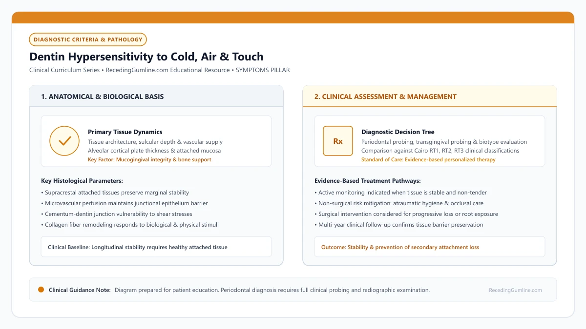
Educational illustration: Dentin Hypersensitivity to Cold, Air & Touch. Clinical management requires comprehensive periodontal evaluation rather than isolated self-assessment.
Source: RecedingGumline.com Clinical Editorial Team (Proprietary educational diagram for RecedingGumline.com)
Hydrodynamic Fluid Movement & Pulpal Neural Activation
Dentin is a living hard tissue composed of thousands of parallel microscopic tubules extending radially from the dental pulp chamber outward to the cementoenamel junction. In coronal dentin, these tubules are permanently sealed by a thick cap of mineralized enamel; along the root, they are protected by a thin layer of cellular and acellular cementum.
When gingival recession occurs, the fragile cementum (often less than 50 microns thick) is quickly worn away by mechanical brushing abrasion, dietary acid dissolution, or professional scaling. This uncaps the tubule apertures, exposing vital dentinal fluid directly to the mouth.
According to Martin Brännström's classic hydrodynamic theory (1966), external physical stimuli induce rapid displacement of fluid within these open tubules. Cold temperatures cause the fluid to contract, drawing it rapidly outward toward the surface at velocities up to 2 to 3 millimeters per second. This sudden hydrodynamic shear stress exerts mechanical tension on the odontoblastic process and deforms myelinated A-delta and A-beta nerve fibers in the subodontoblastic plexus of Raschkow, firing an immediate, sharp pain signal.
[object Object]
Key Clinical Insights
- Uncapped dentinal tubules contain vital fluid communicating directly with the dental pulp.
- Cold stimuli contract tubule fluid, triggering rapid outward fluid movement toward the tooth surface.
- Fluid shear stress deforms A-delta pulpal nerve endings, generating sharp, instantaneous pain.
Thermal, Osmotic & Tactile Triggers: The Role of Dietary Acid
Dentin hypersensitivity is provoked by a wide variety of daily environmental challenges. Cold stimuli—such as drinking ice water, eating ice cream, or breathing cold winter air—are by far the most prevalent and intense triggers due to the rapid rate of thermal contraction.
Osmotic triggers, including concentrated sweet solutions or acidic salad dressings, draw fluid outward via high osmotic gradients across the semi-permeable dentin membrane. Tactile triggers, such as running a fingernail or toothbrush bristle across the cervical notch, physically agitate the exposed tubule fluid.
Crucially, dietary acids act as a powerful co-factor in maintaining sensitivity. Consuming citrus fruits, soft drinks, wine, and sports beverages (pH 2.5 to 3.5) dissolves the natural organic smear layer and mineral plugs that saliva attempts to deposit within tubules. Frequent acid exposure keeps tubules wide open and hyper-reactive, preventing natural desensitization.
Key Clinical Insights
- Cold temperatures, sweet foods, and physical touch induce rapid fluid shifts within tubules.
- Dietary acids dissolve protective smear layers and salivary mineral plugs, maintaining sensitivity.
- Managing dietary acid frequency is essential for allowing dentinal tubules to naturally seal.
Biological Defense: Dentinal Sclerosis & Tertiary Dentin
The human tooth possesses remarkable intrinsic defense mechanisms designed to combat dentin exposure. When exposed to mild, chronic stimulation over months and years, odontoblasts and mesenchymal pulp cells respond by depositing mineralized repair tissue.
Within the lumen of exposed tubules, mineral crystals (hydroxyapatite and whitlockite) slowly precipitate, progressively narrowing the tubule diameter in a process known as dentinal sclerosis. Sclerosis significantly reduces dentin permeability, dampening hydrodynamic fluid displacement and naturally eliminating sensitivity over time.
Simultaneously, odontoblasts lining the pulp wall deposit a protective layer of tertiary (reactionary) dentin along the pulp chamber ceiling, increasing the physical distance between the receded root surface and the vital nerve core. This biological remodeling explains why older adults with severe, extensive recession frequently report zero pain.
Key Clinical Insights
- Dentinal sclerosis gradually narrows open tubule lumina through mineral precipitation.
- Odontoblasts deposit tertiary dentin on the pulp chamber wall, shielding internal nerve fibers.
- Intrinsic biological remodeling explains why mature, long-standing recession is often completely painless.
Clinical Evaluation: Hypersensitivity vs. Pulpal Pathology
Accurate clinical evaluation of dentin hypersensitivity requires differentiating it from more serious dental pathologies that present with similar symptoms. The hallmark characteristic of dentin hypersensitivity is that pain is strictly transient: it strikes immediately upon stimulus application and ceases within 1 to 2 seconds after the stimulus is removed.
If cold stimulus produces pain that lingers for 15 to 30 seconds or longer, if the tooth throbs continuously, or if pain awakens the patient at night, clinicians evaluate for irreversible pulpitis rather than simple hypersensitivity. Irreversible pulpitis indicates that bacterial toxins have deeply infected the pulp tissue, requiring endodontic root canal therapy or tooth extraction.
Other lookalike conditions include cracked tooth syndrome (sharp pain upon chewing release), defective restoration margins, and cervical root caries. A professional examination employing thermal testing, percussion, transillumination, and periapical radiographs is required to confirm an uncomplicated dentin hypersensitivity etiology.
Key Clinical Insights
- True dentin hypersensitivity ceases immediately (within 1-2 seconds) upon stimulus removal.
- Lingering or spontaneous throbbing pain indicates pulpal infection (pulpitis) requiring root canal therapy.
- Thermal testing, percussion, and radiographs are essential to rule out cracked teeth and root caries.
Evidence-Based Management Hierarchy
Clinical management of dentin hypersensitivity follows a stepped hierarchy, beginning with non-invasive at-home therapies and progressing to chairside interventions when necessary. First-line therapy utilizes desensitizing dentifrices containing potassium nitrate (which depolarizes pulpal sensory nerves) or stannous fluoride / NovaMin (which physically plug tubule lumina).
Patients must be instructed to use desensitizing pastes twice daily without rinsing with water immediately after spitting, allowing active ingredients to remain in contact with root surfaces.
For acute or localized sensitivity that resists at-home therapy, clinicians apply in-office glutaraldehyde-HEMA primers (such as Gluma), fluoride varnishes, or light-cured resin bonding agents that immediately occlude tubule apertures. When hypersensitivity coexists with severe, progressive gingival recession, soft-tissue grafting provides the definitive biological solution by surgically covering the root with vascularized tissue.
Key Clinical Insights
- First-line management utilizes potassium nitrate or stannous fluoride desensitizing toothpastes.
- In-office glutaraldehyde primers and bonding resins provide immediate, durable tubule occlusion.
- Surgical root coverage grafting permanently covers exposed root dentin, eliminating sensitivity.
Tooth Sensitivity During Brushing: Root Exposure vs. Enamel Wear
Experiencing a sharp, electric wince the moment your toothbrush bristles touch a specific tooth is a classic hallmark of exposed root dentin. While brushing is essential for preventing gum disease, touching a receded root with bristles directly excites mechanoreceptors in exposed tubules. Differentiating mechanical bristle sensitivity on roots from generalized enamel wear ensures appropriate intervention.
• Tactile sensitivity during brushing occurs when bristles mechanically drag across open dentinal tubules.
• It is strongly localized to the cervical margin where the gum has receded below the enamel boundary.
• Using medium or hard bristles acts like fine sandpaper, scouring away the protective smear layer.
• Switching to an ultra-soft brush and adjusting hand pressure significantly reduces brushing pain within 7 days.
Mechanics: How Bristles Excite Root Nerves
When you run a toothbrush across your teeth, the bristles exert mechanical friction. On enamel, this friction is harmless. However, on an exposed root surface, the bristles slide directly across thousands of microscopic dentinal tubule openings.
As the flexible bristle tip drags across the tubule opening, it creates mechanical shear stress and microscopic fluid turbulence. According to hydrodynamic theory, this mechanical disturbance instantly activates A-delta mechanosensitive nerve endings, producing an intense, sharp "electric shock" sensation.
Mechanical contact from toothbrush bristles against exposed root surfaces exerts direct tactile friction across open dentinal tubules. Patients experiencing brushing sensitivity often flinch or unconsciously avoid cleaning the cervical margins of affected teeth entirely.
Modern motorized toothbrushes featuring automated pressure sensors immediately alert patients through visual LED indicators or haptic pulses when excessive brushing force is detected, safeguarding exposed radicular dentin from ongoing mechanical micro-abrasion.
Key Clinical Insights
- Bristles exert direct shear stress across microscopic dentinal tubule openings
- Mechanical friction creates fluid displacement that excites pulpal mechanoreceptors
- Produces an instant sharp, electric wince that subsides the second the brush lifts
The Smear Layer and the Dangerous "Fear of Brushing" Cycle
Under normal conditions, microscopic mineral and protein debris called the smear layer naturally covers and plugs exposed tubules. Aggressive brushing with hard bristles or abrasive whitening toothpaste scrubs this natural smear layer away, leaving tubules wide open.
When brushing becomes painful, patients instinctively avoid brushing that sensitive tooth. This triggers a dangerous clinical cycle: undisturbed bacterial plaque produces acidic byproducts that dissolve mineral plugs, widening tubule apertures and making the tooth even more excruciatingly sensitive next time.
Avoiding hygiene around sensitive receded areas allows virulent dental plaque biofilm to accumulate undisturbed along the gingival margin. This biofilm colonization accelerates inflammatory soft-tissue destruction, inadvertently causing further apical recession and worsening nerve sensitivity.
Key Clinical Insights
- Abrasive pastes scrub away the natural mineral smear layer that plugs tubules
- Avoiding brushing allows bacterial plaque to accumulate along the receded margin
- Bacterial acids dissolve deeper tubule plugs, worsening sensitivity and accelerating decay
Clinical Solutions: How to Brush Receded Teeth Without Pain
Breaking the cycle requires immediate modification of your brushing protocol. Switch to an ultra-soft brush featuring micro-tapered bristles (under 0.15 mm diameter). These filaments bend effortlessly over exposed roots without scouring the dentin or jarring the nerves.
Angle the bristles at a 45-degree angle toward the gumline using the Modified Bass technique: make tiny, gentle circular vibrations without scrubbing horizontally. Consider using warm water to soften the bristles before applying toothpaste, which eliminates the thermal shock while cleaning.
Switching to ultra-soft microfiber bristle designs combined with room-temperature water rinses minimizes mechanical and thermal trigger activation during brushing. Professional application of glutaraldehyde-based varnishes provides instant tactile relief so patients can resume plaque removal.
Key Clinical Insights
- Switch immediately to ultra-soft, micro-tapered toothbrush filaments
- Use gentle circular vibrations (Modified Bass technique) rather than back-and-forth scrubbing
- Rinse the brush head under warm water before brushing to soften bristles and prevent thermal shock
Clinical Evaluation: Marginal Mucosal Abrasion vs. Dentinal Shock
Sensitivity felt during toothbrushing can stem from two distinct anatomical origins: mechanical trauma to the exposed root dentin, or physical laceration of inflamed, friable marginal soft tissue. Differentiating between these sources is essential for proper therapy.
When the sensation is a sharp, instantaneous electrical shock that resolves the exact moment bristle contact ceases, the origin is dentinal tubule excitation triggered by direct mechanical bristle friction against open dentin. Conversely, if brushing produces a dull, burning, or throbbing ache accompanied by bleeding, the cause is mechanical injury to ulcerated sulcular epithelium.
Clinicians use a blunt periodontal probe to gently stroke the exposed root and marginal soft tissue separately. Touching the dentin reproduces dentin hypersensitivity, whereas touching the gingival margin reproduces mucosal inflammation.
Key Clinical Insights
- Instantaneous electrical pain during bristle contact indicates dentinal tubule excitation.
- Burning, lingering discomfort accompanied by bleeding points to inflamed marginal epithelium.
- Blunt tactile probing distinguishes between hard-tissue sensitivity and soft-tissue ulceration.
Corrective Cleansing Techniques: The Modified Bass Vibratory Protocol
Patients experiencing brushing sensitivity often enter a destructive cycle: fearing pain, they skip cleaning the receded area, allowing bacterial plaque to accumulate. Subgingival bacteria release acidic byproducts that demineralize dentin and reopen tubules, intensifying sensitivity.
To break this cycle, patients must transition to the modified Bass vibratory technique. Using an ultra-soft brush with rounded bristle filaments (< 0.1 mm diameter), the brush head is angled at 45 degrees toward the gumline with featherweight pressure (no greater than the weight of a coin).
Instead of sweeping or scrubbing horizontally, the patient gently vibrates the filaments in small circular motions. This disrupts bacterial biofilm without displacing the protective smear layer or exciting pulpal nerve endings.
Key Clinical Insights
- Skipping brushing allows acid-producing bacteria to demineralize dentin and worsen sensitivity.
- The modified Bass technique angles extra-soft bristles at 45 degrees using featherweight pressure.
- Gentle vibratory motions disrupt plaque biofilms without removing the protective smear layer.
Clinical Reality Check
Dentin hypersensitivity can be so sharp that patients avoid brushing the affected tooth, inadvertently allowing plaque biofilms to accumulate. This bacterial buildup triggers localized inflammation that worsens sensitivity and accelerates further gum recession.
Questions to Ask Your Dentist or Periodontist
- Is my tooth sensitivity caused by exposed root dentin, a cracked tooth, or a dying nerve?
- Which specific desensitizing toothpaste ingredient would work best for my type of sensitivity?
- Can you apply an in-office desensitizing varnish or bonding agent to seal these sensitive areas today?
- Would covering this receded root with a gum graft permanently resolve my sensitivity?
- Are the acidic foods or drinks in my daily diet preventing my exposed roots from naturally desensitizing?
- Is my brushing sensitivity localized to a specific receded root or generalized across my enamel?
- Do my receded teeth have deep toothbrush abrasion notches that require a protective filling?
- Can you demonstrate the exact Modified Bass brushing motion on my teeth to prevent pain?
- Would switching to an electric toothbrush with a pressure sensor reduce my brushing discomfort?
Unsure What Your Gum Changes Mean?
Take our free, evidence-based Gum Recession Assessment — approximately 3 minutes. Identify potential risk factors, evaluate symptoms, and receive personalized discussion questions for your dentist or periodontist.
Non-diagnostic educational triage. Private, secure, completed in your browser.
Related Educational Topics
Scientific Literature & Clinical Guidelines
4sources · Hide ▲
- Brännström M (1966).
"Sensitivity of dentine." Oral Surgery, Oral Medicine, Oral Pathology.
Clinical relevance: Foundational paper formulating the hydrodynamic theory of dentin hypersensitivity: rapid fluid displacement within patent dentinal tubules physically deforms intradental nerve endings at the pulp-dentin boundary, explaining thermal, mechanical, and evaporative root sensitivity.
- West NX, Seong J, Davies M (2015).
"Management of dentine hypersensitivity: efficacy of professionally and self-administered agents." Journal of Clinical Periodontology.
Clinical relevance: Systematic review evaluating professionally and self-administered desensitizing agents; found evidence supporting tubule-occluding dentifrices and potassium-based nerve desensitizers in providing transient to moderate symptom relief, with substantial heterogeneity across clinical trials.
- Jepsen S, Caton JG, Albandar JM, Bissada NF, Bouchard P, Cortellini P, et al. (2018).
"Periodontal manifestations of systemic diseases and developmental and acquired conditions: Consensus report of workgroup 3 of the 2017 World Workshop on the Classification of Periodontal and Peri-Implant Diseases and Conditions." Journal of Periodontology.
Clinical relevance: Consensus report defining mucogingival conditions, gingival phenotype (replacing biotype), non-carious cervical lesions, and the multifactorial etiology of gingival recession; emphasizes that recession can occur without periodontitis and classifies recession by interdental clinical attachment loss.
- Cairo F, Nieri M, Cincinelli S, Mervelt J, Pagliaro U (2011).
"The interproximal clinical attachment level to classify gingival recessions and predict root coverage outcomes: an explorative and reliability study." Journal of Clinical Periodontology.
Clinical relevance: Exploratory and reliability study establishing the Cairo classification based on interdental clinical attachment level (CAL): RT1 (no interproximal attachment loss; complete root coverage is clinically predictable), RT2 (interproximal attachment loss <= buccal loss; partial coverage predictable), and RT3 (interproximal loss exceeds buccal recession; complete coverage not predictable).
Important Medical Notice
The contents of RecedingGumline.com, including text, graphics, self-assessment calculators, and other materials, are intended solely for educational and informational purposes. This content is not intended to replace professional dental examination, diagnosis, or treatment. Always seek the advice of a qualified dentist, periodontist, or other licensed oral healthcare provider with any questions you may have regarding a medical or dental condition. Never disregard professional medical advice or delay seeking it because of something you read on this website.