Orthodontic Movement and Gum Grafting: Sequencing Interventions for Optimal Stability
Should a gum graft be performed before starting orthodontics or after tooth movement is completed?
The relationship between orthodontic tooth movement and receding gumline options requires careful interdisciplinary coordination. Moving tooth roots through thin alveolar bone plates can cause dehiscence and rapid recession. Clinicians must strategically determine whether soft-tissue grafting should occur before orthodontic treatment to reinforce the phenotype, or after alignment when roots are properly centered.
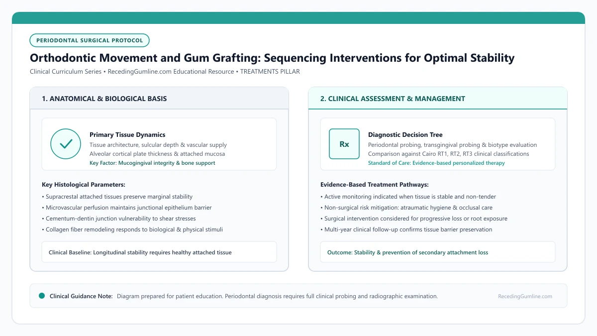
Educational illustration: Orthodontic Movement and Gum Grafting: Sequencing Interventions for Optimal Stability. Clinical management requires comprehensive periodontal evaluation rather than isolated self-assessment.
Source: RecedingGumline.com Clinical Editorial Team (Proprietary educational diagram for RecedingGumline.com)
Key Clinical Distinctions & Diagnostic Boundaries
- Pre-orthodontic grafting focuses on augmenting attached gingival thickness (phenotype modification) rather than cosmetic root coverage.
- Post-orthodontic grafting allows the surgeon to perform root coverage on teeth that have been uprighted and centered within the bone envelope.
- Clear aligners generate different biomechanical forces than fixed brackets but can still cause recession if teeth are tipped beyond cortical boundaries.
- Recession that develops during active orthodontic treatment requires immediate clinical evaluation to modify biomechanical vector forces.
The Bone Envelope: Biological Boundaries of Tooth Movement
Every tooth root is enclosed by the alveolar bone process. When an orthodontist expands dental arches or proinclines lower front teeth to relieve crowding, the tooth root is pushed closer to the paper-thin outer cortical bone plate.
If root movement pushes the root completely outside the cortical plate, an alveolar bone dehiscence (cleft) forms. If the overlying gingiva is thin and delicate, the soft tissue collapses into the cleft, manifesting as severe, progressive gingival recession.
Pre-orthodontic periodontal assessment evaluates whether tooth roots are being moved outside the protective cortical bone plates of the alveolar ridge. Expanding dental arches beyond the natural skeletal envelope dramatically elevates the risk of progressive buccal bone dehiscence.
Clinical Considerations:
- Orthodontic arch expansion pushes roots toward the outer cortical bone plate
- Moving roots beyond the bone plate creates structural bone dehiscences
- Thin gingival tissue over a dehiscence rapidly breaks down into recession
When to Graft Before Orthodontics: Prophylactic Phenotype Augmentation
A pre-orthodontic gum graft is recommended when a patient presents with a very thin gingival biotype (translucent tissue where the probe shines through) and less than 1 to 2 mm of attached keratinized tissue on teeth scheduled for labial expansion or proinclination.
In this scenario, a free gingival graft or connective tissue graft is performed prior to placing brackets or aligners. The primary goal is not root coverage, but converting the delicate biotype into a thick, resilient fibrotic cuff capable of withstanding orthodontic stress without tearing.
When thin phenotypes are identified prior to clear aligner or fixed appliance therapy, prophylactic soft-tissue grafting thickens the gingival biotype. This preemptive surgical reinforcement shields delicate marginal tissue from subsequent orthodontic forces and rotational stress.
Clinical Considerations:
- Indicated when attached tissue is < 1 mm and labial movement is planned
- Converts thin tissue into a dense, protective phenotype
- Typically requires 6 to 8 weeks of healing before starting tooth movement
When to Graft After Orthodontics: Centering Roots for Complete Coverage
In other situations, delaying the gum graft until after orthodontic treatment is completed offers distinct advantages. If a tooth is currently tilted forward (proclined) and already has recession, attempting root coverage surgery while the root is protruding from the arch has a low success rate.
By having the orthodontist first upright the tooth and move the root lingually back inside the alveolar housing, the periodontist can later perform a coronally advanced flap and connective tissue graft with substantially higher predictability for complete root coverage.
Close interdisciplinary collaboration between orthodontists and periodontists ensures forces are kept light, continuous, and directed along optimal physiological vectors. Regular three-month periodontal check-ups during active tooth movement catch emerging mucogingival complications early.
Clinical Considerations:
- Lingual root movement reduces root prominence and narrow defect width
- Centering the root within bone creates a stable vascular recipient bed
- Surgical root coverage achieves higher success rates after orthodontic alignment
Orthodontic Decompensation: Re-Centering Roots in the Bone Housing
When past orthodontic treatment has tipped or expanded tooth roots outward through the facial cortical plate, attempting soft-tissue grafting alone often produces disappointing results. Without an underlying bony vascular bed, the grafted tissue lacks the biological support needed for survival.
In these complex cases, interdisciplinary orthodontic retreatment is essential to "decompensate" the teeth. Using customized bracket positioning or clear aligner torque staging, the orthodontist applies light, continuous biomechanical forces to torque the displaced root lingually back into the center of the cancellous bone envelope.
Once the root is centered and surrounded by supportive bone, the recipient bed anatomy is normalized, transforming an unpredictable surgical site into a receptive environment for soft-tissue coverage.
Clinical Considerations:
- Roots positioned outside the cortical plate lack the vascular bed required for graft survival.
- Orthodontic torque mechanics move roots lingually back into the cancellous alveolar envelope.
- Centering the root within the bone housing dramatically increases subsequent graft predictability.
Surgical Decision Framework: Grafting Before vs. After Tooth Movement
Deciding whether to perform soft-tissue grafting before initiating orthodontic retreatment or after completing tooth movement is a critical clinical determination. The decision depends primarily on baseline keratinized tissue width and gingival thickness.
If a tooth has less than 1.0 mm of keratinized tissue or exhibits a paper-thin phenotype (< 0.5 mm), pre-orthodontic grafting is mandatory. Moving a tooth through bone with deficient soft-tissue coverage inevitably triggers rapid, severe recession. In this scenario, prophylactic grafting thickens the phenotype to withstand orthodontic forces.
Conversely, if the tissue biotype is moderate and the primary defect is a prominent root contour, moving the root into the bone first often results in spontaneous tissue rebound, allowing a smaller, more conservative graft procedure after tooth movement is completed.
Clinical Considerations:
- Thin phenotypes (< 1.0 mm KTW) mandate prophylactic grafting before active orthodontic retreatment.
- Prophylactic grafting prevents severe soft-tissue breakdown during tooth repositioning.
- Moving prominent roots into bone first may facilitate spontaneous tissue rebound.
Clinical Reality Check
Always ensure your orthodontist and periodontist review 3D CBCT imaging together before finalizing tooth expansion plans in patients with thin gum phenotypes.
Questions to Ask Your Periodontist or Dentist
- Does my orthodontic treatment plan involve expanding my dental arches or moving my lower incisors forward?
- Do I have a thin gum phenotype that puts me at risk of developing recession during braces or aligners?
- Would it be safer for me to have a soft-tissue graft before starting tooth movement or wait until treatment is completed?
- How frequently should my periodontist evaluate my gum tissue during my orthodontic treatment?
Unsure What Your Gum Changes Mean?
Take our free, evidence-based Gum Recession Assessment — approximately 3 minutes. Identify potential risk factors, evaluate symptoms, and receive personalized discussion questions for your dentist or periodontist.
Non-diagnostic educational triage. Private, secure, completed in your browser.
Related Educational Topics
Clinical Evidence & Claim Traceability (3 Mapped Assertions)
Scientific Literature & Clinical Guidelines
3sources · Hide ▲
- Wennström JL (1987).
"Lack of association between width of attached gingiva and development of soft tissue recession. A 5-year longitudinal study." Journal of Clinical Periodontology.
Clinical relevance: Clinical study demonstrating that in the presence of meticulous plaque control, an extremely narrow zone or absence of attached keratinized gingiva does not inevitably lead to soft-tissue breakdown or recession progression, qualifying historical mandatory width dogmas.
- Jepsen S, Caton JG, Albandar JM, Bissada NF, Bouchard P, Cortellini P, et al. (2018).
"Periodontal manifestations of systemic diseases and developmental and acquired conditions: Consensus report of workgroup 3 of the 2017 World Workshop on the Classification of Periodontal and Peri-Implant Diseases and Conditions." Journal of Periodontology.
Clinical relevance: Consensus report defining mucogingival conditions, gingival phenotype (replacing biotype), non-carious cervical lesions, and the multifactorial etiology of gingival recession; emphasizes that recession can occur without periodontitis and classifies recession by interdental clinical attachment loss.
- Chambrone L, Salinas Ortega MA, Sukekava F, Rotundo R, Kalemaj Z, Buti J, Pini Prato GP (2018).
"Root coverage procedures for treating localised and multiple recession-type defects." Cochrane Database of Systematic Reviews.
Clinical relevance: Cochrane systematic review evaluating root-coverage procedures for localized and multiple recession-type defects. While subepithelial connective tissue grafts (SCTG) combined with coronally advanced flaps demonstrated higher rates of complete root coverage and keratinized tissue gain compared to flap advancement alone, evidence quality varied across outcomes and clinical decisions must balance donor site morbidity and patient-reported outcomes.
Important Medical Notice
The contents of RecedingGumline.com, including text, graphics, self-assessment calculators, and other materials, are intended solely for educational and informational purposes. This content is not intended to replace professional dental examination, diagnosis, or treatment. Always seek the advice of a qualified dentist, periodontist, or other licensed oral healthcare provider with any questions you may have regarding a medical or dental condition. Never disregard professional medical advice or delay seeking it because of something you read on this website.