Orthodontic Movement & Alveolar Bone Architecture in Gum Recession
Orthodontic treatment safely aligns crowded and malpositioned teeth for millions of individuals each year. However, tooth movement occurs within strict biological boundaries defined by the alveolar bone housing. When orthodontic mechanics move roots outside this cortical bone envelope—particularly in patients with a pre-existing thin gingival recession phenotype—the overlying cortical plate can resorb, creating alveolar dehiscences and predisposing the tooth to secondary gingival recession.
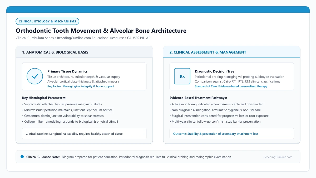
Educational illustration: Orthodontic Tooth Movement & Alveolar Bone Architecture. Clinical management requires comprehensive periodontal evaluation rather than isolated self-assessment.
Source: RecedingGumline.com Clinical Editorial Team (Proprietary educational diagram for RecedingGumline.com)
The Biological Envelope of Alveolar Bone
The alveolar process is a tooth-dependent anatomical structure that develops alongside tooth eruption and resorbs following tooth extraction. Each tooth resides within a bony crypt bounded by buccal and lingual cortical plates and intervening trabecular cancellous bone. The thickness of these cortical plates varies widely: in the mandibular anterior region and around prominent maxillary canines, the facial cortical plate frequently measures less than 0.5 mm in thickness, and may naturally feature congenital fenestrations (isolated windows in bone) or dehiscences (marginal V-shaped clefts).
Orthodontic force induces bone remodeling via coordinated osteoclastic resorption on the pressure side and osteoblastic apposition on the tension side of the periodontal ligament (PDL). However, this biological remodeling can only occur if the tooth remains enveloped within viable bone marrow and periosteum. When a tooth root is moved excessively in a facial direction, the root pushes against the outer cortical plate faster than new bone can be deposited on the periosteal surface.
As a result, the thin cortical bone plate undergoes complete resorption, creating an extensive alveolar bone dehiscence. Although the overlying soft tissue may initially remain intact immediately after debonding, the gingiva is left without underlying skeletal support, anchored only to adjacent periosteum.
Key Etiological Insights
- The facial alveolar bone plate in anterior teeth is often razor-thin (under 0.5 mm).
- Orthodontic tooth movement beyond cortical plate boundaries creates irreversible alveolar bone dehiscences.
- Gingival tissue lacking underlying bone support exhibits extreme vulnerability to subsequent everyday trauma.
Biomechanics: Labial Tipping vs. Bodily Translation
The specific type of orthodontic tooth movement dramatically influences the risk of cortical bone perforation and subsequent recession. Uncontrolled tipping mechanics exert high stress concentrations at the alveolar crest. When anterior teeth are proclined or tipped forward to relieve crowding or camouflage a skeletal discrepancy, the root apex moves lingually while the cervical crown and root neck move sharply labially.
This labial movement compresses the delicate marginal bone crest and supracrestal connective tissue. In contrast, controlled bodily translation (moving root and crown simultaneously in a parallel vector) distributes remodeling forces more evenly across the entire PDL surface area, reducing localized crestal compression.
Rapid maxillary arch expansion (RME) or aggressive non-extraction arch expansion in adult patients poses substantial periodontal risk. Expanding dental arches beyond the genetic baseline pushes posterior root apices against thin buccal cortical bone, frequently inducing dehiscences on maxillary first premolars and first molars.
Key Etiological Insights
- Uncontrolled tipping mechanics concentrate destructive stress directly at the marginal alveolar crest.
- Proclining lower incisors to resolve crowding significantly increases the risk of labial bone loss.
- Aggressive arch expansion in adult dentitions frequently drives posterior root tips through buccal bone.
The Post-Orthodontic Latency Period & Triggering Factors
A perplexing clinical reality for many patients is that gingival recession often does not appear while braces or clear aligners are in place. Instead, recession characteristically manifests 1 to 5 years following the completion of orthodontic therapy. This delayed presentation reflects a two-stage pathological process.
In the first stage, orthodontic movement creates an asymptomatic, subclinical alveolar bone dehiscence. In the second stage, normal daily life factors act upon the unsupported gingival collar. Normal toothbrushing abrasion, mild plaque accumulation, or slight frenal tension—forces that a normal periodontium would easily withstand—overwhelm the delicate, unbacked soft tissue.
Without the vascular supply and mechanical foundation of the underlying cortical plate, the marginal gingiva gradually collapses and recedes apically until it reaches the level of the remaining bone, exposing yellowish root cementum.
Key Etiological Insights
- Recession typically develops years after orthodontic debonding due to secondary mechanical triggers.
- Subclinical bone dehiscences created during treatment leave soft tissue fragile and unsupported.
- Everyday brushing forces easily collapse soft tissue margins that lack underlying alveolar bone.
Interdisciplinary Prevention: Phenotype Modification Therapy
Modern interdisciplinary orthodontics emphasizes pre-treatment periodontal risk assessment and phenotype screening. Patients presenting with a thin periodontal phenotype, minimal keratinized tissue (< 2 mm), and prominent anterior teeth should undergo careful periodontal screening prior to placing active appliances.
In high-risk cases where significant labial tooth movement or arch expansion is planned, periodontists and orthodontists frequently collaborate on Surgically Facilitated Orthodontic Therapy (SFOT) or prophylactic soft-tissue grafting (phenotype modification therapy). By placing an autogenous subepithelial connective tissue graft or particulate bone graft before or during orthodontic movement, clinicians can convert a thin, vulnerable biotype into a robust, thick phenotype.
This proactive tissue augmentation bolsters the soft-tissue barrier, preserves marginal stability during tooth alignment, and dramatically decreases the post-treatment incidence of Cairo RT1 gingival recession.
Key Etiological Insights
- Pre-orthodontic screening identifies thin periodontal biotypes at high risk for treatment-induced recession.
- Prophylactic connective tissue grafting converts vulnerable thin tissue into a thick, protective phenotype.
- Interdisciplinary collaboration between periodontist and orthodontist minimizes iatrogenic complications.
Clinical Reality Check
Orthodontic tooth movement does not inevitably cause recession; it serves as a predisposing factor when roots are positioned outside the bony housing. Patients experiencing recession after orthodontics should consult a periodontist to evaluate bone levels and determine if soft-tissue grafting can re-establish protective attached gingiva.
Questions to Ask Your Dentist or Periodontist
- Did my previous orthodontic treatment push my tooth roots outside the alveolar bone envelope?
- How much underlying bone support remains on the facial surface of my receded teeth?
- Should I have a periodontal evaluation before starting or revising clear aligner or braces treatment?
- Would a gum graft help rebuild lost tissue thickness around teeth that moved forward?
- Is my current retainer or nightguard placing any adverse pressure on my receded gum margins?
Unsure What Your Gum Changes Mean?
Take our free, evidence-based Gum Recession Assessment — approximately 3 minutes. Identify potential risk factors, evaluate symptoms, and receive personalized discussion questions for your dentist or periodontist.
Non-diagnostic educational triage. Private, secure, completed in your browser.
Related Educational Topics
Scientific Literature & Clinical Guidelines
4sources · Hide ▲
- Tonetti MS, Greenwell H, Kornman KS (2018).
"Staging and grading of periodontitis: Framework and proposal of a new classification and case definition." Journal of Clinical Periodontology.
Clinical relevance: Consensus framework establishing the multidimensional staging (severity and extent of periodontal tissue breakdown) and grading (biological rate of disease progression, incorporating smoking and diabetes as grade modifiers) for periodontitis. It addresses periodontitis diagnosis and staging, not the classification of localized gingival recession defects.
- Cairo F, Nieri M, Cincinelli S, Mervelt J, Pagliaro U (2011).
"The interproximal clinical attachment level to classify gingival recessions and predict root coverage outcomes: an explorative and reliability study." Journal of Clinical Periodontology.
Clinical relevance: Exploratory and reliability study establishing the Cairo classification based on interdental clinical attachment level (CAL): RT1 (no interproximal attachment loss; complete root coverage is clinically predictable), RT2 (interproximal attachment loss <= buccal loss; partial coverage predictable), and RT3 (interproximal loss exceeds buccal recession; complete coverage not predictable).
- Wennström JL (1987).
"Lack of association between width of attached gingiva and development of soft tissue recession. A 5-year longitudinal study." Journal of Clinical Periodontology.
Clinical relevance: Clinical study demonstrating that in the presence of meticulous plaque control, an extremely narrow zone or absence of attached keratinized gingiva does not inevitably lead to soft-tissue breakdown or recession progression, qualifying historical mandatory width dogmas.
- Jepsen S, Caton JG, Albandar JM, Bissada NF, Bouchard P, Cortellini P, et al. (2018).
"Periodontal manifestations of systemic diseases and developmental and acquired conditions: Consensus report of workgroup 3 of the 2017 World Workshop on the Classification of Periodontal and Peri-Implant Diseases and Conditions." Journal of Periodontology.
Clinical relevance: Consensus report defining mucogingival conditions, gingival phenotype (replacing biotype), non-carious cervical lesions, and the multifactorial etiology of gingival recession; emphasizes that recession can occur without periodontitis and classifies recession by interdental clinical attachment loss.
Important Medical Notice
The contents of RecedingGumline.com, including text, graphics, self-assessment calculators, and other materials, are intended solely for educational and informational purposes. This content is not intended to replace professional dental examination, diagnosis, or treatment. Always seek the advice of a qualified dentist, periodontist, or other licensed oral healthcare provider with any questions you may have regarding a medical or dental condition. Never disregard professional medical advice or delay seeking it because of something you read on this website.