The Periodontal Evaluation Process: Probing, Radiographs & Clinical Attachment Measurements
How do dental professionals objectively measure and diagnose the severity of gum recession and attachment loss?
In any rigorous receding gums overview assessment, a comprehensive periodontal evaluation forms the diagnostic foundation for all treatment decisions. Rather than relying on visual inspection alone, clinicians utilize calibrated periodontal probes, full-mouth intraoral radiographs, and systematic documentation of mucosal margins to distinguish localized anatomical recession from destructive periodontal attachment loss.
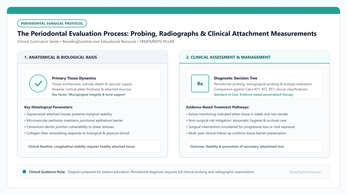
Educational illustration: The Periodontal Evaluation Process: Probing, Radiographs & Clinical Attachment Measurements. Clinical management requires comprehensive periodontal evaluation rather than isolated self-assessment.
Source: RecedingGumline.com Clinical Editorial Team (Proprietary educational diagram for RecedingGumline.com)
Key Clinical Distinctions & Diagnostic Boundaries
- Probing depth measures the distance from the gingival margin to the base of the sulcus, whereas CAL measures the distance from the cementoenamel junction.
- A tooth with 2 mm probing depth can still exhibit severe attachment loss if 4 mm of recession has exposed the root surface.
- Bleeding on probing indicates active inflammatory breakdown within the pocket rather than structural stability.
- Two-dimensional radiographs show interproximal bone levels but cannot accurately depict thin labial or buccal bone plates.
Six-Point Periodontal Probing: Mechanics and Metric Interpretation
During a periodontal examination, the clinician gently inserts a standardized millimeter-marked probe into the gingival sulcus at six distinct anatomical locations around each tooth: distobuccal, mid-buccal, mesiobuccal, distolingual, mid-lingual, and mesiolingual. A gentle probing force of approximately 0.20 to 0.25 Newtons is applied to avoid traumatizing the delicate junctional epithelium.
Healthy sulcular depths typically measure between 1 and 3 millimeters without bleeding upon gentle probing. Depths of 4 millimeters or greater frequently indicate inflammatory swelling or apical migration of the epithelial attachment, defining a periodontal pocket. In cases of pure recession without deep pockets, probing depths may remain 1 to 2 mm, but the margin itself has migrated apically.
Standardized periodontal charting software records these probing depths alongside automated alerts for clinical recession and furcation involvement. Longitudinal comparisons between consecutive annual charts reveal subtle micro-shifts in tissue margins before gross anatomical defect development becomes clinically obvious.
Clinical Considerations:
- Probing force is calibrated to 0.20 to 0.25 N to prevent mechanical tissue puncture
- Six standardized measurement sites per tooth capture circumferential periodontal health
- Bleeding on probing signals active inflammatory cell infiltration in the sulcular lining
Calculating Clinical Attachment Level: The True Measure of Periodontal Support
A critical concept in periodontal diagnosis is the difference between probing pocket depth and Clinical Attachment Level (CAL). Probing depth measures the distance from the moving gingival margin down to the bottom of the sulcus. If the gingival margin has receded 3 mm below the cementoenamel junction (CEJ) and the probing depth is 2 mm, the total clinical attachment loss is 5 mm.
Tracking CAL across sequential recall visits allows the dental team to determine whether recession is structurally arrested or experiencing active, ongoing progression. An increase in CAL over 6 to 12 months provides definitive clinical evidence of active tissue destruction, serving as a primary trigger for clinical intervention.
Calculating attachment levels at regular six-month intervals provides an objective benchmark for therapeutic success or failure. A stable attachment level confirms periodontal disease quiescence even when minor anatomical root exposure persists without symptomatic deterioration.
Clinical Considerations:
- CAL combines marginal recession distance with sulcular probing depth
- CEJ serves as the fixed anatomical landmark for longitudinal attachment measurements
- Sequential CAL increases demonstrate active disease progression rather than historical recession
Radiographic Analysis of Alveolar Bone Architecture
Periapical and bitewing radiographs are indispensable adjuncts to clinical probing. They reveal the height, density, and continuous cortex of the interdental alveolar bone crest. In healthy periodontium, the alveolar crest sits approximately 1.5 to 2.0 millimeters apical to the cementoenamel junction of adjacent teeth.
When horizontal or vertical bone resorption is observed on radiographs, clinicians classify the site according to defect morphology. However, standard intraoral radiographs superimpose facial and lingual cortical plates, meaning thin facial bone dehiscences accompanying recession must be diagnosed primarily through clinical palpation, probing, and direct visual inspection.
High-resolution cone-beam computed tomography (CBCT) may be indicated in complex multi-rooted scenarios where severe buccal fenestrations are suspected. However, traditional bitewings remain the diagnostic standard for routine crestal bone evaluation due to their low radiation dose and high spatial resolution.
Clinical Considerations:
- Radiographs reveal interdental bone height essential for predicting surgical root coverage
- Healthy crestal bone resides 1.5 to 2.0 mm below the cementoenamel junction
- Facial bone plates are often too thin to be resolved on standard dental projection radiographs
Advanced Diagnostic Imaging: CBCT & Periapical Radiographic Analysis
A comprehensive periodontal evaluation integrates advanced radiographic imaging to assess the three-dimensional skeletal architecture beneath the soft tissue. Standard bitewing x-rays provide an overview of coronal relationships but lack the detail necessary to resolve thin facial cortical bone plates.
High-resolution periapical radiographs, exposed using the long-cone paralleling technique with precision positioners, reveal interproximal bone crest height—the critical metric dictating root coverage predictability according to the Cairo classification. In complex cases involving multiple recession defects or planned implant therapy, Cone-Beam Computed Tomography (CBCT) provides cross-sectional volumetric views.
CBCT cross-sections permit precise sub-millimeter measurements of remaining labial bone plate thickness, identifying fenestrations and dehiscences before surgical flaps are elevated, thereby informing surgical technique selection.
Clinical Considerations:
- Paralleling-technique periapicals accurately establish interdental bone crest levels.
- Interproximal bone height dictates the biological ceiling for predictable root coverage.
- CBCT volumetric cross-sections quantify labial bone thickness and detect hidden dehiscences.
Longitudinal Attachment Tracking & Progression Thresholds
Determining whether gingival recession requires active surgical intervention or ongoing conservative monitoring relies on longitudinal clinical attachment level (CAL) tracking. Clinicians measure two distinct parameters: probing depth (from gingival margin to the base of the sulcus) and recession depth (from the cementoenamel junction to the gingival margin).
Summing these measurements provides the total clinical attachment loss. Periodontists consider an increase in recession depth of 1.0 millimeter or more between consecutive recall visits as confirmed disease progression, prompting re-evaluation of the preventive regimen or recommendation of soft-tissue augmentation.
Digital intraoral scanners now generate precise 3D surface mesh superimpositions over time, detecting volumetric tissue loss as subtle as 0.1 millimeters long before it becomes visible to the unaided eye.
Clinical Considerations:
- Total clinical attachment loss equals probing depth plus recession depth.
- Documented apical movement of ≥ 1.0 mm between recalls confirms progressive attachment loss.
- Digital 3D intraoral scan superimposition detects micro-volumetric tissue loss over time.
Clinical Reality Check
A shallow probing depth of 2 mm does not guarantee periodontal health if substantial root exposure has already occurred; attachment loss must be calculated relative to the cementoenamel junction.
Questions to Ask Your Periodontist or Dentist
- What were my deepest probing depth numbers, and were there specific sites that bled during probing?
- Do my radiographs show any bone loss between my teeth or is my recession isolated to the outer facial surfaces?
- How does my clinical attachment loss compare to my previous checkup measurements?
- What specific home care modifications do you recommend based on my plaque distribution and tissue phenotype?
Unsure What Your Gum Changes Mean?
Take our free, evidence-based Gum Recession Assessment — approximately 3 minutes. Identify potential risk factors, evaluate symptoms, and receive personalized discussion questions for your dentist or periodontist.
Non-diagnostic educational triage. Private, secure, completed in your browser.
Related Educational Topics
Clinical Evidence & Claim Traceability (2 Mapped Assertions)
Scientific Literature & Clinical Guidelines
3sources · Hide ▲
- Tonetti MS, Greenwell H, Kornman KS (2018).
"Staging and grading of periodontitis: Framework and proposal of a new classification and case definition." Journal of Clinical Periodontology.
Clinical relevance: Consensus framework establishing the multidimensional staging (severity and extent of periodontal tissue breakdown) and grading (biological rate of disease progression, incorporating smoking and diabetes as grade modifiers) for periodontitis. It addresses periodontitis diagnosis and staging, not the classification of localized gingival recession defects.
- Cairo F, Nieri M, Cincinelli S, Mervelt J, Pagliaro U (2011).
"The interproximal clinical attachment level to classify gingival recessions and predict root coverage outcomes: an explorative and reliability study." Journal of Clinical Periodontology.
Clinical relevance: Exploratory and reliability study establishing the Cairo classification based on interdental clinical attachment level (CAL): RT1 (no interproximal attachment loss; complete root coverage is clinically predictable), RT2 (interproximal attachment loss <= buccal loss; partial coverage predictable), and RT3 (interproximal loss exceeds buccal recession; complete coverage not predictable).
- Pihlstrom BL, Michalowicz BS, Johnson NW (2005).
"Periodontal diseases." The Lancet.
Clinical relevance: Peer-reviewed Lancet seminar review synthesizing global periodontal epidemiology, microbial etiology, host immunopathology, and systemic interactions, emphasizing prevention, biofilm disruption, and early risk factor modification.
Important Medical Notice
The contents of RecedingGumline.com, including text, graphics, self-assessment calculators, and other materials, are intended solely for educational and informational purposes. This content is not intended to replace professional dental examination, diagnosis, or treatment. Always seek the advice of a qualified dentist, periodontist, or other licensed oral healthcare provider with any questions you may have regarding a medical or dental condition. Never disregard professional medical advice or delay seeking it because of something you read on this website.