Minimally Invasive Root Coverage Procedures for Gum Recession
In recent years, receding gums plastic surgery has undergone a transformative shift toward minimally invasive surgical techniques (MIST). Traditional surgical protocols often required extensive vertical releasing incisions, wide flap reflection, and extensive palatal harvesting, resulting in substantial postoperative discomfort and prolonged healing. Minimally invasive techniques for resolving gum recession—including the Coronally Advanced Tunnel (CAT) and vestibular access techniques—prioritize preservation of the interdental papillae, maintenance of the supraperiosteal vascular plexus, and microscopic precision.
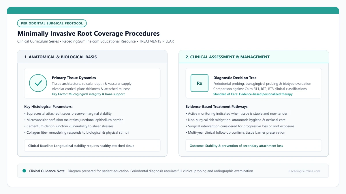
Educational illustration: Minimally Invasive Root Coverage Procedures. Clinical management requires comprehensive periodontal evaluation rather than isolated self-assessment.
Source: RecedingGumline.com Clinical Editorial Team (Proprietary educational diagram for RecedingGumline.com)
The Coronally Advanced Tunnel (CAT) Technique
The Coronally Advanced Tunnel (CAT) technique represents one of the most significant refinements in periodontal plastic surgery. In traditional flap procedures, vertical releasing incisions are placed to mobilize the gingiva, which severs lateral blood vessels and can leave visible mucosal scars. The tunnel approach completely eliminates vertical incisions.
Using specialized micro-instruments, the surgeon creates an intrasulcular continuous subperiosteal or supraperiosteal tunnel beneath the attached gingiva and through the interdental papillae. The papillae are carefully undermined and elevated without cutting their surface connection.
Once the tunnel is fully mobilized, a donor graft (subepithelial CTG or acellular dermal matrix) is guided through the tunnel and positioned over the denuded roots. Continuous sling sutures suspend the entire mucogingival complex coronally, providing complete root coverage with minimal disruption to the microvascular architecture.
[object Object]
Key Clinical Insights
- Tunneling eliminates vertical releasing incisions, completely avoiding unsightly post-surgical scars.
- Interdental papillae remain intact, preserving collateral blood flow to the underlying donor graft.
- Continuous sling sutures coronally reposition the mobilized tunnel without tissue strangulation.
Treating Multiple Adjacent Recession Defects
Gingival recession rarely affects just a single tooth; patients frequently present with recession spanning four to eight adjacent teeth across an entire quadrant or the full anterior sextant. Treating multiple defects with traditional flap surgery requires extensive incisions and multiple surgical stages.
Minimally invasive tunnel techniques excel in multiple-defect scenarios. A single continuous tunnel can span from canine to second premolar or across all six anterior teeth. By inserting an extended connective tissue strip or large-format acellular dermal matrix, the clinician treats the entire aesthetic zone simultaneously.
Clinical trials demonstrate that treating multiple adjacent defects in a single session not only spares the patient from repeated surgeries, but also achieves favorable aesthetic blending, uniform tissue color, and symmetrical gingival margins.
Key Clinical Insights
- A single continuous tunnel can treat up to six to eight adjacent recession defects simultaneously.
- Eliminating repeated surgical episodes substantially reduces cumulative patient recovery time.
- Simultaneous quadrant treatment achieves symmetrical tissue margins and harmonious color blending.
Microsurgical Precision: Magnification, Instruments & Sutures
The success of minimally invasive root coverage relies heavily on microsurgical principles. Surgeons utilize surgical loupes (3.5x to 5.0x magnification) or surgical operating microscopes, paired with micro-scalpels, ophthalmic knives, and micro-forceps.
Magnification allows the surgeon to handle delicate tissues atraumatically, minimizing cellular crushing and preserving the supraperiosteal capillary network. Incisions are executed with sub-millimeter precision, ensuring clean wound edges that adapt with zero dead space.
Furthermore, microsurgery utilizes ultra-fine suture materials (6-0 to 8-0 monofilament polypropylene or polyglycolic acid) with precision micro-needles. These micro-sutures approximate wound margins without tearing delicate papillae, drastically reducing postoperative inflammation and accelerating primary intention healing.
Key Clinical Insights
- High magnification (3.5x to 5.0x) enables sub-millimeter precision and tissue preservation.
- Micro-instruments prevent crushing injuries to the fragile supraperiosteal capillary plexus.
- Ultra-fine 6-0 to 8-0 sutures minimize tissue tension and promote rapid primary intention closure.
Clinical Indications & Anatomical Limitations
While minimally invasive tunneling offers remarkable aesthetic advantages and rapid recovery, it is not universally applicable to all clinical presentations. Tunnel mobilization requires a minimum baseline of gingival thickness; in patients with an ultra-thin biotype (< 0.5 mm), the tunnel instruments can easily perforate the delicate flap, causing tissue necrosis.
In addition, severe root prominence, deep cervical notches, or acute scar tissue from past surgeries can impede tunnel mobilization. When deep clefts or extensive interproximal bone loss (Cairo RT2 or RT3) exist, an open coronally advanced flap with papilla-sparing incisions may provide better surgical access and vascular mobilization than a closed tunnel.
A thorough pre-operative anatomical evaluation is essential to match the patient's unique phenotypic characteristics with the most predictable surgical modality.
Key Clinical Insights
- Tunneling requires sufficient baseline tissue thickness to avoid accidental flap perforation.
- Severe root prominence or dense scar tissue may favor open coronally advanced flap designs.
- Accurate patient selection ensures the benefits of minimally invasive surgery are fully realized.
Post-Operative Recovery & Long-Term Root Coverage Benchmarks
Postoperative care following minimally invasive tunneling procedures requires strict adherence to atraumatic protocols during the critical initial healing phase. Patients must avoid all mechanical toothbrushing, flossing, or direct masticatory pressure on the surgical site for a minimum of 2 to 3 weeks. Instead, chemical plaque biofilm control is maintained using prescribed 0.12% chlorhexidine digluconate rinses applied twice daily to suppress bacterial colonization without disturbing the newly adapted tissues.
Sutures are typically retained for 2 to 4 weeks depending on the tension-free coronal stabilization requirements. Clinical trials evaluating the Coronally Advanced Tunnel technique report mean root coverage outcomes exceeding 85% to 90% in Cairo RT1 sites, with complete root coverage (CRC) achieved in approximately 70% to 80% of treated teeth. Long-term follow-up studies confirm that when patients transition to gentle, atraumatic oral hygiene habits, the newly established root coverage and thickened keratinized tissue remain exceptionally stable over multi-year evaluation periods.
Key Clinical Insights
- Mechanical brushing is strictly prohibited on the surgical site for 2 to 3 weeks post-surgery.
- Chemical plaque biofilm control via chlorhexidine rinse maintains sterile healing conditions.
- Long-term studies demonstrate durable root coverage stability exceeding 85% to 90% in RT1 sites.
Root Coverage Limitations: Cairo Classifications & Anatomical Ceilings
A common misconception among dental patients is that gum graft surgery can always restore 100% of receded tissue back to its original youthful position. In reality, periodontal soft-tissue grafting operates within rigid biological laws. The height of the interdental bone crest and interdental papillae establishes the absolute anatomical ceiling for predictable root coverage.
• The Cairo Classification (RT1, RT2, RT3) replaced the historical Miller system as the clinical benchmark for root coverage predictability.
• Cairo RT1 defects (no interdental bone loss) have high predictability for 100% complete root coverage.
• Cairo RT2 defects (interdental attachment loss less than or equal to buccal loss) allow only partial root coverage up to the interdental level.
• Cairo RT3 defects (interdental loss exceeding buccal loss) are not candidates for predictable root coverage, focusing instead on tissue stabilization.
The Cairo Classification Framework: Understanding RT1, RT2, and RT3
In 2011, Dr. Francesco Cairo and colleagues introduced a modern recession classification system based on objective Clinical Attachment Level (CAL) measurements. This system replaced the older 1985 Miller classification, which relied on the mucogingival junction and was difficult to apply reliably.
Under the Cairo system, recession is divided into three distinct biological categories: Recession Type 1 (RT1) with no interdental attachment loss; Recession Type 2 (RT2) where interdental attachment loss is present but less than or equal to buccal attachment loss; and Recession Type 3 (RT3) where interdental attachment loss is greater than buccal loss.
Interproximal papilla integrity establishes the biological blood supply network required to sustain an advanced surgical flap over avascular denuded root surfaces. When interdental bone and soft tissue are lost, complete coverage cannot be physiologically sustained.
Key Clinical Insights
- RT1: Pure facial recession with pristine interproximal bone and papillae
- RT2: Combined facial and interdental attachment loss where facial loss predominates
- RT3: Severe generalized interdental bone resorption typical of advanced periodontitis
The "Biological Tent-Pole": Why Interdental Bone Dictates Coverage
To understand why complete coverage cannot be achieved in RT2 or RT3 defects, think of interdental bone and papillae as tent poles. When a periodontal flap and connective tissue graft are sutured over a denuded root, the graft receives blood supply from three sides: the apical bed and the two lateral interdental papillae.
If the interdental bone has resorbed due to periodontitis, the "tent poles" are lower. The transplanted tissue cannot survive suspended over an avascular root surface higher than the lateral blood supply that feeds it. As a result, the graft can only heal up to the level of the existing interdental attachment.
Orthodontic torque correction may be required prior to soft-tissue grafting if roots protrude significantly beyond the alveolar housing. Bringing the root back into the cancellous envelope improves the recipient vascular bed and maximizes surgical predictability.
Key Clinical Insights
- Grafts require lateral blood supply from intact interdental papillae to survive over roots
- Soft tissue cannot survive higher than the interdental bone supporting it
- Loss of interdental bone lowers the biological ceiling for all surgical techniques
Redefining Success: Tissue Augmentation vs. Aesthetic Coverage
When patients with RT2 defects undergo grafting, clinicians re-frame the goals of surgery. While covering 100% of the exposed root may be impossible, grafting still achieves vital structural benefits: creating a thick band of attached keratinized tissue, stopping progressive root exposure, reducing severe hypersensitivity, and facilitating plaque control.
Halting recession and stabilizing the tooth within thick, resilient tissue prevents future tooth loss. Patients should appreciate that a graft that covers 60% to 70% of a root while stabilizing the attachment apparatus is an outstanding clinical outcome in compromised anatomy.
Extensive cervical restorations or deep non-carious cervical lesions must be meticulously smoothed or partially removed before flap advancement. Composite resin margins located subgingivally can inhibit hemidesmosomal epithelial attachment and compromise long-term graft stability.
Key Clinical Insights
- Partial root coverage (60% to 80%) remains a highly successful clinical outcome in RT2 defects
- Primary goal shifts from pure cosmetics to stopping progressive attachment loss
- Grafting thickens thin tissue biotype to resist future mechanical brushing trauma
Anatomical Limits: Cairo RT2 and RT3 Predictability Ceilings
The primary biological determinant of root coverage predictability is the height of the interproximal bone crest and soft-tissue papilla adjacent to the recession defect. Cairo RT1 defects exhibit intact interdental attachment; because the interdental bone provides a rich vascular bed, 100% root coverage is achievable.
In Cairo RT2 defects, interproximal attachment loss is present but remains less than or equal to the buccal recession depth. In these cases, complete coverage cannot be achieved; the interproximal bone height establishes the biological ceiling up to which the tissue margin can predictably heal.
Cairo RT3 defects exhibit severe interproximal bone loss exceeding the buccal attachment loss. In RT3 sites, root coverage surgery is generally unpredictable and contraindicated for aesthetic purposes; treatment focuses instead on halting ongoing breakdown and preserving tooth longevity through hygiene and maintenance.
Key Clinical Insights
- Cairo RT1 defects offer predictable 100% root coverage due to intact interproximal bone.
- Cairo RT2 defects permit only partial coverage, bounded by the interdental bone height.
- Cairo RT3 defects have severe interdental loss; treatment aims at stabilization rather than coverage.
Managing Non-Carious Cervical Lesions (NCCLs) in Root Coverage
Gingival recession frequently coincides with Non-Carious Cervical Lesions (NCCLs)—deep wedge-shaped cervical defects resulting from abfraction, toothbrush abrasion, and chemical erosion. When an NCCL extends deep into root dentin, surgical planning requires an integrated restorative-periodontal approach.
Soft-tissue grafts cannot bridge deep hollowed notches without collapsing into the defect, which impairs blood supply and causes graft necrosis. To resolve this dilemma, clinicians perform combined restorative-periodontal therapy.
The deep apical portion of the notch is smoothed or restored with a micro-hybrid composite or resin-modified glass ionomer flush with the original root contour, leaving a shallow, flat, clean root plane over which the connective tissue graft can safely be advanced and vascularized.
Key Clinical Insights
- NCCLs and wedge defects complicate soft-tissue adaptation and jeopardize graft revascularization.
- Restoring the deep apical concavity flush with the root creates a receptive surgical bed.
- Combined restorative-periodontal protocols restore tooth structural contour and soft-tissue coverage.
Clinical Reality Check
Minimally invasive does not mean non-surgical; tunneling is a technically demanding microsurgical procedure that requires specialized training and instrumentation. When performed by experienced periodontists, it offers exceptional aesthetic outcomes and reduced postoperative pain.
Questions to Ask Your Dentist or Periodontist
- Am I a good candidate for the minimally invasive tunneling technique rather than a traditional flap?
- Can you treat all my receded teeth in a single quadrant simultaneously using a continuous tunnel?
- Do you use an operating microscope or high-magnification loupes during this procedure?
- What type of graft material (palatal tissue vs donor matrix) do you recommend for my tunnel procedure?
- What is the expected postoperative swelling and discomfort compared to traditional grafting?
- Which Cairo classification (RT1, RT2, or RT3) applies to my receded teeth?
- Is my interdental bone completely intact, or has some horizontal bone resorption occurred between my teeth?
- What percentage of root coverage should I realistically anticipate from this procedure?
- If 100% coverage is not possible, what structural benefits will the graft provide for tooth longevity?
Unsure What Your Gum Changes Mean?
Take our free, evidence-based Gum Recession Assessment — approximately 3 minutes. Identify potential risk factors, evaluate symptoms, and receive personalized discussion questions for your dentist or periodontist.
Non-diagnostic educational triage. Private, secure, completed in your browser.
Related Educational Topics
Scientific Literature & Clinical Guidelines
5sources · Hide ▲
- Zucchelli G, Mounssif I (2015).
"Periodontal plastic surgery." Periodontology 2000.
Clinical relevance: Peer-reviewed review of periodontal plastic surgery modalities, detailing flap design, coronally advanced flaps, autogenous connective tissue grafting, tunneling techniques, and anatomical factors governing aesthetic and functional root coverage.
- Cortellini P, Bissada NF (2018).
"Mucogingival conditions in the natural dentition: Narrative review, case definitions, and diagnostic considerations." Journal of Periodontology.
Clinical relevance: World Workshop 2017 comprehensive review defining mucogingival conditions, establishing diagnostic criteria for thin vs thick periodontal phenotypes, and detailing surgical indications including progressive recession, hypersensitivity, aesthetic dissatisfaction, and root caries vulnerability.
- Cairo F, Nieri M, Cincinelli S, Mervelt J, Pagliaro U (2011).
"The interproximal clinical attachment level to classify gingival recessions and predict root coverage outcomes: an explorative and reliability study." Journal of Clinical Periodontology.
Clinical relevance: Exploratory and reliability study establishing the Cairo classification based on interdental clinical attachment level (CAL): RT1 (no interproximal attachment loss; complete root coverage is clinically predictable), RT2 (interproximal attachment loss <= buccal loss; partial coverage predictable), and RT3 (interproximal loss exceeds buccal recession; complete coverage not predictable).
- Chambrone L, Salinas Ortega MA, Sukekava F, Rotundo R, Kalemaj Z, Buti J, Pini Prato GP (2018).
"Root coverage procedures for treating localised and multiple recession-type defects." Cochrane Database of Systematic Reviews.
Clinical relevance: Cochrane systematic review evaluating root-coverage procedures for localized and multiple recession-type defects. While subepithelial connective tissue grafts (SCTG) combined with coronally advanced flaps demonstrated higher rates of complete root coverage and keratinized tissue gain compared to flap advancement alone, evidence quality varied across outcomes and clinical decisions must balance donor site morbidity and patient-reported outcomes.
- Tarnow DP, Magner AW, Fletcher P (1992).
"The effect of the distance from the contact point to the crest of bone on the presence or absence of the interproximal dental papilla." Journal of Periodontology.
Clinical relevance: Landmark investigation establishing that when the distance from the interproximal contact point to the alveolar bone crest is 5 mm or less, the dental papilla is present almost 100% of the time, dropping to 56% at 6 mm and 27% at 7 mm or more, defining biological limits of interdental soft-tissue fill.
Important Medical Notice
The contents of RecedingGumline.com, including text, graphics, self-assessment calculators, and other materials, are intended solely for educational and informational purposes. This content is not intended to replace professional dental examination, diagnosis, or treatment. Always seek the advice of a qualified dentist, periodontist, or other licensed oral healthcare provider with any questions you may have regarding a medical or dental condition. Never disregard professional medical advice or delay seeking it because of something you read on this website.