Dentin Hypersensitivity Management for Receding Gums
As detailed throughout our receding gumline clinical guide guide, dentin hypersensitivity is one of the most common and distressing symptoms associated with gingival recession. Characterized by sharp, transient pain elicited by cold, hot, sweet, acidic, or tactile stimuli, hypersensitivity occurs when the loss of marginal gingiva and protective root cementum exposes underlying dentinal tubules to the oral cavity. Understanding the biophysical mechanisms governing fluid movement within dentin enables targeted, highly effective clinical and home management.
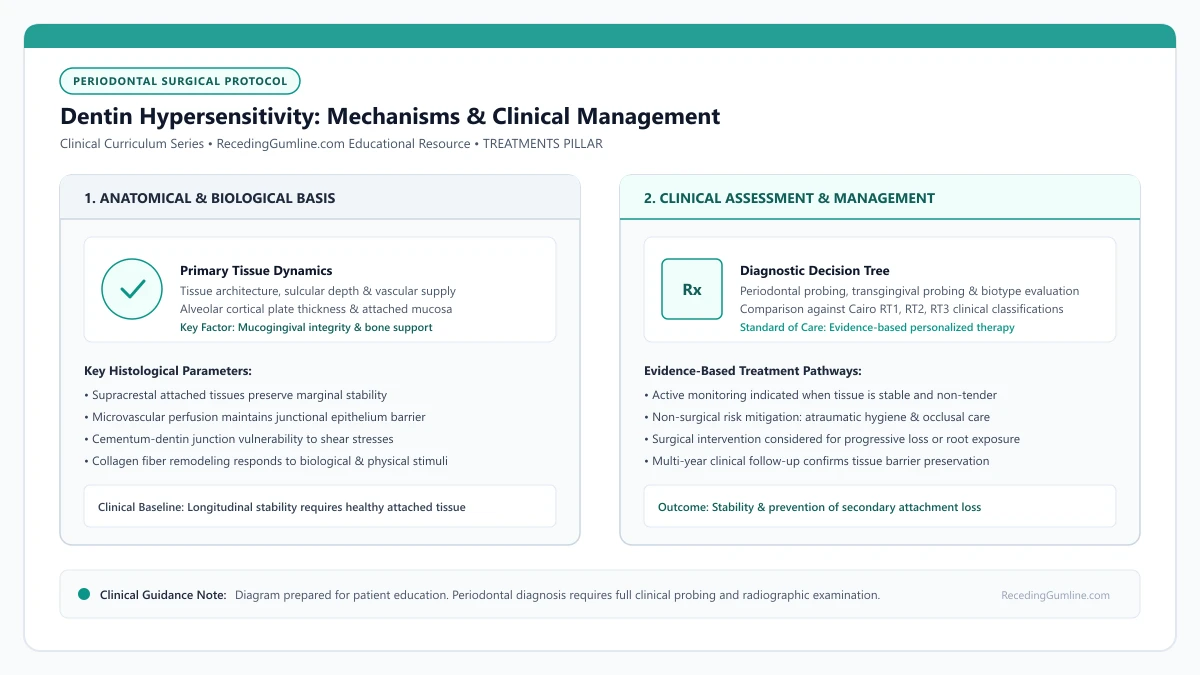
Educational illustration: Dentin Hypersensitivity: Mechanisms & Clinical Management. Clinical management requires comprehensive periodontal evaluation rather than isolated self-assessment.
Source: RecedingGumline.com Clinical Editorial Team (Proprietary educational diagram for RecedingGumline.com)
Brännström's Hydrodynamic Theory & Tubule Biophysics
Human dentin is a vital, permeable hard tissue traversed by approximately 30,000 to 40,000 microscopic tubules per square millimeter near the dental pulp, tapering to roughly 15,000 to 20,000 tubules per square millimeter near the cementoenamel junction. In health, these tubules are completely sealed by coronal enamel and root cementum.
When gingival recession occurs, the thin, fragile root cementum (measuring only 20 to 50 micrometers in thickness) is quickly worn away by toothbrushing friction, dietary acids, or professional instrumentation. This uncaps the tubule apertures, exposing the tubule lumina to the oral environment.
According to the landmark hydrodynamic theory formulated by Martin Brännström in 1966, external stimuli—especially cold air, cold liquids, or tactile contact—cause sudden displacement of the dentinal fluid contained within the tubules. This rapid fluid shift creates shear stress that deforms mechanoreceptive A-delta and A-beta nerve fibers wrapped around the odontoblastic cell bodies in the pulp periphery, triggering a sharp, shooting pain signal.
[object Object]
Key Clinical Insights
- Dentin contains up to 40,000 microscopic fluid-filled tubules per square millimeter.
- Recession and subsequent cementum loss expose patent tubule orifices to the oral cavity.
- Thermal and tactile stimuli trigger rapid fluid movement, exciting mechanoreceptors in the pulp.
At-Home Therapeutic Regimens: Depolarization vs. Occlusion
First-line management of dentin hypersensitivity begins with targeted at-home oral hygiene products. Desensitizing dentifrices function through two distinct pharmacodynamic mechanisms: nerve depolarization and mechanical tubule occlusion.
Potassium nitrate (5%) is the premier nerve-depolarizing agent. Potassium ions penetrate patent dentinal tubules and accumulate in the extracellular fluid surrounding intradental nerves. Elevated extracellular potassium prevents axonal repolarization, dampening sensory nerve excitability and dulling pain transmission. Because nerve depolarization requires cumulative ionic saturation, patients must use potassium nitrate pastes continuously for 2 to 4 weeks before experiencing maximal relief.
Conversely, tubule-occluding agents work by depositing physical mineral plugs within the tubule openings. Dentifrices containing stannous fluoride, calcium sodium phosphosilicate (NovaMin), arginine with calcium carbonate, or nano-hydroxyapatite interact with saliva to form insoluble crystalline precipitates that seal the tubule lumina, stopping hydrodynamic fluid displacement.
Key Clinical Insights
- Potassium nitrate depolarizes pulpal nerves, requiring 2 to 4 weeks of consistent twice-daily use.
- Stannous fluoride and NovaMin physically occlude open tubules with mineralized precipitates.
- At-home therapeutic toothpastes must be used continuously to maintain their protective mineral plugs.
In-Office Clinical Interventions: Primers, Resins & Lasers
When at-home desensitizing pastes fail to provide adequate relief, chairside in-office therapies offer rapid, potent tubule sealing. In-office treatments employ advanced chemical primers, bonding resins, or laser biostimulation.
Glutaraldehyde and 2-hydroxyethyl methacrylate (HEMA) formulations (such as Gluma) represent a established clinical benchmark for in-office sensitivity relief. Glutaraldehyde induces rapid coagulation of serum proteins within the dentinal fluid, forming cross-linked protein septa that physically block the tubules, while HEMA polymerizes within the tubule orifices, providing long-lasting occlusion.
For deeper cervical notches or refractory sensitivity, clinicians can apply light-cured resin glazes, glass ionomer cements, or flowable composite restorations. Additionally, neodymium-doped yttrium aluminum garnet (Nd:YAG) and erbium lasers are increasingly utilized to melt and recrystallize superficial dentin, sealing tubules without requiring chemical adhesives.
Key Clinical Insights
- Glutaraldehyde-HEMA primers coagulate dentinal fluid proteins, creating immediate biological plugs.
- Flowable composite resins and glass ionomers physically cover deep, sensitive cervical notches.
- Dental laser irradiation melts superficial dentin crystals, permanently sealing patent tubules.
Clinical Evaluation: Hypersensitivity vs. Irreversible Pulpitis
Accurate differential assessment is essential because desensitizing therapies are solely effective for true dentin hypersensitivity; they cannot resolve endodontic pathology. Dentin hypersensitivity is characterized by short, sharp, transient pain provoked by external triggers (cold air, ice water, toothbrush bristles) that subsides immediately (within 1 to 5 seconds) upon stimulus removal.
In contrast, if pain lingers for 30 seconds or longer after stimulus removal, throbs spontaneously without external provocation, awakens the patient from sleep, or radiates along the jaw, clinicians evaluate for irreversible pulpitis rather than simple dentin hypersensitivity. Irreversible pulpitis reflects advanced bacterial infection of the dental pulp and cannot be resolved with desensitizing pastes; it requires endodontic root canal therapy.
A comprehensive dental examination—incorporating cold thermal testing (Endo-Ice), electronic pulp testing (EPT), percussion testing, and high-resolution periapical radiographs—is required to ensure accurate clinical assessment and prevent misdirected treatment.
Key Clinical Insights
- Dentin hypersensitivity produces sharp pain that dissipates immediately upon stimulus removal.
- Lingering, throbbing, or spontaneous pain indicates irreversible pulpal inflammation (pulpitis).
- Thermal testing and radiographic evaluation are required to distinguish sensitivity from tooth infection.
Dietary Acid Erosion Synergy & Preventive Dietary Adjustments
Dietary acid exposure acts as a powerful accelerant of dentin hypersensitivity by continuously dissolving both the natural smear layer and newly deposited mineral precipitates within exposed dentinal tubules. Acidic foods and beverages—including citrus fruits, carbonated soft drinks, sports beverages, wine, and vinegar-based dressings—routinely possess a pH well below the critical pH of dentin (pH 6.2 to 6.7).
When exposed root surfaces encounter erosive acids, the mineral matrix of peritubular and intertubular dentin dissolves within seconds, reopening sealed tubule lumina and rekindling hydrodynamic fluid movement. Furthermore, brushing immediately after acid consumption inflicts rapid abrasive loss on the temporarily softened dentin matrix.
Patients experiencing persistent root sensitivity should implement practical dietary modifications: rinsing with plain water or a basic baking soda solution immediately following acidic food consumption, avoiding toothbrushing for at least 30 to 60 minutes after meals, and utilizing a straw to bypass anterior teeth when drinking acidic liquids.
Key Clinical Insights
- Dietary acids with a pH below 6.2 rapidly demineralize dentin and reopen sealed tubules.
- Brushing immediately after acid exposure inflicts accelerated abrasive wear on softened dentin.
- Delaying brushing for 30 to 60 minutes after meals preserves protective tubule mineral plugs.
In-Office Desensitizing Treatments for Exposed Roots: Varnishes, Glumas & Sealants
Across various receding gumline conditions, when gingival recession exposes the cervical root surface, the protective enamel layer is absent, leaving microscopic dentinal tubules exposed to the oral cavity. Temperature swings, cold air, and brushing can trigger sharp, agonizing pain through hydrodynamic fluid movement. In-office professional desensitizing agents provide rapid, targeted relief by physically occluding open tubules or blocking nerve transmission.
• Exposed root sensitivity is driven by hydrodynamic fluid movement within open microscopic dentinal tubules.
• 5% sodium fluoride varnishes precipitate calcium fluoride globules to physically plug open tubule orifices.
• Glutaraldehyde/HEMA agents (such as Gluma) coagulate plasma proteins inside tubules, creating an instant biological barrier.
• Resin-based dentin bonding agents and glass ionomer sealants provide durable mechanical seals for deep cervical notches.
The Biology of Root Pain: Hydrodynamic Tubule Fluid Shifts
Under healthy conditions, the root surface is covered by cementum and embedded securely beneath attached gingiva. When recession occurs, mechanical abrasion or acid erosion quickly strips away the micro-thin cementum layer, exposing underlying dentin containing thousands of microscopic channels called dentinal tubules.
According to Brännström's Hydrodynamic Theory, cold drinks or air cause the fluid inside these tubules to contract and rush outward. This rapid fluid displacement deforms mechanoreceptors around the pulpal nerve endings (A-beta and A-delta fibers), triggering sharp, sudden pain.
Topical 5% sodium fluoride varnishes and glutaraldehyde-based desensitizing agents precipitate plasma proteins within dentinal tubules, creating physical intratubular barriers. These micro-plugs prevent the hydrodynamic fluid movement that stimulates mechanoreceptors within the dental pulp.
Key Clinical Insights
- Recession exposes dentinal tubules containing odontoblastic processes and fluid
- Thermal changes cause rapid fluid movement that excites pulpal sensory nerves
- Treatment requires occluding tubule orifices to stop fluid displacement
Professional In-Office Treatments: Varnishes vs. Glumas
The first line of professional intervention is 5% Sodium Fluoride Varnish (22,600 ppm fluoride). Applied directly to the dried root with a small brush, it reacts with salivary calcium to precipitate calcium fluoride crystals within the tubule apertures, blocking fluid flow.
For more stubborn sensitivity, dentists utilize glutaraldehyde and HEMA formulations (such as Gluma Desensitizer). Glutaraldehyde reacts with albumin and plasma proteins within the dentinal fluid, causing them to coagulate and form biological "plugs" deep inside the tubules within 60 seconds.
Arginine and calcium carbonate toothpastes work over several weeks by depositing an alkaline mineral layer across exposed cervical dentin surfaces. Daily application during routine oral hygiene reinforces tubule occlusion and dampens thermal shock responses.
Key Clinical Insights
- 5% NaF varnish precipitates mineral crystals to plug outer tubule openings
- Glutaraldehyde formulations coagulate proteins inside tubules for instantaneous relief
- Both procedures are completely painless and require zero local anesthesia or drilling
Resin Bonding Agents and Flowable Restorations
When root sensitivity is severe or accompanied by structural loss—such as a deep V-shaped wedge defect (abfraction) caused by clenching—varnishes may wear off too quickly. In these cases, dentists apply light-cured resin bonding agents or unfilled resins to create an impermeable plastic seal over the root.
If the notch extends deeper than 1 millimeter into the dentin, a tooth-colored flowable resin composite or glass ionomer restoration is placed. This not only eliminates nerve sensitivity permanently but also restores normal tooth contour and shields the root from future toothbrush abrasion.
For severe refractory cervical sensitivity accompanied by physical notch defects, flowable resin-modified glass ionomer restorations may be placed over the exposed root. These restorative materials chemically bond to dentin and release fluoride while protecting vulnerable root structures from acid erosion.
Key Clinical Insights
- Light-cured dentin bonding agents provide long-lasting physical polymer barriers
- Glass ionomer restorations release protective fluoride while sealing cervical notches
- Indicated when significant tooth structure has worn away from grinding or scrubbing
Mechanisms of Action: Tubule Occlusion vs. Neural Depolarization
Professional management of dentin hypersensitivity on receded roots relies on two distinct biological mechanisms: physical dentinal tubule occlusion and sensory nerve depolarization. When root cementum is lost, millions of microscopic dentinal tubules are exposed to thermal and osmotic stimuli.
Tubule-occluding agents (such as potassium oxalate, ferric oxalate, sodium fluoride varnish, and casein phosphopeptide-amorphous calcium phosphate [CPP-ACP]) precipitate mineral crystals within the tubule lumens. This blocks fluid movement, preventing activation of mechanoreceptors in the pulp according to Brännström's hydrodynamic theory.
In contrast, potassium nitrate formulations increase extracellular potassium ion concentrations around pulpal nerve fibers, hyperpolarizing the neural membrane and preventing transmission of pain impulses even if fluid moves.
Key Clinical Insights
- Tubule occluders precipitate crystals within dentinal tubules to stop hydrodynamic fluid flow.
- Potassium nitrate hyperpolarizes pulpal sensory nerve fibers, preventing pain signal transmission.
- Combining physical occlusion with neural dampening provides comprehensive sensitivity relief.
In-Office Clinical Treatments: Glutaraldehyde, Resin Seals & Lasers
When over-the-counter desensitizing toothpastes provide insufficient relief, periodontists employ advanced in-office therapies. Formulations containing 5% glutaraldehyde and 35% HEMA (such as Gluma) act instantaneously by coagulating serum proteins within dentinal tubules, creating immediate protein plug barriers.
For severe, localized hypersensitivity, clinicians apply light-cured resin bonding agents or flowable resin-modified glass ionomers over the exposed root. These durable resin coatings seal the open tubules with a micro-thin, wear-resistant layer that lasts for several months.
Low-level laser therapy (Nd:YAG and diode lasers) can also be used to melt and recrystallize superficial dentin, occluding tubules while simultaneously biostimulating the pulp to deposit protective secondary and tertiary reparative dentin.
Key Clinical Insights
- Glutaraldehyde/HEMA solutions coagulate tubular proteins to form instantaneous physical seals.
- Light-cured resin adhesives provide durable, wear-resistant barriers over sensitive root dentin.
- Dental lasers melt dentin crystals to seal open tubules and stimulate pulp repair mechanisms.
Clinical Reality Check
Desensitizing toothpastes and in-office sealants manage the painful symptom of recession; they do not replace lost gum tissue or alveolar bone. When hypersensitivity co-exists with progressive attachment loss, periodontal surgical grafting provides definitive root coverage while permanently eliminating sensitivity.
Questions to Ask Your Dentist or Periodontist
- Is my tooth pain simple root sensitivity or a sign of deeper pulpal inflammation or infection?
- Which in-office desensitizing treatment would be most effective for my exposed root surfaces?
- What specific toothpaste ingredient (potassium nitrate vs stannous fluoride) do you recommend for my teeth?
- Could surgical gum grafting cover these sensitive roots and permanently eliminate my pain?
- Are dietary acids (citrus, vinegar, carbonated beverages) dissolving the mineral plugs in my dentin?
- Can you apply an in-office fluoride varnish or Gluma treatment to my sensitive receded teeth today?
- Is my sensitivity caused purely by open tubules, or is there a cervical notch that requires a resin restoration?
- How long should I expect in-office desensitizing treatments to last before needing reapplication?
- What specific toothpaste ingredients should I use at home to maintain the tubule seal?
Unsure What Your Gum Changes Mean?
Take our free, evidence-based Gum Recession Assessment — approximately 3 minutes. Identify potential risk factors, evaluate symptoms, and receive personalized discussion questions for your dentist or periodontist.
Non-diagnostic educational triage. Private, secure, completed in your browser.
Related Educational Topics
Scientific Literature & Clinical Guidelines
5sources · Hide ▲
- West NX, Seong J, Davies M (2015).
"Management of dentine hypersensitivity: efficacy of professionally and self-administered agents." Journal of Clinical Periodontology.
Clinical relevance: Systematic review evaluating professionally and self-administered desensitizing agents; found evidence supporting tubule-occluding dentifrices and potassium-based nerve desensitizers in providing transient to moderate symptom relief, with substantial heterogeneity across clinical trials.
- Brännström M (1966).
"Sensitivity of dentine." Oral Surgery, Oral Medicine, Oral Pathology.
Clinical relevance: Foundational paper formulating the hydrodynamic theory of dentin hypersensitivity: rapid fluid displacement within patent dentinal tubules physically deforms intradental nerve endings at the pulp-dentin boundary, explaining thermal, mechanical, and evaporative root sensitivity.
- Jepsen S, Caton JG, Albandar JM, Bissada NF, Bouchard P, Cortellini P, et al. (2018).
"Periodontal manifestations of systemic diseases and developmental and acquired conditions: Consensus report of workgroup 3 of the 2017 World Workshop on the Classification of Periodontal and Peri-Implant Diseases and Conditions." Journal of Periodontology.
Clinical relevance: Consensus report defining mucogingival conditions, gingival phenotype (replacing biotype), non-carious cervical lesions, and the multifactorial etiology of gingival recession; emphasizes that recession can occur without periodontitis and classifies recession by interdental clinical attachment loss.
- Cairo F, Nieri M, Cincinelli S, Mervelt J, Pagliaro U (2011).
"The interproximal clinical attachment level to classify gingival recessions and predict root coverage outcomes: an explorative and reliability study." Journal of Clinical Periodontology.
Clinical relevance: Exploratory and reliability study establishing the Cairo classification based on interdental clinical attachment level (CAL): RT1 (no interproximal attachment loss; complete root coverage is clinically predictable), RT2 (interproximal attachment loss <= buccal loss; partial coverage predictable), and RT3 (interproximal loss exceeds buccal recession; complete coverage not predictable).
- Pihlstrom BL, Michalowicz BS, Johnson NW (2005).
"Periodontal diseases." The Lancet.
Clinical relevance: Peer-reviewed Lancet seminar review synthesizing global periodontal epidemiology, microbial etiology, host immunopathology, and systemic interactions, emphasizing prevention, biofilm disruption, and early risk factor modification.
Important Medical Notice
The contents of RecedingGumline.com, including text, graphics, self-assessment calculators, and other materials, are intended solely for educational and informational purposes. This content is not intended to replace professional dental examination, diagnosis, or treatment. Always seek the advice of a qualified dentist, periodontist, or other licensed oral healthcare provider with any questions you may have regarding a medical or dental condition. Never disregard professional medical advice or delay seeking it because of something you read on this website.