Localized vs. Generalized Gum Recession: Etiological and Diagnostic Distinctions
What is the difference between localized and generalized gum recession, and what causes each pattern?
gum recession does not affect every patient in the same manner. In some individuals, recession is confined strictly to a single isolated tooth or quadrant, while in others, it manifests across nearly every tooth in the dentition. Differentiating localized from generalized recession is the first critical diagnostic step periodontists use to pinpoint the root cause and formulate an effective treatment plan.
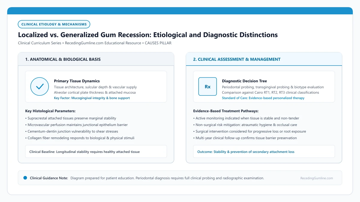
Educational illustration: Localized vs. Generalized Gum Recession: Etiological and Diagnostic Distinctions. Clinical management requires comprehensive periodontal evaluation rather than isolated self-assessment.
Source: RecedingGumline.com Clinical Editorial Team (Proprietary educational diagram for RecedingGumline.com)
Key Clinical Distinctions & Diagnostic Boundaries
- Localized recession on an isolated tooth is almost never caused by generalized periodontitis; it is overwhelmingly mechanical or anatomical.
- Generalized recession with interdental bone loss indicates a history of chronic inflammatory periodontal disease.
- Localized single-tooth defects can often be repaired with a localized soft-tissue graft.
- Generalized recession requires mouth-wide periodontal therapy, scaling, and lifestyle habit overhauls before any grafting is considered.
The 30% Clinical Threshold: Localized vs. Generalized
In periodontal diagnosis, the boundary between localized and generalized conditions is mathematically defined by the 2017 World Workshop. If recession or attachment loss affects fewer than 30% of the teeth present in the mouth, the diagnosis is classified as localized.
If the condition involves 30% or more of the dentition (typically 8 or more teeth in a full dentition of 28 teeth), it is classified as generalized. This numerical distinction immediately alerts the clinician to look for either a localized mechanical/anatomical issue or a generalized biological/systemic disease process.
Localized recession involving only one or two teeth is typically driven by isolated anatomical or mechanical factors such as high frenum pull, prominent root positioning, or localized toothbrush trauma. The surrounding periodontal structures remain completely healthy with zero interdental attachment loss.
Clinical Considerations:
- < 30% of teeth involved = Localized recession (isolated mechanical or anatomical cause)
- ≥ 30% of teeth involved = Generalized recession (systemic periodontitis, phenotype, or mouth-wide habit)
- Determines whether treatment is targeted to an individual site or requires mouth-wide therapy
Drivers of Localized Recession: Anatomical and Mechanical Hot Spots
When recession is localized to one or two teeth, periodontists investigate site-specific factors. The most common culprit is root prominence: a tooth that erupted slightly outside the dental arch has no facial bone plate and thin gingiva.
Other common localized etiologies include high frenal attachments pulling on a specific incisor, a tongue or lip piercing repeatedly hitting one tooth, an ill-fitting restoration crown margin impinging on biologic width, or a habit of picking at a single area with a fingernail.
Generalized recession affecting multiple quadrants across the mouth is predominantly caused by chronic inflammatory periodontitis or systemic conditions like osteoporosis and uncontrolled diabetes. Interdental bone loss is widespread, producing Cairo RT2 or RT3 tissue configurations.
Clinical Considerations:
- Root prominence and developmental alveolar bone dehiscences on isolated teeth
- Aberrant high frenum attachments causing dynamic tension
- Localized mechanical friction from oral piercings or aggressive one-sided brushing
Drivers of Generalized Recession: Periodontitis and Phenotype
When recession occurs across multiple quadrants throughout the mouth, the underlying cause is systemic or pervasive. The most prevalent cause is chronic periodontitis, where bacterial plaque biofilms have triggered generalized bone resorption and pocketing across many teeth over decades.
A second common cause of generalized recession is a mouth-wide thin gingival phenotype combined with decades of aggressive, horizontal scrubbing using medium or hard toothbrushes. Systemic conditions—such as uncontrolled diabetes, heavy cigarette smoking, or severe hormonal shifts—also accelerate generalized attachment loss.
Clinical distinction between localized and generalized patterns directs the clinician toward completely distinct treatment modalities. While localized defects are prime candidates for surgical root coverage, generalized periodontitis requires full-mouth periodontal debridement and strict systemic disease control.
Clinical Considerations:
- Chronic plaque-induced periodontitis causing generalized horizontal bone loss
- Genetically thin periodontal phenotype subjected to lifelong vigorous scrubbing
- Systemic modifying risk factors such as smoking, diabetes, and nutritional deficiencies
Etiological Profiling: Mechanical Friction vs. Microbially Driven Disease
Differentiating between localized and generalized gingival recession is fundamental to formulating an accurate prognosis and treatment strategy. Localized recession (affecting one or two isolated teeth) is predominantly driven by localized anatomical or mechanical factors: prominent root position, isolated bony dehiscence, frenal pulling, or traumatic oral piercings.
In contrast, generalized recession (involving multiple teeth across several quadrants) typically reflects extensive chronic periodontitis, systemic genetic predisposition, age-related cumulative attachment loss, or widespread aggressive toothbrush abrasion. In generalized cases, widespread interdental bone loss is frequently present.
Periodontists utilize the 2017 World Workshop classification to establish staging and grading, determining whether generalized recession is an anatomical manifestation of past periodontitis or active, ongoing disease requiring systemic intervention.
Clinical Considerations:
- Localized recession stems primarily from isolated anatomical defects, piercings, or frenal pull.
- Generalized recession typically reflects extensive chronic periodontitis or systemic biotype vulnerability.
- Accurate staging differentiates active inflammatory disease from stable anatomical recession.
Treatment Sequencing: Targeted Flaps vs. Full-Mouth Stabilization
Therapeutic approaches differ substantially based on recession distribution. For localized defects with intact interdental bone (Cairo RT1), treatment focuses on targeted surgical root coverage via coronally advanced flaps or tunnel grafting, offering high predictability for complete aesthetic restoration.
Conversely, generalized recession demands systematic full-mouth stabilization before any elective surgical plastic procedures can be considered. Phase I non-surgical periodontal therapy (scaling and root planing across all four quadrants) must first resolve all active pocketing and bleeding on probing.
Attempting surgical grafting in a mouth with active, generalized inflammation is strictly contraindicated, as persistent bacterial pathogens and elevated cytokine levels will degrade graft biomaterials and result in surgical failure.
Clinical Considerations:
- Localized RT1 defects can be immediately managed with targeted microsurgical root coverage.
- Generalized recession requires quadrant scaling and root planing to eliminate all inflammation first.
- Elective grafting in the presence of active generalized periodontal disease leads to surgical failure.
Clinical Reality Check
Recession observed on only a single tooth should not be assumed to represent generalized periodontitis; an isolated receded tooth typically reflects an anatomical alignment factor, prominent root, or localized habit rather than widespread infection.
Questions to Ask Your Periodontist or Dentist
- Does my examination show that my gum recession is localized to a few teeth or generalized across my whole mouth?
- If it is localized, what specific anatomical or mechanical factor caused this particular tooth to recede?
- Do I have bone loss between my teeth or is it strictly confined to the outer facial surfaces?
- What different treatment steps are needed for my isolated receded teeth compared to the rest of my mouth?
Unsure What Your Gum Changes Mean?
Take our free, evidence-based Gum Recession Assessment — approximately 3 minutes. Identify potential risk factors, evaluate symptoms, and receive personalized discussion questions for your dentist or periodontist.
Non-diagnostic educational triage. Private, secure, completed in your browser.
Related Educational Topics
Clinical Evidence & Claim Traceability (2 Mapped Assertions)
Scientific Literature & Clinical Guidelines
3sources · Hide ▲
- Tonetti MS, Greenwell H, Kornman KS (2018).
"Staging and grading of periodontitis: Framework and proposal of a new classification and case definition." Journal of Clinical Periodontology.
Clinical relevance: Consensus framework establishing the multidimensional staging (severity and extent of periodontal tissue breakdown) and grading (biological rate of disease progression, incorporating smoking and diabetes as grade modifiers) for periodontitis. It addresses periodontitis diagnosis and staging, not the classification of localized gingival recession defects.
- Jepsen S, Caton JG, Albandar JM, Bissada NF, Bouchard P, Cortellini P, et al. (2018).
"Periodontal manifestations of systemic diseases and developmental and acquired conditions: Consensus report of workgroup 3 of the 2017 World Workshop on the Classification of Periodontal and Peri-Implant Diseases and Conditions." Journal of Periodontology.
Clinical relevance: Consensus report defining mucogingival conditions, gingival phenotype (replacing biotype), non-carious cervical lesions, and the multifactorial etiology of gingival recession; emphasizes that recession can occur without periodontitis and classifies recession by interdental clinical attachment loss.
- Cairo F, Nieri M, Cincinelli S, Mervelt J, Pagliaro U (2011).
"The interproximal clinical attachment level to classify gingival recessions and predict root coverage outcomes: an explorative and reliability study." Journal of Clinical Periodontology.
Clinical relevance: Exploratory and reliability study establishing the Cairo classification based on interdental clinical attachment level (CAL): RT1 (no interproximal attachment loss; complete root coverage is clinically predictable), RT2 (interproximal attachment loss <= buccal loss; partial coverage predictable), and RT3 (interproximal loss exceeds buccal recession; complete coverage not predictable).
Important Medical Notice
The contents of RecedingGumline.com, including text, graphics, self-assessment calculators, and other materials, are intended solely for educational and informational purposes. This content is not intended to replace professional dental examination, diagnosis, or treatment. Always seek the advice of a qualified dentist, periodontist, or other licensed oral healthcare provider with any questions you may have regarding a medical or dental condition. Never disregard professional medical advice or delay seeking it because of something you read on this website.