"Longer Teeth" Appearance & Interdental Spaces in Gum Recession
The visual perception that teeth are becoming "longer" is one of the most prominent aesthetic manifestations of gumline recession. As the marginal gingival collar retreats apically, it uncovers the root surface, distorting the natural golden proportions of the clinical crown. Furthermore, when recession involves the interproximal spaces between adjacent teeth, the triangular interdental papillae can be lost, creating unsightly dark voids known as "black triangles." Understanding the clinical architecture of tooth proportions enables targeted restorative and periodontal planning.
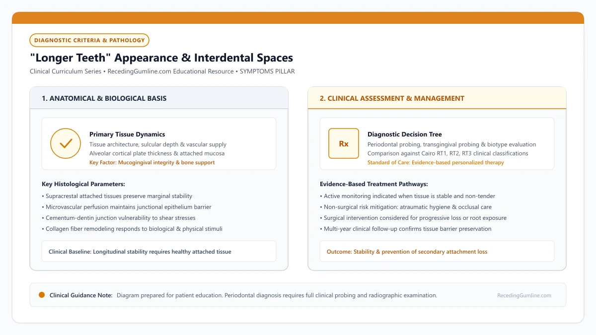
Educational illustration: "Longer Teeth" Appearance & Interdental Spaces. Clinical management requires comprehensive periodontal evaluation rather than isolated self-assessment.
Source: RecedingGumline.com Clinical Editorial Team (Proprietary educational diagram for RecedingGumline.com)
Dental Aesthetics & Ideal Clinical Crown Proportions
Dental aesthetics is governed by precise mathematical and anatomical principles. In a harmonious smile, the maxillary central incisors exhibit an ideal width-to-length ratio between 75% and 80%. For example, an incisor with a width of 8.5 mm typically has an aesthetic clinical crown length of approximately 10.5 to 11.0 mm, bounded superiorly by a symmetrical, scalloped gingival margin.
When gingival recession occurs, the marginal tissue retreats apically, exposing 1 to 4 millimeters of root surface. This increases the vertical height of the visible clinical crown without altering its mesiodistal width. As the width-to-length ratio drops below 70%, the tooth appears unnaturally elongated and narrow, creating an aged or unesthetic appearance.
Furthermore, recession rarely progresses symmetrically across the entire dental arch. When one central incisor recedes while the adjacent incisor remains at its normal level, the resulting gingival zenith asymmetry disrupts the horizontal smile line, drawing immediate visual attention to the defect.
Key Clinical Insights
- Ideal aesthetic tooth proportion exhibits a width-to-length ratio between 75% and 80%.
- Gingival recession elongates the clinical crown, lowering the ratio and making teeth look narrow and aged.
- Asymmetrical gingival margins across anterior teeth create glaring visual discrepancies in the smile line.
Interdental Black Triangles & Tarnow's 5-Millimeter Rule
While facial recession elongates the front of the tooth, interproximal recession creates dark triangular voids between adjacent teeth, universally known in dentistry as "black triangles" (open gingival embrasures). In health, this space is completely filled by the interdental papilla—a pyramidal peak of dense, keratinized gingiva that hugs the contact point.
The presence or absence of the interdental papilla is governed by the underlying interproximal alveolar bone crest. In a landmark 1992 study, Dennis Tarnow and colleagues established the definitive biological rule governing papilla presence: when the vertical distance from the apical extent of the interproximal contact point to the alveolar bone crest is 5 millimeters or less, the interdental papilla is present in nearly 100% of cases.
However, when that distance increases to 6 millimeters (due to periodontal bone resorption or tooth divergence), the papilla fills the space only 56% of the time. When the distance reaches 7 millimeters or greater, the papilla is present in only 27% of cases, leaving a permanent black triangle that allows air and saliva to escape during speech.
Key Clinical Insights
- Black triangles develop when interdental papillae recede, creating open triangular embrasures.
- Tarnow's 1992 study proved that papillae fill the embrasure 100% of the time when bone-to-contact distance is <= 5 mm.
- When bone-to-contact distance reaches 7 mm or more, papillae fill the space only 27% of the time.
Functional Consequences: Food Impaction, Speech & Hygiene
Black triangles and elongated clinical crowns represent far more than cosmetic concerns; they inflict significant functional and hygienic challenges. The primary functional complaint is persistent food impaction: fibrous food particles (such as meat and vegetables) are forcefully wedged into open embrasures during chewing, causing localized discomfort and pressure.
In addition, open interdental spaces disrupt normal airflow and phonetics during speech. Patients frequently experience minor lisping or involuntary saliva spraying (sibilant distortion) when pronouncing "s" and "th" sounds, as air escapes through the open voids between anterior teeth.
Hygienically, open embrasures expose complex root concavities and furcation entrances that cannot be cleaned with ordinary toothbrushing. Plaque biofilms readily stagnate in these protected areas, increasing the patient's risk for secondary interproximal caries and accelerated periodontitis.
Key Clinical Insights
- Open embrasures trap fibrous food debris, causing chronic interproximal irritation and discomfort.
- Air leakage through black triangles can cause phonetic distortion (lisping) on sibilant sounds.
- Exposed root concavities in open spaces collect bacterial plaque, elevating the risk of interdental decay.
Clinical Solutions: Restoration, Orthodontics & Papilla Preservation
Rebuilding lost interdental papillae is one of the most challenging frontiers in periodontal surgery, because unlike flat facial surfaces, the interdental space lacks an underlying bony wall to support a soft-tissue graft. Consequently, managing black triangles requires an interdisciplinary approach.
Bioclear Matrix Composite Bonding is a highly effective, minimally invasive restorative solution. Specially contoured anatomical clear matrices allow clinicians to inject heated, flowable composite resin interproximally, closing the triangular space and lowering the contact point closer to the bone crest without grinding down healthy tooth structure.
Orthodontic interproximal reduction (IPR) can also be utilized: slenderizing the contact surfaces of triangular teeth allows the orthodontist to move the crowns closer together, lowering the contact point to satisfy Tarnow's 5-millimeter rule. For facial crown elongation, periodontal connective tissue grafting remains the definitive standard to reposition the facial margin coronally.
Key Clinical Insights
- Surgically rebuilding lost interdental papillae is anatomically difficult due to lack of bone support.
- Bioclear anatomical composite bonding closes black triangles without aggressive tooth drilling.
- Orthodontic space closure and IPR lower contact points, restoring natural papilla fill.
Black Triangles Between Teeth: Interproximal Gum Recession and Papilla Loss
Few aesthetic and functional dental changes cause more patient distress than the appearance of dark, triangular gaps between the teeth near the gumline. Known clinically as "open gingival embrasures" and colloquially as black triangles, this condition represents interproximal gum recession—the loss of the delicate pink papilla tissue that fills the space between adjacent teeth.
• Black triangles occur when the interdental papilla recedes, leaving an open triangular space below the contact point.
• Tarnow's landmark 5mm rule demonstrates that papillae are present 98% of the time when the distance from bone crest to contact point is ≤ 5 mm.
• When the distance from bone to contact point increases to 6 mm or 7 mm, papillae recede, creating open black triangles.
• Surgical gum grafting cannot predictably regrow lost interdental papillae; treatment relies on composite bonding or orthodontic contact reshaping.
Tarnow's 5mm Law: The Biology of the Interdental Papilla
In 1992, Dr. Dennis Tarnow and colleagues published landmark periodontal research that decoded the mystery of the interdental papilla. Measuring hundreds of interdental sites, they discovered that the presence of the pink gum triangle is strictly dictated by the distance from the tooth contact point down to the underlying alveolar bone crest.
When this distance is 5.0 millimeters or less, the papilla completely fills the interdental space in 98% of cases. When the distance increases to 6.0 mm (due to bone resorption or tooth divergence), the papilla fills the space only 56% of the time. At 7.0 mm, the papilla is present in only 27% of cases, creating an open black triangle.
The loss of the interdental papilla creates unsightly triangular dark spaces between adjacent teeth, commonly referred to as black triangles. These open embrasures cause food impaction, speech difficulties including minor phonetic lisping, and significant cosmetic dissatisfaction.
Key Clinical Insights
- Distance ≤ 5 mm from contact point to bone crest = 98% papilla presence
- Distance of 6 mm = 56% papilla presence; distance of 7 mm = 27% presence
- Bone crest height is the absolute biological determinant of papilla height
The Surgical Reality: Why Black Triangles Cannot Be Grafted Away
A frequent heartbreak for patients is learning that periodontal gum grafting cannot regrow a missing interdental papilla. On the facial surface of a tooth, a graft survives because it is nourished by blood supply from the surrounding periosteum and adjacent papillae.
Between two teeth, however, there is no broad vascular bed. If a surgeon attempts to graft tissue into an open triangle, the transplanted cells sit suspended in thin air over an avascular space, lacking capillary blood flow. The graft tissue inevitably undergoes necrosis and disappears.
Black triangle formation occurs when interdental alveolar bone crest height resorbs due to periodontitis, exceeding the biological 5 mm distance from bone crest to contact point. Once the underlying interdental bone support is lost, soft tissue inevitably collapses apically.
Key Clinical Insights
- Soft-tissue grafts require an underlying vascular bed to survive
- Interdental spaces lack the broad blood supply required for graft revascularization
- No surgical technique in modern dentistry can predictably regrow lost papillae between teeth
Proven Solutions: Bioclear Bonding and Orthodontic IPR
Because surgery cannot recreate the tissue, clinicians solve black triangles by modifying the teeth themselves to close the distance. The most popular cosmetic solution is Bioclear composite bonding: the dentist uses anatomical clear mylar matrices to mold smooth, flowable composite resin between the teeth, seamlessly closing the triangle.
Another effective method is orthodontic Interproximal Reduction (IPR). The orthodontist gently polishes a fraction of a millimeter off the contact sides of the triangular teeth and uses aligners to bring them flush together, moving the contact point closer to the bone and allowing the papilla to fill the remaining gap.
Reconstructing lost interdental papillae represents one of the most challenging frontiers in periodontics due to limited collateral blood supply. Interdisciplinary solutions often combine subtle restorative composite bonding to close contact points with orthodontic root re-alignment.
Key Clinical Insights
- Bioclear composite bonding molds seamless tooth-colored resin to close triangular gaps
- Orthodontic contact reshaping (IPR) moves the contact point closer to the bone crest
- Ceramic porcelain veneers can be contoured with extended interproximal wings to mask gaps
Biological Ceilings: Tarnow's 5 mm Rule of Papillary Fill
Loss of the interdental papilla—the triangular soft-tissue wedge filling the space between adjacent teeth—results in open interproximal voids termed "black triangles." The biological presence or absence of the papilla is governed by Tarnow's landmark 5-millimeter rule.
In healthy human dentition, when the distance from the apical contact point between adjacent teeth to the crest of the interdental alveolar bone is 5.0 millimeters or less, the interdental papilla is present in virtually 100% of cases.
However, when this vertical distance increases to 6.0 millimeters (due to periodontal bone resorption), the papilla fills the space only 56% of the time. When the distance expands to 7.0 millimeters or more, the papilla is present in only 27% of cases, creating visible dark voids that impair phonetics and aesthetics.
Key Clinical Insights
- Tarnow's rule establishes that a bone-to-contact distance of ≤ 5 mm guarantees complete papillary fill.
- At 6 mm distance, complete papillary fill drops to 56%; at ≥ 7 mm, it drops to 27%.
- Interdental bone loss is the primary biological cause of permanent black triangles.
Interdisciplinary Solutions: Hyaluronic Injections, Orthodontics & IPR
Reconstructing a lost interdental papilla is one of the most demanding challenges in periodontics because the papilla lacks an isolated vascular supply once interdental bone is lost. Periodontists and restorative dentists deploy interdisciplinary strategies to minimize black triangles.
Minimally invasive hyaluronic acid gel micro-injections can be placed into deficient papillae to volumize soft tissues and reduce interdental voids for 6 to 12 months. For permanent correction, interproximal reduction (IPR) combined with orthodontic closure moves adjacent tooth contact points apically, reducing the bone-to-contact distance below 5 mm.
Restorative dentists can also place micro-thin composite additions or ceramic veneers with specialized emergence profiles that broaden the interdental contact point, closing the aesthetic gap while maintaining hygiene accessibility.
Key Clinical Insights
- Surgical reconstruction of lost papillae is limited by the height of underlying interdental bone.
- Hyaluronic acid micro-injections temporarily volumize interdental soft tissues.
- Orthodontic contact repositioning (IPR) and contoured composite bonding effectively close black triangles.
Clinical Reality Check
While facial root coverage grafting can predictably restore a receded gumline back to the CEJ (Cairo RT1), reconstructing lost interproximal papillae between teeth remains biologically unpredictable. Preventing interdental bone loss through timely periodontal care is far more effective than attempting surgical papilla reconstruction.
Questions to Ask Your Dentist or Periodontist
- Are my teeth looking longer due to active bone loss or simple toothbrush abrasion?
- Can my open black triangles be closed using minimally invasive composite bonding (such as Bioclear)?
- What is the vertical distance between my contact points and my interproximal bone crest (Tarnow rule)?
- Would a gum graft help shorten the appearance of my front teeth and restore my natural smile line?
- What specialized interdental brushes do you recommend to keep these open spaces clean and plaque-free?
- What is the measured distance from my tooth contact point to the bone crest at my black triangles?
- Would Bioclear composite bonding or porcelain veneers be the best option to close these gaps?
- Are these black triangles trapping plaque and food debris that could threaten my tooth roots?
- Could gentle orthodontic tooth reshaping (IPR) help close these spaces naturally?
Unsure What Your Gum Changes Mean?
Take our free, evidence-based Gum Recession Assessment — approximately 3 minutes. Identify potential risk factors, evaluate symptoms, and receive personalized discussion questions for your dentist or periodontist.
Non-diagnostic educational triage. Private, secure, completed in your browser.
Related Educational Topics
Scientific Literature & Clinical Guidelines
5sources · Hide ▲
- Tarnow DP, Magner AW, Fletcher P (1992).
"The effect of the distance from the contact point to the crest of bone on the presence or absence of the interproximal dental papilla." Journal of Periodontology.
Clinical relevance: Landmark investigation establishing that when the distance from the interproximal contact point to the alveolar bone crest is 5 mm or less, the dental papilla is present almost 100% of the time, dropping to 56% at 6 mm and 27% at 7 mm or more, defining biological limits of interdental soft-tissue fill.
- Cairo F, Nieri M, Cincinelli S, Mervelt J, Pagliaro U (2011).
"The interproximal clinical attachment level to classify gingival recessions and predict root coverage outcomes: an explorative and reliability study." Journal of Clinical Periodontology.
Clinical relevance: Exploratory and reliability study establishing the Cairo classification based on interdental clinical attachment level (CAL): RT1 (no interproximal attachment loss; complete root coverage is clinically predictable), RT2 (interproximal attachment loss <= buccal loss; partial coverage predictable), and RT3 (interproximal loss exceeds buccal recession; complete coverage not predictable).
- Tonetti MS, Greenwell H, Kornman KS (2018).
"Staging and grading of periodontitis: Framework and proposal of a new classification and case definition." Journal of Clinical Periodontology.
Clinical relevance: Consensus framework establishing the multidimensional staging (severity and extent of periodontal tissue breakdown) and grading (biological rate of disease progression, incorporating smoking and diabetes as grade modifiers) for periodontitis. It addresses periodontitis diagnosis and staging, not the classification of localized gingival recession defects.
- Chapple ILC, Mealey BL, Van Dyke TE, Bartold PM, Dommisch H, Eickholz P, et al. (2018).
"Periodontal health and gingival diseases and conditions on an intact and a reduced periodontium: Consensus report of workgroup 1 of the 2017 World Workshop on the Classification of Periodontal and Peri-Implant Diseases and Conditions." Journal of Clinical Periodontology.
Clinical relevance: Consensus report establishing diagnostic criteria for periodontal health and gingivitis across intact and reduced periodontia, defining clinical gingival health as <10% bleeding on probing without attachment loss and strictly differentiating gingivitis from periodontitis.
- Chambrone L, Salinas Ortega MA, Sukekava F, Rotundo R, Kalemaj Z, Buti J, Pini Prato GP (2018).
"Root coverage procedures for treating localised and multiple recession-type defects." Cochrane Database of Systematic Reviews.
Clinical relevance: Cochrane systematic review evaluating root-coverage procedures for localized and multiple recession-type defects. While subepithelial connective tissue grafts (SCTG) combined with coronally advanced flaps demonstrated higher rates of complete root coverage and keratinized tissue gain compared to flap advancement alone, evidence quality varied across outcomes and clinical decisions must balance donor site morbidity and patient-reported outcomes.
Important Medical Notice
The contents of RecedingGumline.com, including text, graphics, self-assessment calculators, and other materials, are intended solely for educational and informational purposes. This content is not intended to replace professional dental examination, diagnosis, or treatment. Always seek the advice of a qualified dentist, periodontist, or other licensed oral healthcare provider with any questions you may have regarding a medical or dental condition. Never disregard professional medical advice or delay seeking it because of something you read on this website.