Receding Gums on One Tooth: Differential Factors & Clinical Evaluation
Why can gum recession occur on just one tooth, and how do clinicians differentiate localized contributors from periodontal disease?
Noticing an isolated receding gumline on a single tooth often prompts concern, but localized recession is a distinct clinical presentation that warrants careful differential evaluation. When marginal tissue recedes on an isolated tooth—such as a mandibular central incisor, canine, or premolar—clinicians examine localized anatomical vulnerabilities, site-specific mechanical forces, and tissue tension, while also verifying that underlying periodontal breakdown is not present.
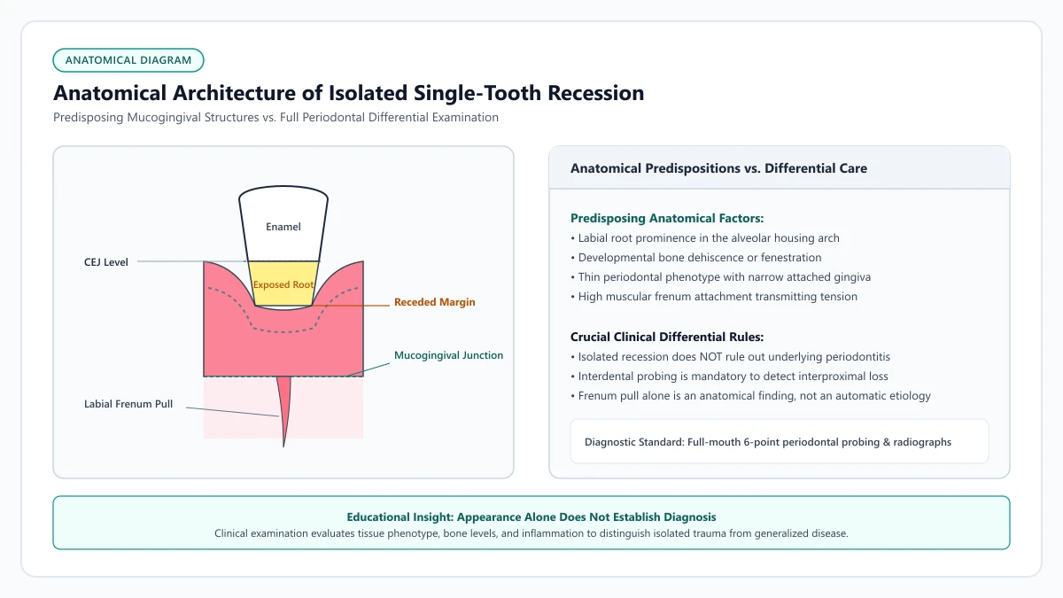
Educational illustration: Localized gingival recession on an individual tooth. Contributing factors such as root position, bone dehiscence, and frenal tension are evaluated through clinical examination and periodontal probing.
Source: RecedingGumline.com Clinical Editorial Team (Proprietary educational diagram for RecedingGumline.com)
Key Clinical Distinctions & Diagnostic Boundaries
- Localized recession can occur on an anatomically vulnerable tooth without generalized periodontitis, but clinical probing is required to confirm whether interdental attachment is intact.
- Anatomical root prominence and alveolar bone dehiscence represent predisposing structural vulnerabilities rather than isolated conclusions.
- Tension from a high labial frenum attachment may pull on the marginal gingiva of an isolated tooth during oral movements, acting as a potential local contributing factor.
- Differentiating between plaque-induced localized periodontitis and non-inflammatory mechanical or anatomical recession requires standardized clinical charting.
Root Prominence & Alveolar Bone Dehiscence
Anatomical tooth position is a recognized predisposing factor in single-tooth recession. In many individuals, specific teeth—especially mandibular central incisors, maxillary canines, and first premolars—are positioned slightly facially (toward the outside of the arch) relative to neighboring teeth.
When a tooth root is prominent within the dental arch, the overlying buccal cortical bone plate can be naturally thin or anatomically absent, a structural condition termed an alveolar dehiscence. In these locations, the marginal gingiva lacks rigid skeletal backing and is supported primarily by soft tissue.
The 2018 World Workshop consensus on periodontal classifications (Jepsen et al., 2018) notes that teeth with thin periodontal phenotypes and underlying bony dehiscences are biologically vulnerable to marginal recession when subjected to mechanical forces or inflammatory challenges. However, root prominence is a predisposing anatomical feature, not a standalone diagnosis.
Clinical Considerations:
- Prominent root position can be associated with thin or absent facial cortical bone (alveolar dehiscence)
- Thin periodontal phenotypes possess less soft-tissue volume to buffer against daily mechanical stresses
- Anatomical predisposition explains why one tooth may recede while adjacent teeth remain unaffected
- Clinical evaluation is necessary to distinguish anatomical vulnerability from active inflammatory attachment loss
Frenum Attachments & Local Soft-Tissue Tension
Another localized factor that clinicians evaluate is tension from an oral frenum—a fold of mucous membrane connecting the lips or cheeks to the alveolar mucosa. The mandibular and maxillary labial frenums are the most frequently examined.
When a frenum attaches unusually close to the gingival margin (often described as a high frenum attachment), routine movements during speaking, chewing, and facial expression can exert dynamic mechanical pull on the marginal tissue. In some individuals, this chronic tension may facilitate separation of the marginal tissue from the tooth surface.
This dynamic pull is often noted on mandibular central incisors, where a prominent lower labial frenum may act as a contributing factor to localized apical tissue movement. Nevertheless, frenum tension alone does not necessarily cause recession; its clinical impact depends on the width of attached keratinized gingiva and overall tissue phenotype.
Clinical Considerations:
- A frenum attaching close to the gingival margin can exert dynamic tension during routine facial movements
- Lower central incisors and upper canines are common locations for high frenum attachments
- The clinical significance of frenum tension is evaluated in conjunction with attached gingival width
- Tension is considered a potential localized contributor rather than an isolated cause
Localized Mechanical Forces & Habitual Factors
Localized recession can also reflect site-specific mechanical friction. For instance, right-handed individuals may inadvertently apply heavier horizontal scrubbing force to the left maxillary canine and premolars, where the toothbrush first contacts the arch.
Oral piercings represent another potential source of localized mechanical trauma. Lower lip studs (labrets) or tongue jewelry can repeatedly contact the facial or lingual gingival margin of specific anterior teeth, leading to localized tissue irritation and recession over time.
Foreign object habits—such as habitual pen-chewing, fingernail manipulation against a specific gum margin, or aggressive use of rigid toothpicks—can similarly contribute to localized marginal damage. Clinicians carefully review these habits during examination.
Clinical Considerations:
- Disproportionate toothbrushing force may concentrate at specific arch entry points
- Oral jewelry contacting the gingival margin can produce localized mechanical trauma
- Habitual manipulation with fingernails, pens, or rigid tools can irritate isolated margins
- Addressing mechanical habits is an essential initial step before considering corrective therapies
Comprehensive Clinical Evaluation & Differential Assessment
Because localized recession can arise from varied contributors, accurate identification requires a professional clinical evaluation. Clinicians perform periodontal probing around all six surfaces of the affected tooth to measure probing depth and clinical attachment loss.
Under the 2018 classification framework (Tonetti et al., 2018), assessing interdental attachment loss and radiographic bone levels distinguishes isolated mucogingival recession from localized periodontitis. Periodontitis involves inflammatory breakdown of connective tissue attachment and alveolar bone, which presents differently from isolated mechanical recession.
Furthermore, the clinician assesses occlusal contacts during lateral jaw movements to determine whether traumatic occlusal forces are concentrating stress on the tooth. By synthesizing probing depths, radiographic findings, tissue phenotype, and patient habits, the dental team establishes an accurate differential picture.
Clinical Considerations:
- Six-point periodontal probing verifies whether pocketing or active inflammation is present
- Interdental attachment assessment distinguishes localized periodontitis from isolated recession
- Radiographs evaluate interdental bone crest height and rule out hidden bone loss
- Occlusal analysis identifies potential premature contacts or lateral grinding interferences
Clinical Reality Check
Discovering isolated recession on a single tooth does not justify assuming that periodontal disease is absent. While localized anatomical and mechanical factors frequently contribute, a dental professional must evaluate full-mouth probing depths and radiographs to rule out site-specific or generalized periodontitis before deciding on appropriate management.
Questions to Ask Your Periodontist or Dentist
- Are the probing depths and attachment levels around this specific tooth normal, or is there localized bone loss?
- Does this tooth have an anatomical root prominence, thin tissue phenotype, or alveolar bone dehiscence?
- Is my labial frenum exerting tension on the gum margin when I move my lips or speak?
- Could my toothbrushing technique, oral habits, or the way my teeth contact during chewing be contributing to this isolated site?
Unsure What Your Gum Changes Mean?
Take our free, evidence-based Gum Recession Assessment — approximately 3 minutes. Identify potential risk factors, evaluate symptoms, and receive personalized discussion questions for your dentist or periodontist.
Non-diagnostic educational triage. Private, secure, completed in your browser.
Related Educational Topics
Clinical Evidence & Claim Traceability (4 Mapped Assertions)
Scientific Literature & Clinical Guidelines
3sources · Hide ▲
- Jepsen S, Caton JG, Albandar JM, Bissada NF, Bouchard P, Cortellini P, et al. (2018).
"Periodontal manifestations of systemic diseases and developmental and acquired conditions: Consensus report of workgroup 3 of the 2017 World Workshop on the Classification of Periodontal and Peri-Implant Diseases and Conditions." Journal of Periodontology.
Clinical relevance: Consensus report defining mucogingival conditions, gingival phenotype (replacing biotype), non-carious cervical lesions, and the multifactorial etiology of gingival recession; emphasizes that recession can occur without periodontitis and classifies recession by interdental clinical attachment loss.
- Tonetti MS, Greenwell H, Kornman KS (2018).
"Staging and grading of periodontitis: Framework and proposal of a new classification and case definition." Journal of Clinical Periodontology.
Clinical relevance: Consensus framework establishing the multidimensional staging (severity and extent of periodontal tissue breakdown) and grading (biological rate of disease progression, incorporating smoking and diabetes as grade modifiers) for periodontitis. It addresses periodontitis diagnosis and staging, not the classification of localized gingival recession defects.
- Cairo F, Nieri M, Cincinelli S, Mervelt J, Pagliaro U (2011).
"The interproximal clinical attachment level to classify gingival recessions and predict root coverage outcomes: an explorative and reliability study." Journal of Clinical Periodontology.
Clinical relevance: Exploratory and reliability study establishing the Cairo classification based on interdental clinical attachment level (CAL): RT1 (no interproximal attachment loss; complete root coverage is clinically predictable), RT2 (interproximal attachment loss <= buccal loss; partial coverage predictable), and RT3 (interproximal loss exceeds buccal recession; complete coverage not predictable).
Important Medical Notice
The contents of RecedingGumline.com, including text, graphics, self-assessment calculators, and other materials, are intended solely for educational and informational purposes. This content is not intended to replace professional dental examination, diagnosis, or treatment. Always seek the advice of a qualified dentist, periodontist, or other licensed oral healthcare provider with any questions you may have regarding a medical or dental condition. Never disregard professional medical advice or delay seeking it because of something you read on this website.