Gum Recession With Pink, Healthy Gums: The Non-Inflammatory Presentation
Can your gums recede if they are completely healthy, pink, and free of gum disease?
One of the most confusing clinical scenarios for dental patients is being told their gums are receding despite their dentist confirming they have excellent oral hygiene and zero gum disease. The gums appear coral pink, firm, and stippled, with zero bleeding upon probing. Understanding non-inflammatory receding gumline etiology reveals how mechanical friction, thin anatomy, and past tooth movement cause tissue loss in completely healthy mouths.
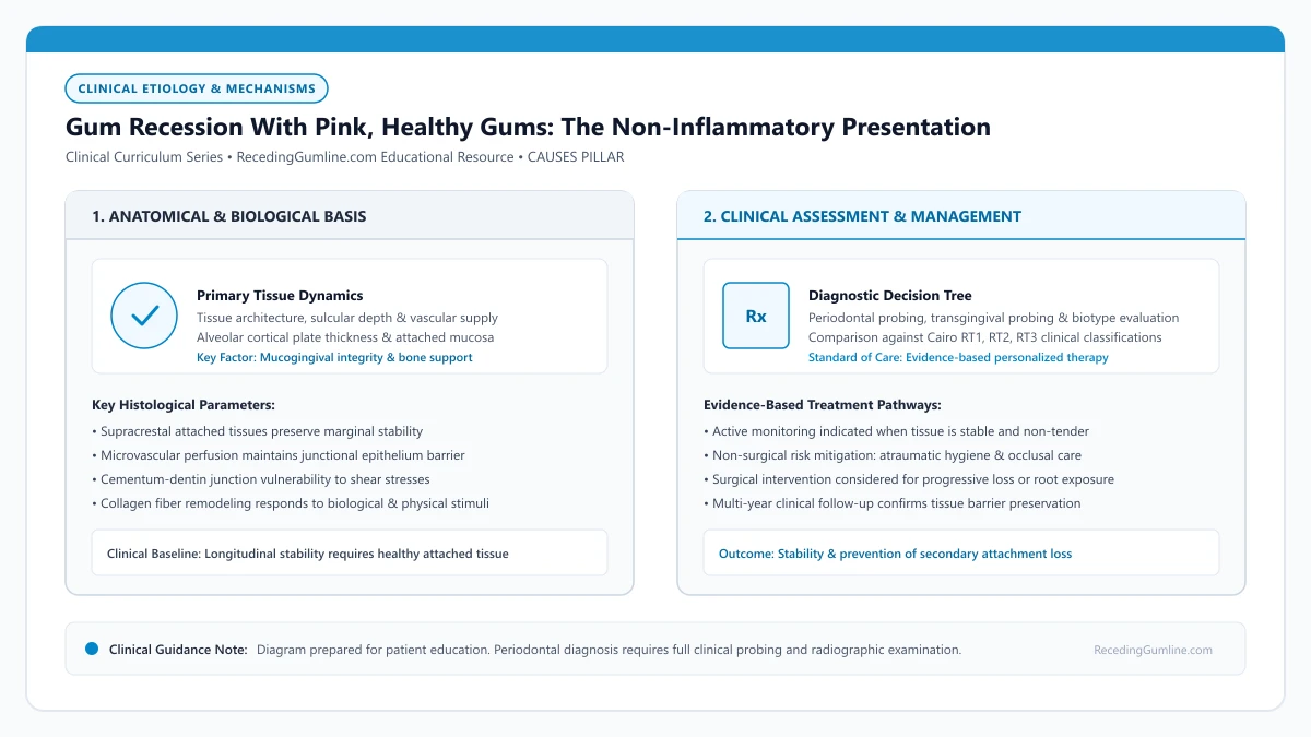
Educational illustration: Gum Recession With Pink, Healthy Gums: The Non-Inflammatory Presentation. Clinical management requires comprehensive periodontal evaluation rather than isolated self-assessment.
Source: RecedingGumline.com Clinical Editorial Team (Proprietary educational diagram for RecedingGumline.com)
Key Clinical Distinctions & Diagnostic Boundaries
- Periodontitis recession is driven by bacterial plaque, deep pockets, and bleeding; non-inflammatory recession has zero deep pockets and zero bleeding.
- Patients with non-inflammatory recession do NOT require scaling and root planing (deep cleaning); doing so can unnecessarily traumatize healthy tissue.
- Pink receded gums indicate that active destruction is not currently occurring from bacteria, but the root remains exposed to wear and decay.
- Monitoring clinical attachment levels every 6 months determines whether non-inflammatory recession is stable or progressing.
Clinical Definition: Periodontal Health on a Reduced Periodontium
Under the 2017 AAP/EFP World Workshop diagnostic criteria, clinicians recognize an important official classification: "Clinical Periodontal Health on a Reduced Periodontium." This describes a mouth where attachment loss or recession has previously occurred, but the tissues are currently 100% healthy.
In this state, probing depths measure a shallow 1 to 2 mm, bleeding on probing occurs at fewer than 10% of sites, and there is no active inflammatory breakdown. The exposed root is a historical anatomical scar, not an active infection.
It is entirely possible to have receded gums while maintaining completely healthy, non-inflamed periodontal tissues with zero bleeding on probing. This clinical phenomenon occurs when recession is initiated by anatomical thinness or toothbrush abrasion rather than active bacterial infection.
Clinical Considerations:
- Recognized officially as "Clinical Periodontal Health on a Reduced Periodontium"
- Probing depths remain shallow (1 to 2 mm) with zero active pocketing
- Bleeding on probing is absent; the tissue is structurally stable and healthy
Why Healthy Gums Recede: The Anatomy-Trauma Nexus
How can healthy gums recede without disease? The answer lies in the interaction between delicate anatomy and physical friction. If an individual inherits a thin gingival phenotype with razor-thin facial bone, the tissue margin has very little physical volume.
When this patient practices diligent oral hygiene—often brushing multiple times a day with firm pressure or an abrasive whitening toothpaste—the mechanical friction simply wears away the thin soft-tissue margin. The patient is literally brushing away their gums in an effort to keep them clean.
In these clinically quiescent scenarios, probing pocket depths remain shallow (1 to 2 mm), and the gingival tissues appear firm, pale pink, and firmly bound down. The primary clinical challenges are root dentin hypersensitivity, aesthetic dissatisfaction, and vulnerability to cervical root caries.
Clinical Considerations:
- Genetically thin tissue biotype provides minimal physical resistance against abrasion
- High frequency of brushing combined with excessive hand pressure wears tissue away
- Abrasive whitening toothpastes scour away root cementum and thin marginal tissue
Management Strategy: Protect, Desensitize & Monitor
When gums are pink and healthy, the clinical strategy is conservative and protective. Deep cleanings (scaling and root planing) are strictly contraindicated because there are no deep bacterial pockets to treat.
Instead, the dental team focuses on three interventions: modifying hygiene habits (switching to ultra-soft brushes and gentle circular motions), applying desensitizing varnishes or sealants if exposed roots are sensitive to cold, and capturing baseline photographs to monitor stability over annual checkups.
Dentists adopt a conservative monitoring approach for healthy receded sites, measuring clinical attachment levels annually to ensure the margin remains stable. Active surgical intervention is reserved for sites with progressive attachment loss, severe hypersensitivity, or high aesthetic demand.
Clinical Considerations:
- Deep cleaning is contraindicated when probing depths are shallow and healthy
- Switch immediately to ultra-soft manual or pressure-sensing electric brushes
- Apply fluoride varnishes or bonding agents if exposed roots are sensitive to cold
Non-Carious Acquired Mucogingival Defects: Anatomy Without Pathology
Many patients are surprised to discover visible gum recession despite receiving exemplary dental reports confirming zero plaque, zero calculus, and zero bleeding on probing. This clinical presentation represents a non-inflammatory acquired mucogingival defect, where recession develops entirely independently of bacterial periodontitis.
In these clinically healthy mouths, recession is usually the consequence of past mechanical trauma (such as vigorous horizontal brushing), prior orthodontic expansion through thin cortical bone, or physiological remodeling around anatomically prominent roots. Because the tissue is pink, firm, and free of inflammatory cells, it is classified as periodontal health on a reduced periodontium.
Understanding that recession does not necessarily mean "infection" provides immense psychological relief to fastidious dental patients who mistakenly believe their recession is caused by personal hygiene failure.
Clinical Considerations:
- Recession can occur in completely plaque-free, healthy mouths with zero bleeding on probing.
- Etiologies include past brushing trauma, prior orthodontic arch leveling, or anatomical root prominence.
- Periodontal health on a reduced periodontium is a recognized, stable clinical diagnosis.
Structured Monitoring vs. Prophylactic Soft-Tissue Protection
When healthy gums recede, the primary clinical objective is determining whether the defect is active and progressive or inactive and stable. Structured monitoring requires baseline digital photography, intraoral scanning, and calibrated millimeter probe measurements from the cementoenamel junction to the soft-tissue margin.
If repeat assessments over 12 to 24 months demonstrate dimensional stability, absence of root sensitivity, and easy patient cleansability, surgical intervention is generally not required. Conservative management consists of ongoing monitoring and reinforcement of non-abrasive brushing habits.
However, if sequential measurements document progressive apical migration, or if the band of keratinized attached tissue drops below 1.0 millimeter, prophylactic grafting may be recommended to halt further loss before root exposure reaches advanced levels.
Clinical Considerations:
- Calibrated measurements and photographic records establish whether healthy recession is progressive.
- Stable, asymptomatic non-inflammatory recession can be safely monitored without surgery.
- Documented progressive apical movement justifies prophylactic soft-tissue augmentation.
Clinical Reality Check
Do not let anyone perform a "deep cleaning" on pink, healthy, non-bleeding receded gums with 2 mm probing depths; deep cleaning is only indicated for active periodontal pockets.
Questions to Ask Your Periodontist or Dentist
- Does my recession represent active gum disease or stable health on a reduced periodontium?
- Are my probing depths shallow (1 to 3 mm) around these receded teeth?
- What changes to my toothbrush, bristle stiffness, and technique should I make to stop this wear?
- Do we need to treat these exposed roots surgically or can we safely monitor them with photographs?
Unsure What Your Gum Changes Mean?
Take our free, evidence-based Gum Recession Assessment — approximately 3 minutes. Identify potential risk factors, evaluate symptoms, and receive personalized discussion questions for your dentist or periodontist.
Non-diagnostic educational triage. Private, secure, completed in your browser.
Related Educational Topics
Clinical Evidence & Claim Traceability (3 Mapped Assertions)
Scientific Literature & Clinical Guidelines
3sources · Hide ▲
- Jepsen S, Caton JG, Albandar JM, Bissada NF, Bouchard P, Cortellini P, et al. (2018).
"Periodontal manifestations of systemic diseases and developmental and acquired conditions: Consensus report of workgroup 3 of the 2017 World Workshop on the Classification of Periodontal and Peri-Implant Diseases and Conditions." Journal of Periodontology.
Clinical relevance: Consensus report defining mucogingival conditions, gingival phenotype (replacing biotype), non-carious cervical lesions, and the multifactorial etiology of gingival recession; emphasizes that recession can occur without periodontitis and classifies recession by interdental clinical attachment loss.
- Chapple ILC, Mealey BL, Van Dyke TE, Bartold PM, Dommisch H, Eickholz P, et al. (2018).
"Periodontal health and gingival diseases and conditions on an intact and a reduced periodontium: Consensus report of workgroup 1 of the 2017 World Workshop on the Classification of Periodontal and Peri-Implant Diseases and Conditions." Journal of Clinical Periodontology.
Clinical relevance: Consensus report establishing diagnostic criteria for periodontal health and gingivitis across intact and reduced periodontia, defining clinical gingival health as <10% bleeding on probing without attachment loss and strictly differentiating gingivitis from periodontitis.
- Löe H, Theilade E, Jensen SB (1965).
"Experimental gingivitis in man." The Journal of Periodontology.
Clinical relevance: Classic experimental gingivitis study demonstrating that withdrawal of oral hygiene leads to bacterial plaque accumulation and reversible marginal gingival inflammation within 10 to 21 days, establishing the microbial etiology of gingival inflammation. It serves as foundational evidence for plaque-induced gingivitis, not modern comprehensive models of periodontitis or gingival recession.
Important Medical Notice
The contents of RecedingGumline.com, including text, graphics, self-assessment calculators, and other materials, are intended solely for educational and informational purposes. This content is not intended to replace professional dental examination, diagnosis, or treatment. Always seek the advice of a qualified dentist, periodontist, or other licensed oral healthcare provider with any questions you may have regarding a medical or dental condition. Never disregard professional medical advice or delay seeking it because of something you read on this website.