Attached Gingiva in Gum Recession: Structural Barrier, Mucogingival Junction & Functional Role
What is attached gingiva, how does it differ from movable mucosa, and why is it essential for tooth stability?
As noted in our management of receding gums guide, not all oral gum tissue is created equal. While the inner lips and cheeks are lined by loose, elastic, movable mucosa, the tissue immediately surrounding the teeth is a specialized, rigid, keratinized band known as attached gingiva. Firmly bound to the underlying alveolar bone and cementum, attached gingiva serves as an indispensable physical shock absorber against mechanical wear and bacterial invasion.
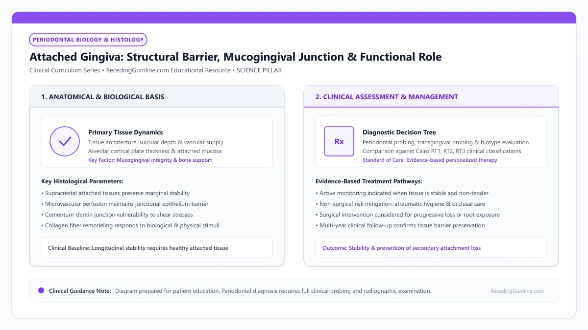
Educational illustration: Attached Gingiva: Structural Barrier, Mucogingival Junction & Functional Role. Clinical management requires comprehensive periodontal evaluation rather than isolated self-assessment.
Source: RecedingGumline.com Clinical Editorial Team (Proprietary educational diagram for RecedingGumline.com)
Key Clinical Distinctions & Diagnostic Boundaries
- Attached gingiva is completely immobile because it lacks a submucosa; alveolar mucosa is loose and highly elastic due to abundant elastin fibers.
- Attached gingiva displays stippling (an "orange-peel" texture) in health, reflecting deep connective tissue rete pegs.
- The palate has no mucogingival junction because the entire hard palate is composed of immobile masticatory mucosa.
- Surgical free gingival grafting takes tissue from the palate specifically to recreate missing attached gingiva around teeth.
Histological Architecture: Masticatory Mucosa vs. Alveolar Mucosa
The oral cavity features two fundamentally different mucosal types meeting at the mucogingival junction (MGJ). Attached gingiva is categorized as masticatory mucosa: its surface consists of a thick, orthokeratinized or parakeratinized stratified squamous epithelium supported by dense, collagenous lamina propria.
Crucially, attached gingiva possesses zero submucosal layer; its deep collagen fibers insert directly into the periosteum of the alveolar bone and the supra-alveolar root cementum. This dense anchoring makes it completely immobile. In contrast, alveolar mucosa is non-keratinized, thin, red, and rich in elastic fibers, allowing free facial movement.
Attached gingiva is firmly bound to the underlying alveolar periosteum and root cementum by dense networks of coarse type-I collagen fiber bundles. This immovable fibrous anchor prevents dynamic muscle forces from the lips, cheeks, and tongue from pulling directly on the free marginal cuff.
Clinical Considerations:
- Attached gingiva lacks a submucosa, inserting directly into bone periosteum
- Heavily keratinized surface epithelium protects against abrasive food and brushes
- Alveolar mucosa contains abundant elastin fibers that make it loose and stretchable
The Mucogingival Junction: The Critical Anatomical Landmark
The demarcation line between immobile pink attached gingiva and dark red, movable alveolar mucosa is the Mucogingival Junction (MGJ). Clinicians identify this boundary by gently rolling the patient's lip; the mucosa moves freely, while the attached gingiva remains stationary.
The MGJ is genetically determined and remains relatively constant in position throughout adult life. When gingival recession progresses past the MGJ—leaving zero millimeters of attached tissue—the movable mucosa pulls directly on the marginal sulcus, causing rapid pocket deepening and tooth instability.
A stippled orange-peel surface texture frequently characterizes healthy attached gingiva, reflecting the microscopic anchoring of collagen rete pegs within the underlying connective tissue papillae. The presence of dense keratinization provides formidable resistance to frictional shear during mastication.
Clinical Considerations:
- MGJ represents the sharp physiological boundary between fixed and movable tissues
- Remains in a constant anatomical position relative to the basal bone
- Recession extending past the MGJ allows facial muscles to pull open the gum pocket
Functional Purpose: Shock Absorber and Hygiene Buffer
Attached gingiva performs two critical protective functions. First, it serves as a mechanical shock absorber. When you chew tough foods or brush your teeth, the forces are absorbed by this tough, keratinized collar without pulling the tissue away from the root.
Second, attached gingiva facilitates plaque removal. Because the tissue is immobile and firm, toothbrush bristles can sweep across it effectively without causing pain or bruising. If attached tissue is missing, brushing movable mucosa is intensely uncomfortable, causing patients to avoid cleaning the area.
When the zone of attached gingiva is lost or reduced to zero millimeters, the movable alveolar mucosa directly borders the tooth. Without an attached collar, routine functional movements of the vestibule pull open the sulcular seal, predisposing the site to progressive recession and inflammation.
Clinical Considerations:
- Absorbs muscular traction from lips, cheeks, and tongue during chewing and speaking
- Provides a painless, firm foundation that allows comfortable daily toothbrushing
- Prevents bacterial plaque from migrating apically into deep connective tissue spaces
The Mucogingival Junction: Histochemical & Elastic Properties
The mucogingival junction (MGJ) marks the definitive biological boundary between attached gingiva and movable alveolar mucosa. This demarcation is readily visualized clinically using Schiller's iodine solution; the non-keratinized alveolar mucosa stains dark brown due to abundant intracellular glycogen, whereas glycogen-deficient keratinized gingiva remains unstained.
Histologically, attached gingiva is dense, fibrous, and tightly bound to the underlying alveolar periosteum and cementum via collagenous Sharpey's fibers, containing zero elastic fibers. In stark contrast, the alveolar mucosa is loose, highly vascular, and rich in elastin fibers, permitting wide mobility during speech and chewing.
The attached gingiva acts as a rigid buffer that absorbs the dynamic pull of facial muscles, preventing mechanical forces from being transmitted to the delicate junctional epithelial attachment.
Clinical Considerations:
- Schiller's iodine staining differentiates glycogen-rich alveolar mucosa from attached gingiva.
- Attached gingiva is dense and inelastic, anchored directly to periosteum via collagen bundles.
- The attached tissue band acts as a static shock absorber against facial and lip muscular pull.
Historical Thresholds vs. Contemporary Phenotype Perspectives
In 1972, Lang and Löe published a landmark investigation concluding that at least 2.0 mm of keratinized gingiva (corresponding to ≥ 1.0 mm of attached gingiva) is biologically necessary to maintain clinical gingival health and prevent inflammation under standard oral hygiene.
While this 2 mm rule served as a universal surgical guideline for decades, contemporary periodontal consensus—formalized in the 2017 World Workshop—takes a more nuanced, individualized view. In individuals with exemplary plaque control and non-abrasive brushing habits, sites with less than 1 mm of attached gingiva can remain stable without progressive recession for years.
However, when thin attached tissue coincides with restorative margin placement, subgingival preparation, or planned orthodontic expansion, surgical augmentation remains strongly indicated to prevent rapid marginal breakdown.
Clinical Considerations:
- Lang & Löe (1972) historically proposed 2 mm of keratinized tissue (≥ 1 mm attached) as a health threshold.
- Modern consensus recognizes that plaque-free sites with narrow attached gingiva can remain stable.
- Augmentation is indicated when narrow tissue faces restorative margins or orthodontic expansion forces.
Clinical Reality Check
You can check your own attached gingiva in a mirror: the firm, paler pink, stippled band above your teeth is attached gingiva; the glossy, loose, dark red tissue further up inside your lip is movable mucosa.
Questions to Ask Your Periodontist or Dentist
- How many millimeters of attached keratinized gingiva do I have around my receded teeth?
- Has my recession extended past my mucogingival junction into the movable mucosa?
- Is my remaining band of attached tissue wide enough to keep my tooth stable without surgery?
- Would a soft-tissue graft be necessary to recreate attached gingiva in this area?
Unsure What Your Gum Changes Mean?
Take our free, evidence-based Gum Recession Assessment — approximately 3 minutes. Identify potential risk factors, evaluate symptoms, and receive personalized discussion questions for your dentist or periodontist.
Non-diagnostic educational triage. Private, secure, completed in your browser.
Related Educational Topics
Clinical Evidence & Claim Traceability (3 Mapped Assertions)
Scientific Literature & Clinical Guidelines
3sources · Hide ▲
- Lang NP, Löe H (1972).
"The relationship between the width of keratinized gingiva and gingival health." Journal of Periodontology.
Clinical relevance: Landmark clinical investigation observing that gingival sites with less than 2 mm of keratinized gingiva (corresponding to less than 1 mm of attached gingiva) frequently exhibited clinical signs of persistent marginal inflammation despite plaque control, historically establishing the 2 mm keratinized tissue reference point.
- Wennström JL (1987).
"Lack of association between width of attached gingiva and development of soft tissue recession. A 5-year longitudinal study." Journal of Clinical Periodontology.
Clinical relevance: Clinical study demonstrating that in the presence of meticulous plaque control, an extremely narrow zone or absence of attached keratinized gingiva does not inevitably lead to soft-tissue breakdown or recession progression, qualifying historical mandatory width dogmas.
- Jepsen S, Caton JG, Albandar JM, Bissada NF, Bouchard P, Cortellini P, et al. (2018).
"Periodontal manifestations of systemic diseases and developmental and acquired conditions: Consensus report of workgroup 3 of the 2017 World Workshop on the Classification of Periodontal and Peri-Implant Diseases and Conditions." Journal of Periodontology.
Clinical relevance: Consensus report defining mucogingival conditions, gingival phenotype (replacing biotype), non-carious cervical lesions, and the multifactorial etiology of gingival recession; emphasizes that recession can occur without periodontitis and classifies recession by interdental clinical attachment loss.
Important Medical Notice
The contents of RecedingGumline.com, including text, graphics, self-assessment calculators, and other materials, are intended solely for educational and informational purposes. This content is not intended to replace professional dental examination, diagnosis, or treatment. Always seek the advice of a qualified dentist, periodontist, or other licensed oral healthcare provider with any questions you may have regarding a medical or dental condition. Never disregard professional medical advice or delay seeking it because of something you read on this website.