Free Gingival Graft (FGG) Surgery for Gum Recession
Why do periodontists recommend a free gingival graft instead of a connective tissue graft, and what does it accomplish?
The Free Gingival Graft (FGG) is one of the earliest and most extensively documented surgical procedures in periodontics (see our receding gumline etiology). First described in the 1960s, its primary clinical purpose in the management of receding gums is not cosmetic root coverage, but the predictable creation of a wide, dense band of attached, keratinized gingiva to arrest progressive recession and establish periodontal stability.
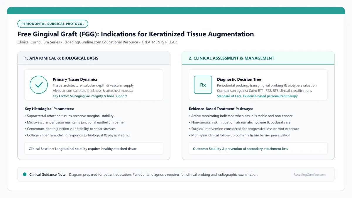
Educational illustration: Free Gingival Graft (FGG): Indications for Keratinized Tissue Augmentation. Clinical management requires comprehensive periodontal evaluation rather than isolated self-assessment.
Source: RecedingGumline.com Clinical Editorial Team (Proprietary educational diagram for RecedingGumline.com)
Key Clinical Distinctions & Diagnostic Boundaries
- FGG includes the superficial epithelium, whereas subepithelial connective tissue grafts (SCTG) harvest only the internal collagen layer.
- FGG is the most predictable procedure in periodontics for widening attached tissue, but less predictable for complete root coverage.
- FGG leaves an open secondary wound on the palate that requires 2 to 3 weeks to heal, producing more post-operative discomfort than SCTG.
- FGG is preferred in non-aesthetic zones (like lower front teeth), whereas SCTG is preferred in the smile zone due to more harmonious color blending.
When Is a Free Gingival Graft Indicated? Halting Progressive Recession
A free gingival graft is recommended when a tooth has zero or minimal remaining attached keratinized tissue—a condition where the fragile, movable alveolar mucosa extends directly to the gingival margin. Without an attached collar, daily toothbrushing and lip movements pull the margin away from the tooth, facilitating bacterial penetration and progressive attachment loss.
FGG is most commonly performed on the facial surfaces of lower incisors and premolars. In these areas, aesthetic appearance is rarely a primary concern, but structural preservation against strong mentalis muscle attachments or high frenal pulls is paramount for keeping the teeth.
The primary objective of a free gingival graft is to establish a dense, resilient collar of non-movable keratinized tissue that resists mechanical brushing abrasion. While root coverage is often minimal, halting progressive apical migration of the mucosal margin protects underlying alveolar bone.
Clinical Considerations:
- Indicated when movable alveolar mucosa extends directly to the tooth margin
- Commonly performed on lower incisors where strong muscle pull accelerates recession
- Arrests progressive recession even if exposed roots are not fully recovered
Surgical Protocol: Recipient Bed Preparation and Palatal Harvest
The procedure begins at the recipient site, where the clinician performs a partial-thickness dissection to separate movable mucosa and muscle fibers from the underlying periosteum, creating a stable, vascular recipient bed firmly bound to the alveolar bone.
At the palate, the surgeon harvests a graft consisting of both the keratinized oral epithelium and the dense underlying lamina propria (approximately 1.0 to 1.5 mm thick). The graft is trimmed and meticulously sutured onto the periosteal bed, immobilizing it completely to allow revascularization.
Patients with severe high frenum pull and minimal attached gingiva benefit substantially from the deepening of the vestibular fornix provided by the graft. The transplanted fibrous tissue halts the transmission of lip tension directly to the fragile marginal tissue.
Clinical Considerations:
- Recipient bed is anchored to fixed periosteum to eliminate muscle movement
- Full-thickness graft includes epithelium and dense lamina propria from the palate
- Immobilization with compressive sutures is vital to establish plasmatic diffusion
Tissue Maturation: Why FGG Leaves a Distinct Cosmetic "Patch"
Because the free gingival graft retains its palatal epithelial genetics, it heals with the histology of the roof of the mouth. This means the grafted area becomes thick, dense, and heavily keratinized, but appears noticeably paler and whiter than the adjacent pink gingiva.
This "tire-patch" appearance is completely functional and healthy, providing an impenetrable barrier against recession. However, because of this stark aesthetic difference, periodontists rarely place free gingival grafts on upper front teeth where they would be visible when smiling.
Palatal donor harvesting for free gingival grafts involves taking both surface epithelium and underlying dense lamina propria. The resulting palatal donor site heals via secondary intention over three to four weeks, necessitating protective stents and gentle oral hygiene.
Clinical Considerations:
- Palatal genetic memory results in a paler, firmer, heavily keratinized band
- Provides unmatched structural resilience against brushing and plaque trauma
- Rarely used in the maxillary aesthetic smile zone due to visible color contrast
Clinical Indications: Eliminating High Muscle Pull in Mandibular Sites
The free gingival graft (FGG) is primarily indicated when the clinical objective is widening the zone of keratinized, attached gingiva rather than achieving primary cosmetic root coverage. It is most frequently deployed in the anterior mandible, where shallow vestibular depth and aberrant frenal attachments create dynamic mechanical pull on the gingival margin.
When teeth exhibit thin, movable alveolar mucosa extending directly to the margin, even minor lip movements pull the sulcus open, facilitating plaque accumulation and rapid recession. An FGG surgically arrests this breakdown by creating a wide, dense, immovable band of keratinized tissue anchored to the periosteum.
By establishing a static, collagenous mucogingival barrier and deepening the vestibular fornix, the FGG halts further apical attachment loss and provides a maintainable foundation for lifelong hygiene.
Clinical Considerations:
- FGG is the established benchmark procedure for increasing the width of keratinized, attached tissue.
- Highly effective in the anterior mandible where shallow vestibules and muscle pull drive recession.
- Creates an immovable, dense collagenous barrier that halts further clinical attachment loss.
Donor Palatal Harvest & Secondary Intention Epithelialization
Unlike a subepithelial connective tissue graft, a free gingival graft includes both the lamina propria connective tissue and the overlying stratum corneum epithelium. The donor tissue is harvested from the premolar region of the hard palate at a calibrated uniform thickness of 1.0 to 1.5 millimeters.
Because the surface epithelium is excised, the palatal donor site is left as an open wound that must heal by secondary intention. Epithelial cells must migrate across the bare connective tissue bed from the wound margins, a process requiring 14 to 21 days for complete re-epithelialization.
At the recipient site, the grafted epithelium undergoes complete desquamation during the first four days as the graft survives on plasmatic circulation, followed by re-epithelialization from the graft margins to form durable, pale keratinized mucosa.
Clinical Considerations:
- FGG harvests include both surface epithelium and underlying dense connective tissue (1.0-1.5 mm thick).
- The donor palatal wound heals by secondary intention, requiring 2-3 weeks for full re-epithelialization.
- Grafted tissue undergoes initial surface desquamation before establishing a durable keratinized zone.
Clinical Reality Check
Do not expect a free gingival graft to make your gums look uniform in color; its goal is structural defense and saving the tooth, not cosmetic enhancement.
Questions to Ask Your Periodontist or Dentist
- Is the primary objective of this procedure to cover my exposed root or to create protective attached tissue?
- Why did you choose a free gingival graft over a connective tissue graft for this specific tooth?
- What measures will be taken to manage pain and protect the open donor site on my palate?
- How many millimeters of attached keratinized tissue do you expect this graft to produce?
Unsure What Your Gum Changes Mean?
Take our free, evidence-based Gum Recession Assessment — approximately 3 minutes. Identify potential risk factors, evaluate symptoms, and receive personalized discussion questions for your dentist or periodontist.
Non-diagnostic educational triage. Private, secure, completed in your browser.
Related Educational Topics
Clinical Evidence & Claim Traceability (3 Mapped Assertions)
Scientific Literature & Clinical Guidelines
3sources · Hide ▲
- Lang NP, Löe H (1972).
"The relationship between the width of keratinized gingiva and gingival health." Journal of Periodontology.
Clinical relevance: Landmark clinical investigation observing that gingival sites with less than 2 mm of keratinized gingiva (corresponding to less than 1 mm of attached gingiva) frequently exhibited clinical signs of persistent marginal inflammation despite plaque control, historically establishing the 2 mm keratinized tissue reference point.
- Kassab MM, Cohen RE (2003).
"The etiology and prevalence of gingival recession." The Journal of the American Dental Association.
Clinical relevance: Comprehensive epidemiological and etiological review establishing that gingival recession affects more than 50% of adults aged 18 to 64 and over 88% of individuals aged 65 and older, driven by interactions between predisposing anatomical factors and precipitating mechanical or inflammatory stimuli.
- Chambrone L, Salinas Ortega MA, Sukekava F, Rotundo R, Kalemaj Z, Buti J, Pini Prato GP (2018).
"Root coverage procedures for treating localised and multiple recession-type defects." Cochrane Database of Systematic Reviews.
Clinical relevance: Cochrane systematic review evaluating root-coverage procedures for localized and multiple recession-type defects. While subepithelial connective tissue grafts (SCTG) combined with coronally advanced flaps demonstrated higher rates of complete root coverage and keratinized tissue gain compared to flap advancement alone, evidence quality varied across outcomes and clinical decisions must balance donor site morbidity and patient-reported outcomes.
Important Medical Notice
The contents of RecedingGumline.com, including text, graphics, self-assessment calculators, and other materials, are intended solely for educational and informational purposes. This content is not intended to replace professional dental examination, diagnosis, or treatment. Always seek the advice of a qualified dentist, periodontist, or other licensed oral healthcare provider with any questions you may have regarding a medical or dental condition. Never disregard professional medical advice or delay seeking it because of something you read on this website.