Gingival Phenotype Classification in Gum Recession: AAP/EFP 2018 Criteria for Thin vs Thick Biotypes
How do periodontists officially classify periodontal phenotypes into thin vs. thick categories?
For evaluating diverse gum recession conditions, the 2018 AAP/EFP global classification updated diagnostic criteria across the periodontium. Among the most impactful changes was replacing the subjective term "biotype" with the standardized, multidimensional concept of periodontal phenotype. This classification provides objective criteria to stratify patients into thin or thick categories, directly predicting recession risk and surgical outcomes.
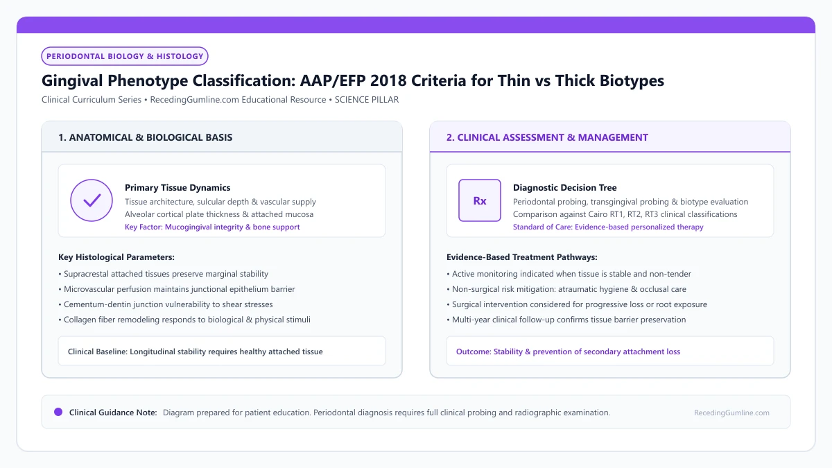
Educational illustration: Gingival Phenotype Classification: AAP/EFP 2018 Criteria for Thin vs Thick Biotypes. Clinical management requires comprehensive periodontal evaluation rather than isolated self-assessment.
Source: RecedingGumline.com Clinical Editorial Team (Proprietary educational diagram for RecedingGumline.com)
Key Clinical Distinctions & Diagnostic Boundaries
- "Biotype" implied an unchangeable genetic destiny; "phenotype" recognizes that surgical grafting can permanently convert thin tissue to thick.
- Probe transparency (whether a probe is visible through the sulcus) is the primary validated clinical method to diagnose phenotype.
- Thin phenotypes respond to plaque by receding apically; thick phenotypes respond by forming deep, fibrotic periodontal pockets.
- Phenotype determines the choice of surgical technique: thin phenotypes require connective tissue grafting alongside flap advancement.
Terminology Evolution: Why "Phenotype" Replaced "Biotype"
For decades, dentists used the term "gingival biotype," coined in the 1970s. However, in genetics and biology, a biotype represents a genetically fixed, immutable profile. This was scientifically inaccurate because dental surgeons regularly alter soft-tissue dimensions through grafting.
At the 2017 World Workshop in Chicago, the AAP and EFP officially adopted periodontal phenotype. Phenotype accurately describes the observable physical characteristics resulting from the interaction between genetic predisposition and environmental factors, acknowledging that clinicians can modify tissue thickness surgically.
The 2017 AAP/EFP World Workshop standardized the terminology of periodontal phenotype, recognizing it as a composite of gingival thickness, keratinized tissue width, and alveolar bone morphotype. Clinicians assess phenotype objectively using standard periodontal probe transparency through the sulcular margin.
Clinical Considerations:
- Replaced "biotype" with "periodontal phenotype" in the 2018 global classification
- Acknowledges that tissue thickness can be modified through periodontal plastic surgery
- Integrates soft-tissue dimensions with underlying alveolar bone architecture
The Three Phenotypic Categories: Thin, Thick, and Scalloped
The 2018 classification stratifies periodontal architecture into three primary categories: Thin Scalloped, Thick Flat, and Thick Scalloped.
The Thin Scalloped phenotype features delicate, translucent soft tissue (thickness < 1.0 mm), a narrow zone of keratinized tissue, pronounced architectural scalloping, triangular tooth crowns, and a paper-thin underlying cortical bone plate. The Thick Flat phenotype features dense, fibrous tissue (thickness ≥ 1.0 mm), a broad keratinized band, flatter gingival contours, square tooth crowns, and a thick cortical plate.
Patients exhibiting a thin scalloped phenotype possess delicate, highly translucent gingiva, narrow zones of keratinized tissue, and thin, scalloped cortical bone plates. In contrast, thick flat phenotypes feature dense, fibrotic tissue, broad keratinized collars, and thick, square alveolar bone architecture.
Clinical Considerations:
- Thin Scalloped: Delicate tissue (< 1 mm), triangular crowns, thin cortical bone, high recession risk
- Thick Flat: Dense fibrous tissue (≥ 1 mm), square crowns, thick bone, low recession risk
- Thick Scalloped: Thick tissue with pronounced architectural scalloping and intermediate bone thickness
Clinical Diagnosis and Phenotype Modification Therapy
To determine phenotype chairside, clinicians rely on the probe transparency method: inserting a periodontal probe into the sulcus. If the probe outline shines through the tissue, it is thin (< 1 mm); if invisible, it is thick (≥ 1 mm). High-frequency ultrasound and CBCT can also measure thickness to the tenth of a millimeter.
Identifying a thin phenotype allows periodontists to implement Phenotype Modification Therapy. By performing a connective tissue graft or placing a collagen matrix, the clinician thickens the delicate margin to > 1.5 mm, permanently converting a high-risk thin phenotype into a durable, recession-resistant thick phenotype.
Recognizing phenotypic characteristics is paramount before initiating implant placement, orthodontic movement, or cosmetic crown preparations. Thin phenotypes carry a significantly heightened risk of rapid recession and bone fenestration when subjected to surgical or mechanical trauma.
Clinical Considerations:
- Probe transparency test provides instantaneous chairside diagnosis of thickness
- Thin phenotypes require ultra-gentle hygiene and caution during orthodontics
- Phenotype modification surgery thickens tissue to permanently resist recession
The 2017 AAP/EFP Paradigm: GT, KTW & Bone Morphotype Integration
The 2017 World Workshop on Periodontal and Peri-Implant Diseases formally retired the historical term "biotype" in favor of "periodontal phenotype." This update reflects that while genetic factors establish baseline anatomy, the phenotype is dynamic and modifiable by environmental factors and clinical interventions.
The standardized classification assesses three core components: (1) Gingival Thickness (GT), clinically categorized as thin (≤ 1.0 mm) or thick (> 1.0 mm) using the probe transparency test or ultrasonic pulse-echo measurement; (2) Keratinized Tissue Width (KTW); and (3) Bone Morphotype, evaluated via CBCT.
Patients with a thin scalloped phenotype combine thin translucent gingiva, narrow KTW, and delicate underlying cortical bone with frequent dehiscences, representing the highest risk category for progressive recession under physical stress.
Clinical Considerations:
- The 2017 World Workshop replaced "biotype" with the dynamic, modifiable "periodontal phenotype."
- Diagnostic criteria integrate gingival thickness (≤ 1 mm thin; > 1 mm thick), KTW, and bone morphotype.
- Thin scalloped phenotypes combine delicate soft tissue with thin cortical bone, maximizing recession risk.
Phenotype Conversion: Biology of Connective Tissue Integration
One of the most powerful capabilities of periodontal plastic surgery is phenotype conversion—the surgical transformation of a thin, vulnerable phenotype into a thick, protective tissue band. Placing a subepithelial connective tissue graft (SCTG) under the thin margin achieves this objective.
Histologic studies reveal that the donor connective tissue harvested from the palate retains its genetically programmed phenotypic memory. When grafted into the recipient bed, donor fibroblasts dictate epithelial differentiation, instructing the overlying mucosal cells to express cytokeratins characteristic of thick, parakeratinized gingiva.
This biological conversion permanently increases mucosal thickness beyond 1.5 to 2.0 mm, creating a dense collagenous barrier that resists mechanical toothbrush trauma and shields the underlying alveolar bone from inflammatory resorption.
Clinical Considerations:
- Surgical grafting transforms a fragile thin phenotype into a thick, durable protective tissue band.
- Grafted palatal fibroblasts retain phenotypic memory, instructing overlying cells to keratinize.
- Thickened tissue (> 1.5 mm) provides lifelong resistance against mechanical abrasion and inflammation.
Clinical Reality Check
Knowing your phenotype is crucial before getting braces, veneers, or implants; thin phenotypes require pre-treatment soft-tissue reinforcement to prevent rapid gum recession during treatment.
Questions to Ask Your Periodontist or Dentist
- Which official 2018 periodontal phenotype (thin scalloped, thick flat, or thick scalloped) describes my mouth?
- Did the probe transparency test confirm that my gingival thickness is under 1.0 millimeter?
- Does my phenotype put me at elevated risk for recession during future dental work?
- Would phenotype modification therapy (tissue grafting) benefit my long-term tooth preservation?
Unsure What Your Gum Changes Mean?
Take our free, evidence-based Gum Recession Assessment — approximately 3 minutes. Identify potential risk factors, evaluate symptoms, and receive personalized discussion questions for your dentist or periodontist.
Non-diagnostic educational triage. Private, secure, completed in your browser.
Related Educational Topics
Clinical Evidence & Claim Traceability (3 Mapped Assertions)
Scientific Literature & Clinical Guidelines
3sources · Hide ▲
- Jepsen S, Caton JG, Albandar JM, Bissada NF, Bouchard P, Cortellini P, et al. (2018).
"Periodontal manifestations of systemic diseases and developmental and acquired conditions: Consensus report of workgroup 3 of the 2017 World Workshop on the Classification of Periodontal and Peri-Implant Diseases and Conditions." Journal of Periodontology.
Clinical relevance: Consensus report defining mucogingival conditions, gingival phenotype (replacing biotype), non-carious cervical lesions, and the multifactorial etiology of gingival recession; emphasizes that recession can occur without periodontitis and classifies recession by interdental clinical attachment loss.
- Cortellini P, Bissada NF (2018).
"Mucogingival conditions in the natural dentition: Narrative review, case definitions, and diagnostic considerations." Journal of Periodontology.
Clinical relevance: World Workshop 2017 comprehensive review defining mucogingival conditions, establishing diagnostic criteria for thin vs thick periodontal phenotypes, and detailing surgical indications including progressive recession, hypersensitivity, aesthetic dissatisfaction, and root caries vulnerability.
- Cairo F, Nieri M, Cincinelli S, Mervelt J, Pagliaro U (2011).
"The interproximal clinical attachment level to classify gingival recessions and predict root coverage outcomes: an explorative and reliability study." Journal of Clinical Periodontology.
Clinical relevance: Exploratory and reliability study establishing the Cairo classification based on interdental clinical attachment level (CAL): RT1 (no interproximal attachment loss; complete root coverage is clinically predictable), RT2 (interproximal attachment loss <= buccal loss; partial coverage predictable), and RT3 (interproximal loss exceeds buccal recession; complete coverage not predictable).
Important Medical Notice
The contents of RecedingGumline.com, including text, graphics, self-assessment calculators, and other materials, are intended solely for educational and informational purposes. This content is not intended to replace professional dental examination, diagnosis, or treatment. Always seek the advice of a qualified dentist, periodontist, or other licensed oral healthcare provider with any questions you may have regarding a medical or dental condition. Never disregard professional medical advice or delay seeking it because of something you read on this website.