Root Cementum Biology & Exposure in Gum Recession
What is root cementum, how thick is it, and what happens to it when gums recede?
Essential to understanding receding gums, covering the entire anatomical root surface of the human tooth is a micro-thin, specialized calcified tissue known as root cementum. Functioning as the biological glue of the periodontium, cementum anchors the principal periodontal ligament fibers to the tooth. When gum recession exposes cementum to the harsh oral cavity, this fragile layer undergoes rapid physical wear and bacterial contamination.
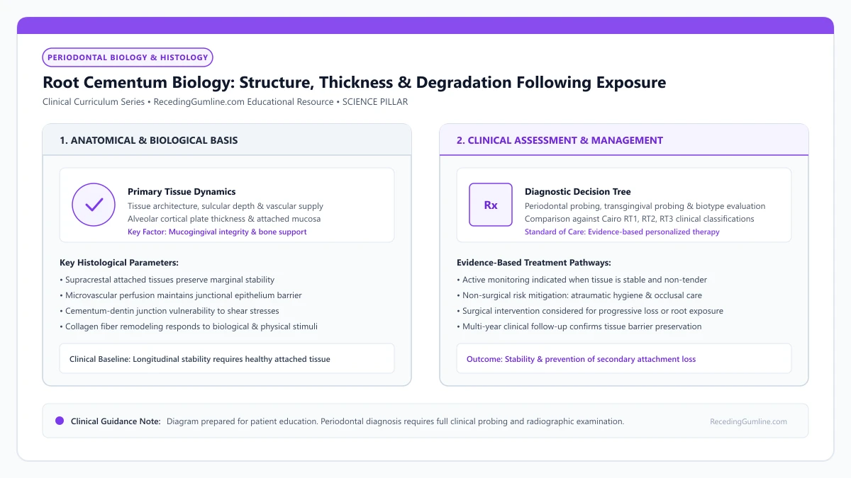
Educational illustration: Root Cementum Biology: Structure, Thickness & Degradation Following Exposure. Clinical management requires comprehensive periodontal evaluation rather than isolated self-assessment.
Source: RecedingGumline.com Clinical Editorial Team (Proprietary educational diagram for RecedingGumline.com)
Key Clinical Distinctions & Diagnostic Boundaries
- Enamel is 96% mineralized and white; cementum is only 45% to 50% mineralized, softer, and yellowish-brown.
- Cementum in the cervical neck of the tooth is paper-thin (thinner than a single human hair: 20–50 µm).
- Ordinary toothbrushing with abrasive toothpaste can scrub away the entire cementum layer in a matter of months.
- Contaminated, endotoxin-soaked cementum inhibits periodontal ligament fibroblast reattachment, necessitating root instrumentation.
Histological Architecture: Acellular vs. Cellular Cementum
Root cementum is classified into two major histological varieties based on the presence of embedded cells (cementocytes). Acellular extrinsic fiber cementum (AEFC) covers the coronal and middle third of the root. It contains zero cells and forms slowly during tooth eruption, providing the primary anchor for inserting Sharpey's fibers.
Crucially, AEFC at the cervical cementoenamel junction is razor-thin, measuring only 20 to 50 micrometers in thickness (a human hair is approximately 70 micrometers). Cellular intrinsic fiber cementum (CIFC) covers the apical third of the root, containing cementocytes that continue depositing layers throughout life to compensate for occlusal wear.
Root cementum is a specialized, avascular, mineralized connective tissue covering the anatomical roots of teeth, categorized into acellular extrinsic fiber cementum and cellular intrinsic fiber cementum. Acellular cementum covers the coronal root third and serves as the critical anchor for inserting principal periodontal fibers.
Clinical Considerations:
- Acellular extrinsic fiber cementum (AEFC) covers the vulnerable cervical third of the root
- Cervical cementum thickness is only 20 to 50 µm—thinner than a human hair
- Cellular cementum is restricted to the root tip and contains living cementocytes
Degradation Upon Exposure: Abrasion and Endotoxin Saturation
Under healthy conditions, cementum is shielded beneath attached gingiva. When gum recession exposes cervical cementum to the oral environment, two destructive processes occur almost simultaneously: mechanical abrasion and biochemical contamination.
Because cementum is only 45% to 50% inorganic hydroxyapatite, it is significantly softer than enamel. Daily toothbrushing with standard toothpaste scrubs away this 30-micrometer layer within weeks. Concurrently, bacterial biofilms saturate the porous cementum with lipopolysaccharides (endotoxins), creating a toxic surface that prevents soft tissue from reattaching.
Because cementum contains only about 50% hydroxyapatite mineral content by weight, it is significantly softer and more porous than dental enamel. When gingival recession exposes the cementum to the oral environment, the thin layer is easily worn away by toothbrush friction or dissolved by dietary acids within weeks.
Clinical Considerations:
- Low mineral content (50%) makes cementum soft and easily abraded by toothbrushing
- Ordinary brushing can completely wear through cervical cementum in under six months
- Bacterial endotoxins permeate porous cementum, creating a biologically toxic root surface
Clinical Management: Root Planing and Chemical Decontamination
The rapid loss of cementum explains why receded roots develop acute cold sensitivity: once cementum is abraded, underlying dentinal tubules are exposed. Furthermore, when periodontists perform root coverage surgery, they must thoroughly debride the root surface.
Surgeons gently plane the root to remove cytotoxic endotoxins and often condition the surface with EDTA (ethylenediaminetetraacetic acid) or citric acid. This chemical etching dissolves smear plugs, exposes native collagen fibers, and creates a biocompatible surface that allows newly transplanted gum grafts to attach securely.
Once cementum is lost, the underlying radicular dentin is exposed, opening thousands of microscopic dentinal tubules to the oral cavity. Exposed dentin is highly vulnerable to rapid cervical root caries, erosion, and excruciating hydrodynamic thermal hypersensitivity.
Clinical Considerations:
- Loss of cementum denudes underlying dentin, triggering hydrodynamic cold sensitivity
- Root planing removes necrotic, endotoxin-impregnated cementum prior to surgery
- EDTA chemical conditioning decontaminates the root to promote new soft-tissue adhesion
Histological Diversity: Acellular vs. Cellular Intrinsic Fiber Cementum
Root cementum is a specialized, avascular mineralized tissue covering the anatomical root. Histologically, it exists in two primary functional varieties: Acellular Extrinsic Fiber Cementum (AEFC) and Cellular Intrinsic Fiber Cementum (CIFC).
AEFC covers the coronal two-thirds of the root, forming a thin, delicate layer (20 to 50 micrometers thick) that contains densely packed Sharpey's fibers. AEFC is the primary tissue responsible for anchoring the tooth to alveolar bone in the cervical region where recession occurs.
Because AEFC is exceptionally thin and completely devoid of living cementocytes, once it is destroyed by toothbrush abrasion or aggressive root planing, it cannot regenerate spontaneously from within. Regeneration requires recruitment of new cementoblast precursors from the periodontal ligament space.
Clinical Considerations:
- Acellular Extrinsic Fiber Cementum (AEFC) covers the cervical two-thirds of roots where recession occurs.
- AEFC is paper-thin (20-50 micrometers) and contains dense inserting Sharpey's fibers.
- Acellular cementum possesses no living cells; once stripped away, it cannot spontaneously regenerate.
The Denuded Root: Toxic Endotoxin Adsorption & Smear Layers
When gingival recession exposes acellular cementum to the oral environment, the tissue undergoes rapid physical and chemical degradation. Exposure to oral fluids and saliva leads to alternating cycles of surface demineralization and hypermineralization.
Simultaneously, subgingival gram-negative bacteria release lipopolysaccharides (endotoxins) that adsorb directly into the porous organic matrix of the superficial cementum. This endotoxin-impregnated cementum is cytotoxic, actively preventing periodontal ligament fibroblasts from attaching to the root.
During root coverage surgery, periodontists must detoxify this contaminated root layer using ultrasonic scaling, sharp curettes, and chemical conditioning agents (such as citric acid or EDTA) to expose clean collagen fibrils receptive to new soft-tissue attachment.
Clinical Considerations:
- Exposed cementum adsorbs cytotoxic bacterial lipopolysaccharides (endotoxins).
- Endotoxin contamination prevents fibroblast adhesion and prohibits soft-tissue reattachment.
- Chemical and mechanical root conditioning detoxifies the root to enable surgical graft integration.
Clinical Reality Check
Root cementum has zero nerve fibers; you feel pain on an exposed root only because the cementum has worn away, allowing hot or cold liquids to reach the underlying dentinal tubules.
Questions to Ask Your Periodontist or Dentist
- Has the protective cementum layer worn away on my receded teeth, exposing root dentin?
- Are my exposed roots showing signs of mechanical toothbrush abrasion into the cementum?
- Will chemical conditioning (such as EDTA) be used during my gum graft to clean the root surface?
- What gentle, non-abrasive toothpaste will help preserve my remaining root cementum?
Unsure What Your Gum Changes Mean?
Take our free, evidence-based Gum Recession Assessment — approximately 3 minutes. Identify potential risk factors, evaluate symptoms, and receive personalized discussion questions for your dentist or periodontist.
Non-diagnostic educational triage. Private, secure, completed in your browser.
Related Educational Topics
Clinical Evidence & Claim Traceability (2 Mapped Assertions)
Scientific Literature & Clinical Guidelines
3sources · Hide ▲
- Pihlstrom BL, Michalowicz BS, Johnson NW (2005).
"Periodontal diseases." The Lancet.
Clinical relevance: Peer-reviewed Lancet seminar review synthesizing global periodontal epidemiology, microbial etiology, host immunopathology, and systemic interactions, emphasizing prevention, biofilm disruption, and early risk factor modification.
- Brännström M (1966).
"Sensitivity of dentine." Oral Surgery, Oral Medicine, Oral Pathology.
Clinical relevance: Foundational paper formulating the hydrodynamic theory of dentin hypersensitivity: rapid fluid displacement within patent dentinal tubules physically deforms intradental nerve endings at the pulp-dentin boundary, explaining thermal, mechanical, and evaporative root sensitivity.
- Sculean A, Nikolidakis D, Schwarz F (2008).
"Regeneration of periodontal tissues: combinations of barrier membranes and grafting materials - biological foundation and preclinical evidence: a systematic review." Journal of Clinical Periodontology.
Clinical relevance: Comprehensive systematic review analyzing clinical and histological outcomes of biomaterials in periodontal regeneration; confirmed that enamel matrix derivative (EMD) and barrier membranes for guided tissue regeneration achieve histological evidence of true regeneration (new cementum, periodontal ligament, and bone).
Important Medical Notice
The contents of RecedingGumline.com, including text, graphics, self-assessment calculators, and other materials, are intended solely for educational and informational purposes. This content is not intended to replace professional dental examination, diagnosis, or treatment. Always seek the advice of a qualified dentist, periodontist, or other licensed oral healthcare provider with any questions you may have regarding a medical or dental condition. Never disregard professional medical advice or delay seeking it because of something you read on this website.