Dentinal Tubules & Smear Layer in Gum Recession Sensitivity
What is the smear layer on teeth, and how does it block dentinal tubules to stop sensitivity?
When roots become exposed by receding gumline factors, the primary biological barrier preventing excruciating pain is a micro-thin layer of mineralized debris known as the smear layer. Composed of microscopic hydroxyapatite particles, denatured collagen, and salivary glycoproteins, this natural coating forms a protective plug inside open dentinal tubules. Understanding smear layer dynamics explains why sensitivity flares up, calms down, and vanishes.
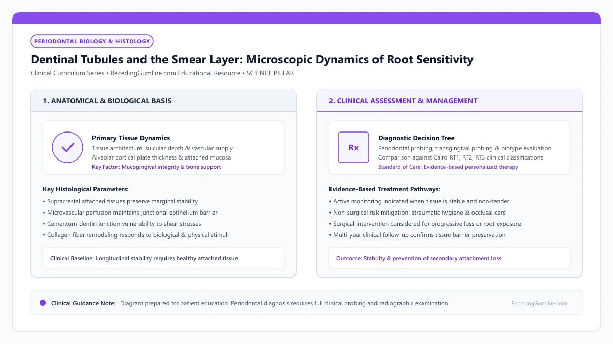
Educational illustration: Dentinal Tubules and the Smear Layer: Microscopic Dynamics of Root Sensitivity. Clinical management requires comprehensive periodontal evaluation rather than isolated self-assessment.
Source: RecedingGumline.com Clinical Editorial Team (Proprietary educational diagram for RecedingGumline.com)
Key Clinical Distinctions & Diagnostic Boundaries
- An intact smear layer reduces fluid conductance by up to 90%, allowing exposed roots to remain completely pain-free.
- Acidic challenges from citrus, soda, or gastric reflux dissolve smear plugs in seconds, triggering acute sensitivity.
- Brushing with hard bristles scrubs away the smear layer faster than saliva can remineralize it.
- In restorative dentistry, dentists purposefully etch away the smear layer with acid to bond fillings, but re-seal it with resin.
Microscopic Composition: The Smear Layer and Smear Plugs
Whenever tooth structure is cut, scraped, or abraded by dental instruments or daily toothbrushing, microscopic mineral fragments do not simply wash away. Instead, they form an amorphous, adherent coating approximately 1 to 2 micrometers thick known as the smear layer.
Crucially, this mineral debris is packed deep into the openings of the dentinal tubules, forming dense cylindrical stoppers called smear plugs that extend 1 to 5 micrometers into the channels. These plugs act like corks in wine bottles, physically blocking fluid movement and rendering an exposed root completely asymptomatic.
Human root dentin contains between 20,000 and 45,000 microscopic dentinal tubules per square millimeter, each radiating outward from the dental pulp chamber toward the cementoenamel junction. In health, these tubule apertures are hermetically sealed by root cementum and a protective amorphous smear layer.
Clinical Considerations:
- Amorphous 1–2 µm coating formed from abraded mineral crystals and collagen fragments
- Smear plugs penetrate 1–5 µm into tubule mouths, corking the microscopic channels
- Reduces fluid movement by 85% to 90%, preventing thermal and tactile nerve firing
The Dissolution Cycle: How Acids Strip the Smear Layer
The smear layer is in a dynamic, continuous state of turnover. The greatest threat to smear layer stability is dietary acidity. Whenever you drink wine, citrus juices, sports drinks, or carbonated sodas, the acidic pH quickly dissolves the sub-micron calcium phosphate particles that compose the smear plugs.
Within seconds, the tubule openings are un-corked and open to the oral cavity. If you immediately brush your teeth with a stiff brush, the softened surface dentin is scoured away. Conversely, if left undisturbed, natural saliva containing supersaturated calcium and phosphate ions slowly remineralizes and re-plugs the tubules over 24 to 48 hours.
The natural smear layer consists of microcrystalline mineral debris and salivary glycoproteins that occlude tubule orifices, reducing hydraulic fluid conductance by over 80%. When patients consume acidic foods or scrub vigorously with abrasive toothpastes, this fragile smear layer is rapidly stripped away.
Clinical Considerations:
- Dietary acids dissolve smear plugs in seconds, reopening tubules to oral fluids
- Brushing immediately after acidic exposure scrubs away the softened dentin permanently
- Salivary calcium and phosphate naturally precipitate new mineral plugs if protected from acid
Therapeutic Remineralization: Rebuilding Smear Plugs with Modern Pastes
Modern desensitizing toothpastes are engineered to artificially replicate and reinforce the natural smear layer. Formulations containing stannous fluoride, calcium sodium phosphosilicate (NovaMin), or arginine-calcium carbonate precipitate insoluble, acid-resistant mineral crystals inside tubule orifices.
These artificial smear plugs are far more durable than natural salivary plugs, resisting subsequent acid challenges and permanently calming sensitive receded roots. Applying these pastes directly with a fingertip before bed allows uninterrupted overnight mineral precipitation.
Removing the smear layer increases tubular fluid flow exponentially according to Poiseuille’s law, resulting in severe clinical dentin hypersensitivity. Therapeutic interventions focus on re-establishing an artificial smear layer or precipitating mineral complexes to permanently seal patent tubules.
Clinical Considerations:
- NovaMin and arginine-calcium toothpastes deposit synthetic mineral plugs deep into tubules
- Stannous fluoride forms an acid-resistant tin-fluoride barrier that resists dietary acids
- Bedtime topical application without rinsing maximizes tubule occlusion efficiency
The Smear Layer: Composition, Thickness & Natural Hydrodynamic Plugs
The smear layer is a 1.0 to 2.0 micrometer thick microcrystalline debris layer that coats dentin surfaces following mechanical instrumentation, cutting, or abrasive toothbrushing. It consists of pulverized hydroxyapatite crystals, denatured collagen fragments, and saliva proteins.
Importantly, portions of this debris are burnished into the orifices of dentinal tubules, forming dense "smear plugs" that extend 2 to 5 micrometers into the tubule lumens. These smear plugs reduce the hydraulic conductance (permeability) of dentin by up to 85% to 90%.
In health, this natural barrier prevents oral fluids from contacting pulpal nerve endings. When recession occurs, preserving or chemically reinforcing the smear layer is the front-line biological defense against hypersensitivity.
Clinical Considerations:
- The smear layer is a 1-2 micrometer thick microcrystalline coating of hydroxyapatite and collagen.
- Smear plugs extend into tubule lumens, reducing dentin permeability by up to 90%.
- Preserving smear plugs prevents fluid movement and protects against root sensitivity.
Acid Dissolution Kinetics & In-Office Biomimetic Remineralization
While the smear layer provides effective natural insulation, it is acid-labile. Exposure to dietary acids (citrus, soda, vinegar) or bacterial lactic acid with a pH below 6.0 rapidly dissolves the smear layer and dissolves the protective intratubular smear plugs within minutes.
Once dissolved, the functional diameter of tubule lumens triples, dramatically increasing hydrodynamic fluid flow and provoking acute hypersensitivity. Restoring this lost barrier requires biomimetic remineralization.
In-office treatments with calcium phosphate technologies, bioactive glass (NovaMin), and potassium oxalate precipitate insoluble mineral complexes inside the dissolved tubule lumens, recreating artificial mineral plugs that withstand subsequent acid challenges.
Clinical Considerations:
- Dietary and bacterial acids (pH < 6.0) dissolve smear plugs and triple tubule lumen diameter.
- Loss of smear plugs exposes dentin tubules to rapid hydrodynamic fluid shifts and pain.
- Biomimetic agents (bioactive glass, oxalates) precipitate mineral complexes to recreate durable plugs.
Clinical Reality Check
When a dentist applies an in-office fluoride varnish, they are essentially creating an instantaneous, synthetic 10-micrometer smear layer to seal your open tubules.
Questions to Ask Your Periodontist or Dentist
- Has my natural smear layer been stripped away by acidic foods or abrasive brushing?
- Do my receded roots show open dentinal tubules under high-magnification examination?
- Which remineralizing toothpaste technology (stannous fluoride, NovaMin, or nano-hydroxyapatite) is best for rebuilding my smear layer?
- How long does it take for saliva to naturally rebuild smear plugs after an acid attack?
Unsure What Your Gum Changes Mean?
Take our free, evidence-based Gum Recession Assessment — approximately 3 minutes. Identify potential risk factors, evaluate symptoms, and receive personalized discussion questions for your dentist or periodontist.
Non-diagnostic educational triage. Private, secure, completed in your browser.
Related Educational Topics
Clinical Evidence & Claim Traceability (2 Mapped Assertions)
Scientific Literature & Clinical Guidelines
3sources · Hide ▲
- Brännström M (1966).
"Sensitivity of dentine." Oral Surgery, Oral Medicine, Oral Pathology.
Clinical relevance: Foundational paper formulating the hydrodynamic theory of dentin hypersensitivity: rapid fluid displacement within patent dentinal tubules physically deforms intradental nerve endings at the pulp-dentin boundary, explaining thermal, mechanical, and evaporative root sensitivity.
- West NX, Seong J, Davies M (2015).
"Management of dentine hypersensitivity: efficacy of professionally and self-administered agents." Journal of Clinical Periodontology.
Clinical relevance: Systematic review evaluating professionally and self-administered desensitizing agents; found evidence supporting tubule-occluding dentifrices and potassium-based nerve desensitizers in providing transient to moderate symptom relief, with substantial heterogeneity across clinical trials.
- Pihlstrom BL, Michalowicz BS, Johnson NW (2005).
"Periodontal diseases." The Lancet.
Clinical relevance: Peer-reviewed Lancet seminar review synthesizing global periodontal epidemiology, microbial etiology, host immunopathology, and systemic interactions, emphasizing prevention, biofilm disruption, and early risk factor modification.
Important Medical Notice
The contents of RecedingGumline.com, including text, graphics, self-assessment calculators, and other materials, are intended solely for educational and informational purposes. This content is not intended to replace professional dental examination, diagnosis, or treatment. Always seek the advice of a qualified dentist, periodontist, or other licensed oral healthcare provider with any questions you may have regarding a medical or dental condition. Never disregard professional medical advice or delay seeking it because of something you read on this website.