Root Cavities and Receding Gums: Vulnerability of Exposed Cementum and Dentin
Why do exposed roots get cavities so quickly, and how are root surface cavities treated?
When gums recede, the biological protective barrier over the root surface is lost. While tooth enamel is exceptionally hard and acid-resistant, the exposed cementum and dentin of the root are soft, porous, and highly susceptible to dental decay. Root caries (cavities on receded roots) represent one of the most destructive and rapidly progressing complications of receding gums management.
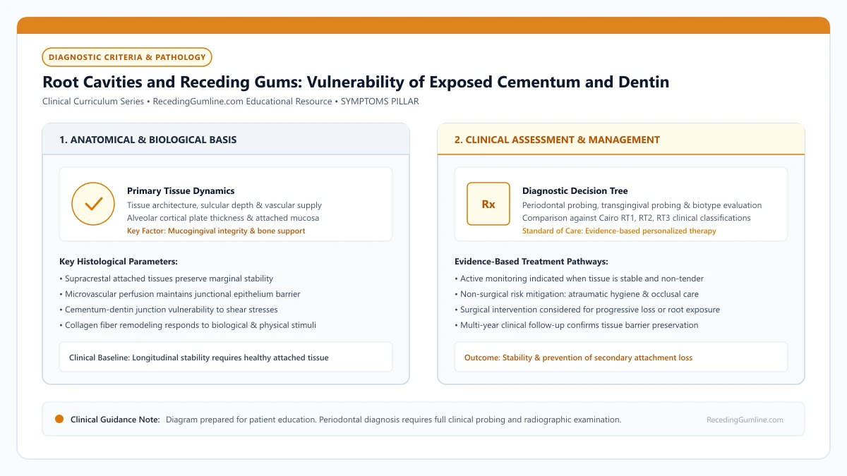
Educational illustration: Root Cavities and Receding Gums: Vulnerability of Exposed Cementum and Dentin. Clinical management requires comprehensive periodontal evaluation rather than isolated self-assessment.
Source: RecedingGumline.com Clinical Editorial Team (Proprietary educational diagram for RecedingGumline.com)
Key Clinical Distinctions & Diagnostic Boundaries
- Enamel decay forms in pits and grooves and takes years to penetrate; root decay spreads horizontally along the gumline and penetrates dentin rapidly.
- An arrested root lesion is hard, dark brown or black, and glassy (stable); an active root cavity is soft, yellow-brown, and sticky to a dental explorer.
- Surgical gum grafting cannot be performed over an active root cavity; the decay must first be excavated and restored with glass ionomer.
- Salivary hypofunction (dry mouth) is the single greatest risk factor multiplying root cavity formation.
Biochemical Vulnerability: Why Roots Melt at pH 6.5
Tooth enamel is a dense crystalline matrix composed of 96% inorganic hydroxyapatite. It remains intact until oral acidity drops to a pH of 5.5. Exposed root dentin, however, contains only 70% inorganic mineral, with the remaining 30% composed of organic collagen fibers and water.
Because of this lower mineral density, dentin begins dissolving at a much higher pH threshold of 6.2 to 6.7. Ordinary dietary carbohydrates, sweetened beverages, or mild plaque acids that would cause zero harm to enamel immediately begin demineralizing exposed root surfaces.
Root cementum and underlying radicular dentin have a significantly higher critical pH threshold (pH 6.2 to 6.7) for demineralization compared to dense enamel (pH 5.5). When recession exposes the root surface, normal dietary acids can trigger rapid biochemical demineralization.
Clinical Considerations:
- Dentin demineralizes at a critical pH of 6.2 to 6.7 (enamel dissolves at 5.5)
- Dentin contains 30% organic collagen, allowing rapid bacterial enzymatic destruction
- Mild dietary acids and plaque sugars that spare enamel actively dissolve roots
Clinical Diagnosis: Differentiating Active from Arrested Root Decay
Dentists diagnose root cavities using visual inspection and gentle tactile exploration. An active, progressing root cavity presents as a soft, mushy, or leathery depression, ranging in color from pale yellow to light brown. A dental explorer sticks in the softened collagen matrix.
In contrast, an arrested (healed) root lesion has successfully remineralized from saliva and fluoride. It appears dark brown or jet black, with a hard, shiny, glassy surface that resists the probe. Arrested lesions require no drilling; they are biological scars that should be left alone and monitored.
Root caries lesions often spread rapidly along the circumferential cementoenamel junction rather than penetrating deeply in a localized pit. Soft, yellow-brown necrotic dentin lesions can encircle the neck of the tooth, undermining the crown and jeopardizing structural integrity.
Clinical Considerations:
- Active decay: Soft, sticky, leathery, yellowish-brown surface requiring treatment
- Arrested decay: Hard, glassy, dark brown or black surface that is stable and safe
- Never drill into an arrested, hard root surface; aggressive drilling weakens the tooth
Treatment Strategies: Prescription Fluoride, SDF & Glass Ionomer
Preventing root cavities requires elevating oral fluoride concentrations. Dentists prescribe 1.1% sodium fluoride toothpaste (5,000 ppm, such as PreviDent), which hardens exposed dentin against acid attacks. Another non-invasive therapy is Silver Diamine Fluoride (SDF), an antimicrobial liquid applied chairside that instantly halts active root decay, though it stains the decay black.
When a root cavity has created a structural hole, the dentist must gently excavate the softened decay and place a restoration. Resin-Modified Glass Ionomer (RMGI) is the material of choice: it bonds chemically to root dentin in moist subgingival environments and continuously releases fluoride to prevent recurrent cavities.
Preventive protocols require high-concentration prescription 5,000 ppm sodium fluoride dentifrices and silver diamine fluoride (SDF) applications to arrest active decay. Restoring root caries requires moisture-tolerant glass ionomer cements that chemically bond to dentin and release fluoride.
Clinical Considerations:
- Prescription 5,000 ppm sodium fluoride toothpaste provides daily high-potency remineralization
- Silver Diamine Fluoride (SDF) arrests active root decay non-invasively without drilling
- Resin-modified glass ionomer (RMGI) is the premier restorative material for root cavities
Demineralization Dynamics: The Critical pH Threshold (pH 6.2 to 6.7)
When gingival recession exposes the tooth root, it bares cementum and dentin—tissues that are far more chemically vulnerable to bacterial decay than enamel. While dense crown enamel resists demineralization until oral pH drops below 5.5, root cementum and dentin demineralize at a critical pH of 6.2 to 6.7.
This narrow buffer means that even mildly acidic dietary exposures or minor bacterial plaque accumulation can initiate root demineralization. Furthermore, root dentin contains approximately 20% organic collagen matrix, which is degraded by bacterial proteolytic enzymes once the mineral crystals are dissolved.
Root caries progresses rapidly and circumferentially around the cervical collar, often invading subgingivally and undermining tooth structure beneath the remaining gumline.
Clinical Considerations:
- Root cementum and dentin demineralize at pH 6.2-6.7, compared to pH 5.5 for enamel.
- Mild dietary acidity or minor plaque easily triggers root surface demineralization.
- Bacterial proteolytic enzymes dissolve the organic collagen matrix, causing rapid circumferential decay.
Chemotherapeutic Prevention: 5,000 ppm Dentifrice & Silver Diamine Fluoride
Preventing and arresting root decay on exposed roots demands aggressive chemotherapeutic intervention beyond standard over-the-counter fluoride toothpaste. The clinical standard of care prescription is 1.1% sodium fluoride dentifrice (5,000 ppm fluoride).
Brushing nightly with 5,000 ppm fluoride creates a dense reservoir of calcium fluoride on the root surface, driving continuous remineralization and forming fluorapatite, which lowers the critical dissolution pH to 4.5.
For active, non-cavitated or cavitated root lesions, 38% Silver Diamine Fluoride (SDF) provides immediate antimicrobial and remineralizing efficacy. Silver ions penetrate dentinal tubules, killing cariogenic bacteria and inhibiting matrix metalloproteinases, while fluoride arrests lesion progression, transforming soft decay into a hardened, glass-like surface.
Clinical Considerations:
- High-concentration 5,000 ppm fluoride dentifrice significantly reduces root caries incidence.
- Fluorapatite formation lowers the critical demineralization pH of root dentin to 4.5.
- Silver Diamine Fluoride (SDF) instantly kills bacteria and hardens active root decay.
Clinical Reality Check
Patients with exposed roots who take prescription medications causing xerostomia face significantly elevated root caries risk; prescription 5,000 ppm fluoride toothpaste may be essential to protect exposed dentin.
Questions to Ask Your Periodontist or Dentist
- Do my exposed root surfaces have active root cavities or are they hard and arrested?
- Would a prescription 5,000 ppm fluoride toothpaste help protect my receded roots from decay?
- Am I a candidate for Silver Diamine Fluoride (SDF) to stop decay without drilling?
- If my root needs a filling, will you use a fluoride-releasing glass ionomer material?
Unsure What Your Gum Changes Mean?
Take our free, evidence-based Gum Recession Assessment — approximately 3 minutes. Identify potential risk factors, evaluate symptoms, and receive personalized discussion questions for your dentist or periodontist.
Non-diagnostic educational triage. Private, secure, completed in your browser.
Related Educational Topics
Clinical Evidence & Claim Traceability (2 Mapped Assertions)
Scientific Literature & Clinical Guidelines
3sources · Hide ▲
- Pihlstrom BL, Michalowicz BS, Johnson NW (2005).
"Periodontal diseases." The Lancet.
Clinical relevance: Peer-reviewed Lancet seminar review synthesizing global periodontal epidemiology, microbial etiology, host immunopathology, and systemic interactions, emphasizing prevention, biofilm disruption, and early risk factor modification.
- Chapple ILC, Mealey BL, Van Dyke TE, Bartold PM, Dommisch H, Eickholz P, et al. (2018).
"Periodontal health and gingival diseases and conditions on an intact and a reduced periodontium: Consensus report of workgroup 1 of the 2017 World Workshop on the Classification of Periodontal and Peri-Implant Diseases and Conditions." Journal of Clinical Periodontology.
Clinical relevance: Consensus report establishing diagnostic criteria for periodontal health and gingivitis across intact and reduced periodontia, defining clinical gingival health as <10% bleeding on probing without attachment loss and strictly differentiating gingivitis from periodontitis.
- Tonetti MS, Greenwell H, Kornman KS (2018).
"Staging and grading of periodontitis: Framework and proposal of a new classification and case definition." Journal of Clinical Periodontology.
Clinical relevance: Consensus framework establishing the multidimensional staging (severity and extent of periodontal tissue breakdown) and grading (biological rate of disease progression, incorporating smoking and diabetes as grade modifiers) for periodontitis. It addresses periodontitis diagnosis and staging, not the classification of localized gingival recession defects.
Important Medical Notice
The contents of RecedingGumline.com, including text, graphics, self-assessment calculators, and other materials, are intended solely for educational and informational purposes. This content is not intended to replace professional dental examination, diagnosis, or treatment. Always seek the advice of a qualified dentist, periodontist, or other licensed oral healthcare provider with any questions you may have regarding a medical or dental condition. Never disregard professional medical advice or delay seeking it because of something you read on this website.