Coronally Advanced Flap (CAF) Surgery for Gum Recession
How does a coronally advanced flap work, and what clinical conditions are required for its success?
For patients seeking root coverage for a gum recession, the Coronally Advanced Flap (CAF) is a foundational periodontal plastic surgery technique designed to cover denuded root surfaces and restore the marginal gingiva. By mobilizing adjacent keratinized soft tissue and advancing it coronally over the exposed root, surgeons can achieve esthetic root coverage without harvesting a large external graft, or combine CAF with an underlying connective tissue graft for maximum predictability.
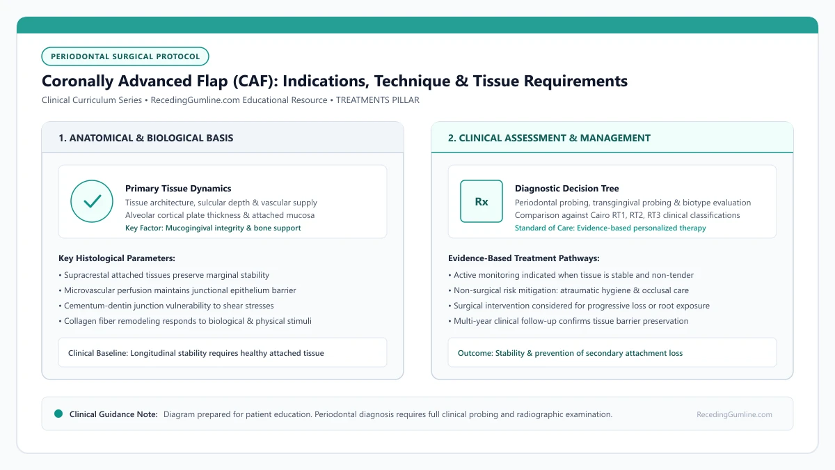
Educational illustration: Coronally Advanced Flap (CAF): Indications, Technique & Tissue Requirements. Clinical management requires comprehensive periodontal evaluation rather than isolated self-assessment.
Source: RecedingGumline.com Clinical Editorial Team (Proprietary educational diagram for RecedingGumline.com)
Key Clinical Distinctions & Diagnostic Boundaries
- CAF alone repositions existing tissue, whereas CAF + SCTG introduces new transplanted donor tissue beneath the flap to increase gingival thickness.
- CAF requires completely intact interdental papillae to serve as receptive vascular beds for suturing.
- Unlike free gingival grafts, CAF maintains the original blood supply from the base of the flap, accelerating early revascularization.
- If keratinized tissue is completely absent (< 1 mm), CAF alone has high recurrence rates and an autogenous graft must be incorporated.
Surgical Mechanics: Incision Design and Tension-Free Flap Release
The success of a coronally advanced flap hinges on micro-surgical precision. The surgeon creates horizontal sulcular and beveled papilla-sparing incisions to mobilize the full-thickness mucoperiosteal flap up to the mucogingival junction, transitioning into a split-thickness dissection in the alveolar mucosa.
The decisive step is the periosteal releasing incision: micro-incisions made through the deep elastic fibers of the periosteum. This relieves all muscle and tissue tension, allowing the flap to passively drape 1 to 2 millimeters coronal to the cementoenamel junction without any retraction force.
Micro-surgical instruments and high-magnification loupes ensure atraumatic handling of thin gingival tissues during incision and split-thickness dissection. Preserving the interdental papillae intact ensures that the lateral vascular supply remains uninterrupted throughout the procedure.
Clinical Considerations:
- Split-thickness dissection in mucosal tissue preserves deep vascular supply
- Periosteal release eliminates muscle tension pulling the flap downward
- The flap must sit passively coronal to the CEJ without requiring suture tension
Anatomical Prerequisites: Tissue Thickness and Keratinized Width
Not every receded tooth is suitable for CAF alone. For a coronally advanced flap to succeed as a monotherapy, the patient must possess a baseline band of keratinized gingiva apical to the recession (ideally at least 1.5 to 2.0 mm) and a flap thickness exceeding 0.8 to 1.0 mm.
If the pre-existing tissue is paper-thin (< 0.8 mm) or lacks keratinization entirely, moving that tissue upward will result in gradual re-recession over time. In such cases, surgeons place a connective tissue graft beneath the flap (CAF + SCTG) to transform the biotype from thin to thick.
Complete release of the underlying periosteum at the base of the flap eliminates all muscular tension from the surrounding facial expressions. When tension is eliminated, the flap passively rests at or coronal to the cementoenamel junction without relying on suture tension.
Clinical Considerations:
- Flap thickness ≥ 0.8 mm is required for predictable root coverage with CAF alone
- At least 2 mm of keratinized tissue must be present apical to the recession defect
- Thin tissues require an underlying connective tissue graft to prevent relapse
Microsurgical Suturing and Vascular Bed Integration
Once mobilized, the flap is secured using ultra-fine microsurgical sutures (6-0 or 7-0 gauge). Sling sutures or suspended mattress sutures anchor the flap securely around the neck of the tooth, compressing the margins against the de-epithelialized interdental papillae.
Because the flap remains pedicled (attached at its base), it maintains its original apical blood supply while simultaneously receiving new collateral circulation from the de-epithelialized papillae, ensuring rapid revascularization within the first 5 to 7 days.
Long-term periodontal trials demonstrate favorable aesthetic color blending and contour harmony with coronally advanced flaps compared to traditional free gingival grafts. Patient satisfaction is consistently high due to the avoidance of open palatal wound harvesting.
Clinical Considerations:
- 6-0 and 7-0 microsurgical sutures minimize tissue trauma and scarring
- De-epithelialized anatomical papillae act as vital vascular recipient beds
- Pedicle base maintains uninterrupted blood supply during initial healing
Surgical Biomechanics: Split-Full-Split Thickness Flap Elevation
The coronally advanced flap (CAF) is a cornerstone of periodontal plastic surgery for single and multiple recession defects. Achieving predictable, tension-free coronal advancement requires the meticulous "split-full-split" flap elevation technique.
Flap elevation commences with a split-thickness incision at the interdental surgical papillae, preserving periosteum over the bone. Over the denuded root and marginal bone crest, a full-thickness mucoperiosteal flap is elevated to include the periosteum, protecting the marginal vascular plexus.
Apical to the mucogingival junction, the dissection converts back to split-thickness, severing muscle insertions and elastic fibers within the alveolar mucosa. This releases all basal tension, allowing the flap to passively drape coronally past the cementoenamel junction with zero elastic recoil.
Clinical Considerations:
- Split-thickness papilla dissection preserves periosteal vascular beds on interdental bone.
- Full-thickness dissection over marginal bone crest preserves the periosteal microvascular plexus.
- Apical muscle release allows the flap to passively drape coronally with zero tissue tension.
Suture Materials & Papillary Anchorage Dynamics
Securing the coronally advanced flap in a stable, coronal position during the critical 14-day healing window depends on suture selection and microsurgical anchoring techniques. Surgeons utilize ultra-fine 6-0 or 7-0 monofilament sutures (polypropylene or polyvinylidene fluoride) with precision micro-needles.
Monofilament sutures eliminate the "wicking effect" characteristic of braided silk or vicryl sutures, preventing the migration of oral bacteria through the suture tract into the healing graft bed. The sutures are placed using sling or suspended mattress configurations that anchor around tooth contact points.
Tension-free coronal stabilization positioned 1.0 to 1.5 millimeters coronal to the cementoenamel junction compensates for minor postoperative tissue contraction, ensuring stable long-term root coverage.
Clinical Considerations:
- Ultra-fine 6-0 or 7-0 monofilament sutures eliminate capillary bacterial wicking.
- Sling and suspended mattress sutures anchor securely around interproximal tooth contact points.
- Positioning the flap 1.0-1.5 mm coronal to the CEJ anticipates physiological contraction during healing.
Clinical Reality Check
Any residual tension in a coronally advanced flap will cause the margin to pull away from the root during facial muscle movement, leading to flap retraction and surgical failure.
Questions to Ask Your Periodontist or Dentist
- Is my receded tooth a candidate for a coronally advanced flap alone, or does it require an underlying graft?
- How thick is my tissue flap, and do I have enough keratinized tissue to prevent recession from returning?
- What size and type of sutures will be used to secure the flap, and when will they be removed?
- Will this technique provide a natural color and texture match with my surrounding gums?
Unsure What Your Gum Changes Mean?
Take our free, evidence-based Gum Recession Assessment — approximately 3 minutes. Identify potential risk factors, evaluate symptoms, and receive personalized discussion questions for your dentist or periodontist.
Non-diagnostic educational triage. Private, secure, completed in your browser.
Related Educational Topics
Clinical Evidence & Claim Traceability (3 Mapped Assertions)
Scientific Literature & Clinical Guidelines
3sources · Hide ▲
- Chambrone L, Salinas Ortega MA, Sukekava F, Rotundo R, Kalemaj Z, Buti J, Pini Prato GP (2018).
"Root coverage procedures for treating localised and multiple recession-type defects." Cochrane Database of Systematic Reviews.
Clinical relevance: Cochrane systematic review evaluating root-coverage procedures for localized and multiple recession-type defects. While subepithelial connective tissue grafts (SCTG) combined with coronally advanced flaps demonstrated higher rates of complete root coverage and keratinized tissue gain compared to flap advancement alone, evidence quality varied across outcomes and clinical decisions must balance donor site morbidity and patient-reported outcomes.
- Cortellini P, Bissada NF (2018).
"Mucogingival conditions in the natural dentition: Narrative review, case definitions, and diagnostic considerations." Journal of Periodontology.
Clinical relevance: World Workshop 2017 comprehensive review defining mucogingival conditions, establishing diagnostic criteria for thin vs thick periodontal phenotypes, and detailing surgical indications including progressive recession, hypersensitivity, aesthetic dissatisfaction, and root caries vulnerability.
- Zucchelli G, Mounssif I (2015).
"Periodontal plastic surgery." Periodontology 2000.
Clinical relevance: Peer-reviewed review of periodontal plastic surgery modalities, detailing flap design, coronally advanced flaps, autogenous connective tissue grafting, tunneling techniques, and anatomical factors governing aesthetic and functional root coverage.
Important Medical Notice
The contents of RecedingGumline.com, including text, graphics, self-assessment calculators, and other materials, are intended solely for educational and informational purposes. This content is not intended to replace professional dental examination, diagnosis, or treatment. Always seek the advice of a qualified dentist, periodontist, or other licensed oral healthcare provider with any questions you may have regarding a medical or dental condition. Never disregard professional medical advice or delay seeking it because of something you read on this website.