Periodontal Ligament Principal Fibers: Sharpey's Fibers and Functional Architecture
How are periodontal ligament fibers organized, and how do they suspend teeth in the jaw?
Critical to the stability of teeth susceptible to receding gumline clinical guide, suspended within the microscopic 0.2 mm space between the tooth root and the alveolar bone is one of the most mechanically sophisticated tissues in the human body: the Periodontal Ligament (PDL). Composed of specialized collagen fiber bundles arranged in precise mathematical orientations, the PDL absorbs masticatory shocks, cushions biting forces, and provides sensory proprioception.
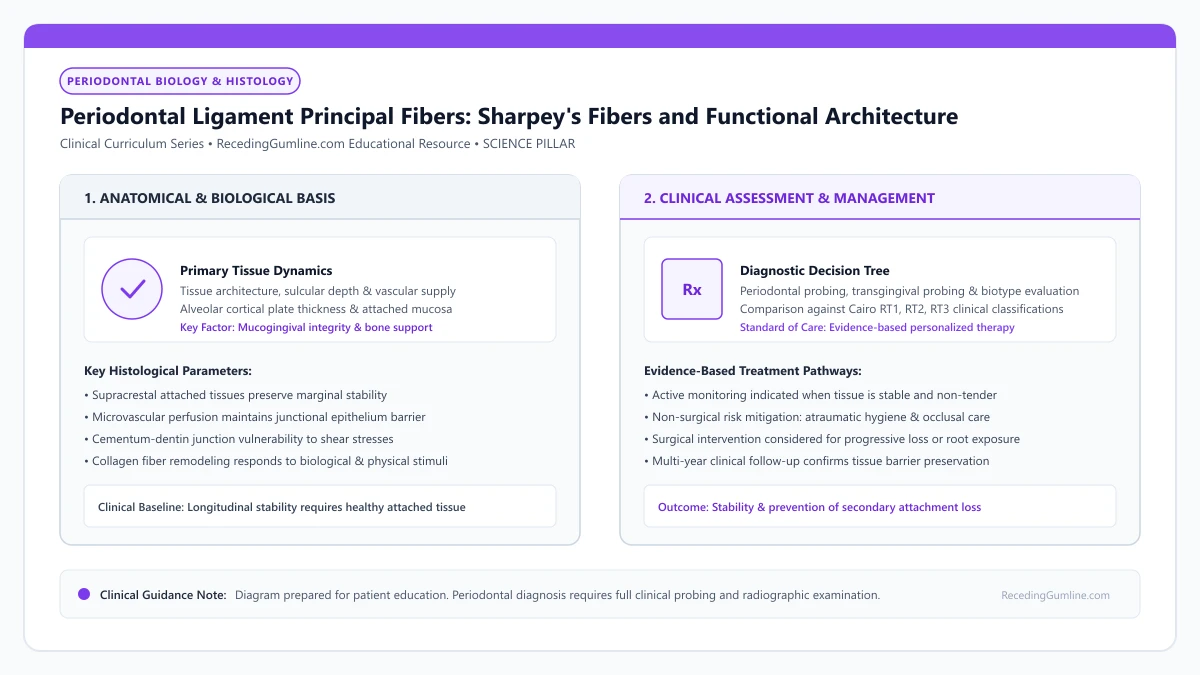
Educational illustration: Periodontal Ligament Principal Fibers: Sharpey's Fibers and Functional Architecture. Clinical management requires comprehensive periodontal evaluation rather than isolated self-assessment.
Source: RecedingGumline.com Clinical Editorial Team (Proprietary educational diagram for RecedingGumline.com)
Key Clinical Distinctions & Diagnostic Boundaries
- Gingival fibers attach gum tissue to the tooth neck (above bone); PDL fibers anchor the tooth root directly to alveolar bone (below bone).
- The PDL contains unmyelinated Ruffini-like mechanoreceptors that sense tiny biting pressures as small as 10 micrometers.
- When an implant is placed, it has zero PDL fibers, meaning it lacks shock absorption and proprioceptive "bite feel."
- Severed PDL fibers cannot reattach across exposed root surfaces without specialized regenerative surgical membranes or biomaterials.
The Functional Geometry: Five Principal Fiber Groups
The collagen fibers of the PDL are not arranged randomly; they are organized into five distinct anatomical groups designed to counter forces from every three-dimensional vector. The Alveolar Crest fibers extend from the cervical cementum downward to the alveolar crest, resisting lateral tilting forces.
Horizontal fibers run perpendicularly from cementum to bone to resist horizontal loads. Oblique fibers represent the largest and most powerful group: running diagonally upward from bone to cementum, they suspend the tooth like a hammock, converting heavy downward chewing shocks into tensile bone stimulation. The Apical fibers stabilize the root tip, while Interradicular fibers stabilize the furcation of multi-rooted molars.
The periodontal ligament is a complex, vascularized cellular connective tissue that surrounds the tooth root, connecting root cementum to the alveolar bone socket. Its principal collagen fibers are arranged in distinct anatomical groups: alveolar crest, horizontal, oblique, periapical, and interradicular bundles.
Clinical Considerations:
- Alveolar crest and horizontal fibers resist lateral tilting and rotational forces
- Oblique fibers form a diagonal hammock that converts vertical chewing into bone tension
- Apical and interradicular fibers stabilize root tips and multi-rooted molar furcations
Destruction in Periodontal Disease: The Coronal-to-Apical Unzipping
In periodontal disease and severe recession, tissue destruction follows an "unzipping" pattern from coronal to apical. Plaque-induced matrix metalloproteinases first degrade the alveolar crest and horizontal fibers.
As bone resorbs, the oblique fiber bundles are progressively severed. With fewer fibers remaining to suspend the tooth, occlusal loads place excessive mechanical stress on the remaining apical attachment, leading to progressive tooth hypermobility (looseness) and eventual tooth loss if clinical treatment is not rendered.
In active periodontal breakdown, matrix metalloproteinases enzymatically sever these principal fibers near the alveolar crest. The loss of ligamentous attachment eliminates the tensile suspension mechanism, accelerating alveolar bone resorption and progressive tooth hypermobility.
Clinical Considerations:
- Enzymes degrade fibers in a progressive coronal-to-apical direction
- Loss of oblique fibers destroys the tooth's shock-absorbing suspension hammock
- Excessive strain on remaining fibers produces tooth looseness and mobility
Principal Fiber Groups: From Alveolar Crest to Apical Bundles
The periodontal ligament (PDL) is dominated by dense, wavy bundles of Type I collagen organized into five distinct principal fiber groups: alveolar crest, horizontal, oblique, periapical, and interradicular fibers.
The alveolar crest and horizontal fibers reside in the most coronal portion of the PDL, directly beneath the junctional epithelium. These fibers are the first biological line of defense against lateral rotational forces and marginal bacterial invasion.
The oblique fibers constitute the largest group, running obliquely downward from alveolar bone to root cementum. This hammock-like configuration suspends the tooth within its osseous socket, converting vertical axial masticatory impact into lateral tensile strain on the alveolar bone.
Clinical Considerations:
- PDL collagen fibers are organized into five anatomically distinct principal bundle groups.
- Alveolar crest and horizontal fibers form the coronal defense barrier against mechanical displacement.
- Oblique fibers suspend the tooth like a biological hammock, absorbing vertical biting forces.
Sensory Proprioception & Neuromuscular Bite Regulation
In addition to its structural role, the periodontal ligament is a highly specialized sensory receptor organ. Mechanoreceptors (Ruffini-like endings) distributed throughout the PDL fibers detect minute physical displacements and directional loads as small as 1 to 3 grams.
These mechanoreceptors transmit rapid afferent impulses via the trigeminal nerve to the central nervous system, coordinating the jaw-opening reflex and regulating the intensity of masseter and temporalis muscle contraction.
When severe gum recession and bone loss destroy coronal PDL fibers, periodontal proprioception is diminished. The loss of sensory feedback can lead to uncoordinated bite forces and traumatic occlusal overload, accelerating further attachment loss.
Clinical Considerations:
- Ruffini-like mechanoreceptors in PDL fibers detect microscopic tooth movements as small as 1 gram.
- Sensory signals regulate masticatory muscle force and trigger protective jaw-opening reflexes.
- Loss of coronal PDL fibers impairs sensory proprioception, increasing vulnerability to bite trauma.
Clinical Reality Check
You can never regrow lost periodontal ligament fibers with toothpastes, oils, or home remedies; true ligament regeneration requires complex specialist surgical procedures using regenerative proteins or bone grafts.
Questions to Ask Your Periodontist or Dentist
- Are my periodontal ligament fibers showing signs of widening or damage on my x-rays?
- Has my gum recession severed the horizontal and oblique fiber bundles around my receded teeth?
- Are my teeth showing any measurable mobility resulting from lost ligament attachment?
- Can periodontal regenerative therapies (such as Emdogain or bone grafting) restore these fibers?
Unsure What Your Gum Changes Mean?
Take our free, evidence-based Gum Recession Assessment — approximately 3 minutes. Identify potential risk factors, evaluate symptoms, and receive personalized discussion questions for your dentist or periodontist.
Non-diagnostic educational triage. Private, secure, completed in your browser.
Related Educational Topics
Clinical Evidence & Claim Traceability (2 Mapped Assertions)
Scientific Literature & Clinical Guidelines
3sources · Hide ▲
- Pihlstrom BL, Michalowicz BS, Johnson NW (2005).
"Periodontal diseases." The Lancet.
Clinical relevance: Peer-reviewed Lancet seminar review synthesizing global periodontal epidemiology, microbial etiology, host immunopathology, and systemic interactions, emphasizing prevention, biofilm disruption, and early risk factor modification.
- Melcher AH (1976).
"On the repair potential of periodontal tissues." Journal of Periodontology.
Clinical relevance: Foundational biological treatise describing the four distinct cellular compartments during periodontal wound healing (lamina propria, periodontal ligament, bone, and cementum); established the biological premise of guided tissue regeneration by excluding rapidly migrating gingival epithelium to permit PDL cell repopulation.
- Gargiulo AW, Wentz FM, Orban B (1961).
"Dimensions and relations of the dentogingival junction in humans." Journal of Periodontology.Peer-Reviewed Study doi:10.1902/jop.1961.32.3.261
Clinical relevance: Foundational morphometric human autopsy investigation defining average histological dimensions of the dentogingival junction: sulcus depth of 0.69 mm, junctional epithelium of 0.97 mm, and supracrestal connective tissue attachment of 1.07 mm, establishing the biological concept of supracrestal tissue attachment.
Important Medical Notice
The contents of RecedingGumline.com, including text, graphics, self-assessment calculators, and other materials, are intended solely for educational and informational purposes. This content is not intended to replace professional dental examination, diagnosis, or treatment. Always seek the advice of a qualified dentist, periodontist, or other licensed oral healthcare provider with any questions you may have regarding a medical or dental condition. Never disregard professional medical advice or delay seeking it because of something you read on this website.