Signs Your Gum Recession Is Getting Worse: Subtle Indicators of Active Disease
How can you tell if your receding gums are actively progressing or stable?
Gingival recession is often an insidious, slow-moving process (see our receding gumline factors). Because tissue detachment typically occurs without acute pain, patients frequently assume their condition is stable when in fact active, destructive attachment loss is quietly progressing. When treating a receding gumline, recognizing the subtle clinical indicators of worsening recession empowers patients to seek timely intervention before teeth become loose.
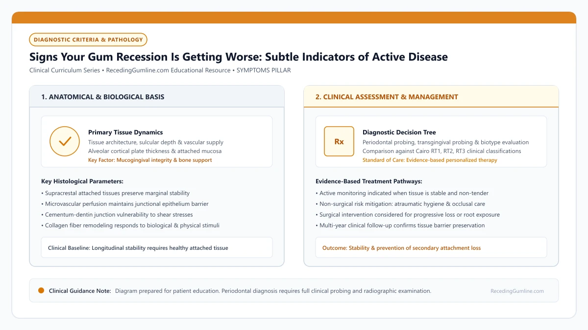
Educational illustration: Signs Your Gum Recession Is Getting Worse: Subtle Indicators of Active Disease. Clinical management requires comprehensive periodontal evaluation rather than isolated self-assessment.
Source: RecedingGumline.com Clinical Editorial Team (Proprietary educational diagram for RecedingGumline.com)
Key Clinical Distinctions & Diagnostic Boundaries
- Stable recession looks unchanged for years with firm, pink, non-bleeding margins; progressing recession displays shifting margins and recurring inflammation.
- Sudden onset of tooth sensitivity on a tooth that was previously comfortable is a prime indicator of recent marginal breakdown.
- Slight tooth mobility or shifting indicates advanced loss of periodontal attachment apparatus.
- Standardized periodontal probing charts comparing measurements across 6 to 12 months are the only definitive clinical proof of stability vs. progression.
Visual and Tactile Clues: The Expanding Root Margin
The most obvious sign of progressing recession is an expanding zone of exposed root. Root cementum has a distinct dull, yellowish, or darker hue compared to translucent, reflective white enamel. If you notice the boundary line (the cementoenamel junction) sitting further away from the gumline in comparison to older photos, active recession is occurring.
Another tactile indicator is running your fingernail or dental floss along the margin. If you detect a new ledge, groove, or notch that was not present six months ago, mechanical brushing friction or tooth flexure (abfraction) is actively carving into your newly exposed root dentin.
Subtle visual warning signs of active recession progression include increasing root exposure, loosening of previously tight crown margins, and deepening cervical notches. Patients may notice teeth appearing progressively longer or feel a distinct sharp ridge along the gumline with their tongue.
Clinical Considerations:
- Expanding yellow or darker root surface visible below the white crown enamel
- Comparison with baseline intraoral photographs reveals apical margin movement
- Emergence of new cervical ledges or grooves detectable by touch or dental floss
Sensory and Inflammatory Signals: Sensitivity and Bleeding
Sensory changes frequently announce recent tissue migration. When gum tissue recedes to expose fresh dentin that has never been exposed to saliva, the unoccluded dentinal tubules react violently to cold drinks or toothbrush contact. A sudden flare-up of sharp sensitivity on a specific tooth strongly suggests recent tissue loss.
Concurrently, pay close attention to your dental floss. If flossing around a receded tooth consistently draws blood or reveals pink saliva, the sulcular epithelial lining is ulcerated. Bleeding on probing (BOP) is a definitive biological sign that inflammatory cytokines are actively breaking down connective tissue fibers.
Regular clinical documentation tracking clinical attachment levels in millimeters provides definitive scientific evidence of progression. An increase of 1 mm or more in attachment loss over consecutive annual exams signals an active, uncontrolled pathological process requiring immediate intervention.
Clinical Considerations:
- Sudden onset of sharp cold sensitivity indicates freshly exposed, unoccluded tubules
- Bleeding when flossing signals active inflammatory breakdown of junctional epithelium
- Swelling, redness, or rolled tissue margins indicate active chronic periodontitis
Objective Clinical Verification: The 6-Month Probing Comparison
While self-checks are valuable, the definitive clinical test of progression is comparative periodontal charting. At every dental checkup, your hygienist or dentist records millimeter measurements of probing depth and recession distance for every tooth.
If your clinical attachment level (CAL) increases by 1 to 2 millimeters between visits, active disease progression is confirmed. This quantitative jump serves as the decisive trigger for clinical intervention—indicating that conservative monitoring has failed and that scaling, grafting, or bite adjustment is immediately required.
Ignoring progressive recession warning signs can lead to extensive alveolar bone loss, tooth mobility, and secondary root canal infections as the recession reaches lateral canals. Early periodontal consultation allows conservative intervention before complex regenerative surgeries are needed.
Clinical Considerations:
- Sequential periodontal probing charts provide definitive quantitative proof of progression
- An increase in CAL ≥ 1 to 2 mm confirms active, destructive attachment loss
- Triggers immediate transition from conservative monitoring to active clinical intervention
Clinical Surveillance: CEJ-to-Margin Probing & 3D Optical Scanning
Accurately identifying active progression in gum recession requires systematic clinical measurement rather than visual estimation. Periodontists measure the distance from the fixed, anatomical cementoenamel junction (CEJ) to the crest of the soft-tissue margin using a calibrated periodontal probe.
Recording this measurement at four- to six-month intervals provides an objective longitudinal record. An increase in recession depth of 1.0 mm or more between visits confirms active attachment loss.
Modern practices increasingly utilize intraoral 3D optical scanners. By superimposing digital surface meshes captured at different appointments, specialized software computes volumetric color-coded heat maps that detect marginal loss as subtle as 0.1 millimeters, alerting clinicians to active recession long before it becomes clinically obvious.
Clinical Considerations:
- Calibrated probing from the CEJ to the margin establishes an objective longitudinal record.
- A 1.0 mm increase in recession depth between recalls confirms active tissue breakdown.
- 3D intraoral scan superimposition detects micro-volumetric tissue loss as small as 0.1 mm.
The Clinical Tipping Point: When Conservative Monitoring Transitions to Surgery
A critical decision in periodontal therapy is identifying the exact "tipping point" where conservative monitoring is no longer appropriate and surgical intervention becomes mandatory. Structured clinical criteria guide this determination.
Surgery is indicated when: (1) sequential recall visits document progressive apical movement of the margin; (2) the remaining band of attached keratinized tissue drops below 1.0 mm, creating an unstable mucosal margin; or (3) root exposure causes debilitating thermal sensitivity that resists desensitizing agents.
Intervening at this tipping point—while the interproximal bone remains intact (Cairo RT1)—guarantees the highest probability of 100% root coverage and prevents the defect from degenerating into an irreversible RT2 or RT3 condition.
Clinical Considerations:
- Documented progressive apical movement is a primary indicator for surgical intervention.
- A remaining keratinized tissue band under 1.0 mm creates an unstable, vulnerable margin.
- Surgical intervention while interdental bone is intact (RT1) ensures optimal coverage predictability.
Clinical Reality Check
Recession does not progress at a constant, steady speed; it often remains quiet for months and then progresses in rapid bursts during periods of high stress, clenching, or aggressive brushing.
Questions to Ask Your Periodontist or Dentist
- How do my current recession and probing depth measurements compare to my chart from one year ago?
- Are there specific teeth showing active clinical attachment loss since my last checkup?
- Can you take standardized intraoral photographs today to establish an exact visual baseline?
- Is my recession currently at a stage where a gum graft is recommended, or is it safe to continue monitoring?
Unsure What Your Gum Changes Mean?
Take our free, evidence-based Gum Recession Assessment — approximately 3 minutes. Identify potential risk factors, evaluate symptoms, and receive personalized discussion questions for your dentist or periodontist.
Non-diagnostic educational triage. Private, secure, completed in your browser.
Related Educational Topics
Clinical Evidence & Claim Traceability (2 Mapped Assertions)
Scientific Literature & Clinical Guidelines
3sources · Hide ▲
- Tonetti MS, Greenwell H, Kornman KS (2018).
"Staging and grading of periodontitis: Framework and proposal of a new classification and case definition." Journal of Clinical Periodontology.
Clinical relevance: Consensus framework establishing the multidimensional staging (severity and extent of periodontal tissue breakdown) and grading (biological rate of disease progression, incorporating smoking and diabetes as grade modifiers) for periodontitis. It addresses periodontitis diagnosis and staging, not the classification of localized gingival recession defects.
- Cairo F, Nieri M, Cincinelli S, Mervelt J, Pagliaro U (2011).
"The interproximal clinical attachment level to classify gingival recessions and predict root coverage outcomes: an explorative and reliability study." Journal of Clinical Periodontology.
Clinical relevance: Exploratory and reliability study establishing the Cairo classification based on interdental clinical attachment level (CAL): RT1 (no interproximal attachment loss; complete root coverage is clinically predictable), RT2 (interproximal attachment loss <= buccal loss; partial coverage predictable), and RT3 (interproximal loss exceeds buccal recession; complete coverage not predictable).
- Jepsen S, Caton JG, Albandar JM, Bissada NF, Bouchard P, Cortellini P, et al. (2018).
"Periodontal manifestations of systemic diseases and developmental and acquired conditions: Consensus report of workgroup 3 of the 2017 World Workshop on the Classification of Periodontal and Peri-Implant Diseases and Conditions." Journal of Periodontology.
Clinical relevance: Consensus report defining mucogingival conditions, gingival phenotype (replacing biotype), non-carious cervical lesions, and the multifactorial etiology of gingival recession; emphasizes that recession can occur without periodontitis and classifies recession by interdental clinical attachment loss.
Important Medical Notice
The contents of RecedingGumline.com, including text, graphics, self-assessment calculators, and other materials, are intended solely for educational and informational purposes. This content is not intended to replace professional dental examination, diagnosis, or treatment. Always seek the advice of a qualified dentist, periodontist, or other licensed oral healthcare provider with any questions you may have regarding a medical or dental condition. Never disregard professional medical advice or delay seeking it because of something you read on this website.