Receding Gums on Front Teeth: Aesthetic Impact, Causes & Conservative vs Surgical Care
How can you fix receding gums on your front teeth and achieve a completely natural cosmetic result?
Receding gums on the front teeth (the maxillary anterior "smile zone") carries intense cosmetic, emotional, and psychological weight for patients experiencing a receding gumline. Unlike recession on hidden molar teeth, front-tooth recession is immediately visible when smiling, laughing, or speaking. Understanding the unique aesthetic dynamics, high surgical root coverage predictability, and conservative options helps patients restore smile harmony.
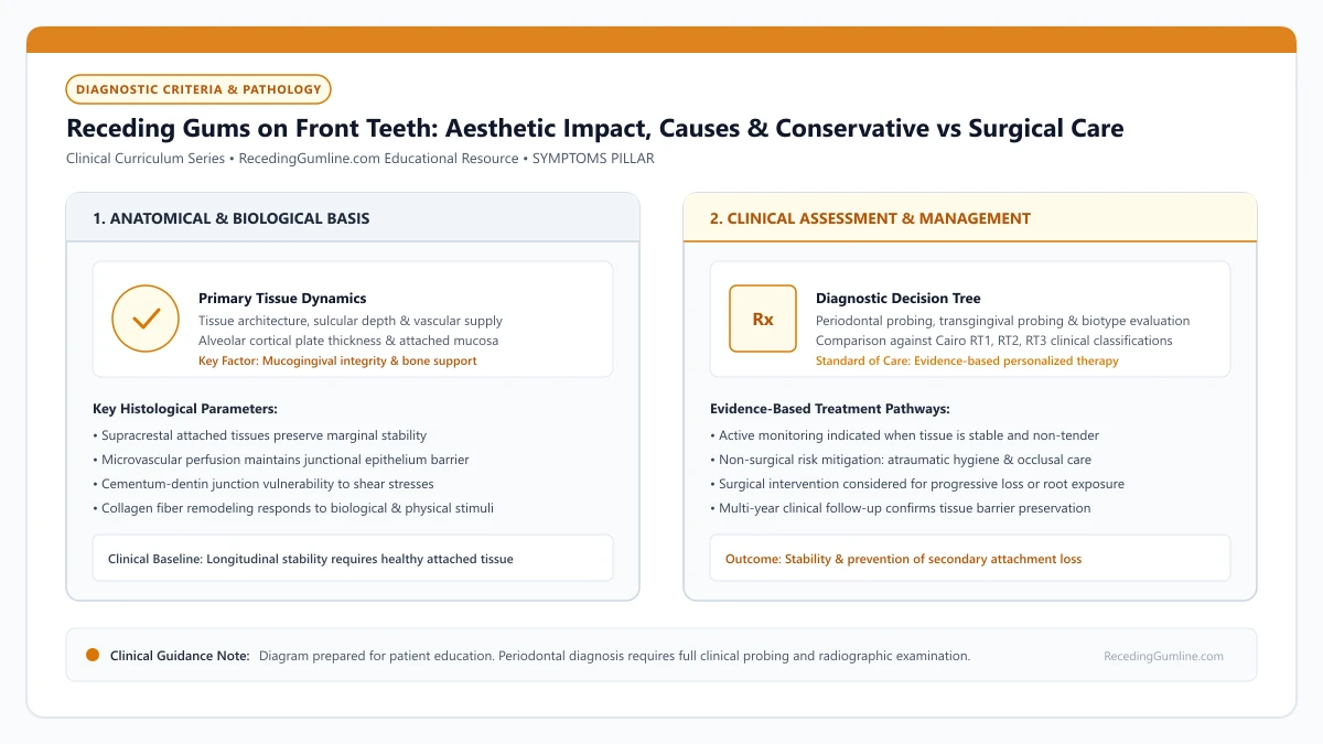
Educational illustration: Receding Gums on Front Teeth: Aesthetic Impact, Causes & Conservative vs Surgical Care. Clinical management requires comprehensive periodontal evaluation rather than isolated self-assessment.
Source: RecedingGumline.com Clinical Editorial Team (Proprietary educational diagram for RecedingGumline.com)
Key Clinical Distinctions & Diagnostic Boundaries
- Free gingival grafts are contraindicated on upper front teeth due to a pale, patchy cosmetic appearance; connective tissue grafts or tunneling are standard of care.
- Aesthetic smile evaluation requires assessing the patient's lip line (high vs. low smile line) and gingival symmetry.
- Recession on a single central incisor creates prominent gingival margin asymmetry that is visually distracting.
- Root coverage on front teeth requires pristine microsurgical suturing to prevent visible scarring or keloid tissue formation.
Aesthetic Dynamics: High Lip Lines and Gingival Zenith Symmetry
In dental aesthetics, the gingival zenith is the most apical point of the gumline contour on each tooth. Under ideal symmetry, the zeniths of the two central incisors should be at the exact same height, with the lateral incisors sitting about 1 mm lower, and the canines matching the central incisors.
When recession strikes a front tooth—even by just 1 to 2 millimeters—this balance is destroyed. In patients with a high smile line (showing gums when smiling), the receded tooth looks noticeably longer and darker, creating an asymmetrical, prematurely aged appearance.
Gum recession on visible maxillary incisors creates significant aesthetic distress, often described by patients as elongated or uneven teeth. The loss of symmetrical gingival scallop contours can compromise the natural harmony of the smile line during social interactions.
Clinical Considerations:
- Gingival zenith symmetry is essential for a harmonious, balanced smile
- Recession of even 1 to 2 mm on an incisor creates prominent visual asymmetry
- Patients with high smile lines experience the greatest cosmetic and emotional distress
High Surgical Predictability: Why Front Teeth Heal Exceptionally Well
Fortunately, front teeth are anatomically favorable candidates for surgical repair. Because front-tooth recession is frequently caused by toothbrush abrasion or orthodontic alignment rather than destructive periodontitis, the interdental bone between the teeth is usually 100% intact (Cairo RT1).
When interdental bone and papillae are intact, periodontists can predictably achieve 90% to 100% complete root coverage. The intact lateral papillae provide robust collateral blood supply that nourishes the newly transplanted connective tissue graft as it heals across the root.
Anterior recession defects are frequently accompanied by localized root prominence and paper-thin labial cortical bone plates. Even minor episodes of aggressive flossing or vigorous brushing can rapidly shear through the delicate anterior attached tissue collar.
Clinical Considerations:
- Front teeth usually have intact interdental bone (Cairo RT1), maximizing coverage success
- Collateral blood supply from adjacent papillae nourishes the transplanted tissue
- Achieving 100% complete root coverage is realistic and clinically routine in RT1 cases
Aesthetic Techniques: Connective Tissue Grafts and Tunneling
In the smile zone, choice of surgical technique is critical. Free gingival grafts are strictly avoided because they heal with a pale, whitish "patch" appearance. Instead, periodontists use subepithelial connective tissue grafts (SCTG) or allograft matrices.
Surgeons often employ minimally invasive tunneling techniques (such as the VISTA procedure). Instead of cutting open the gums with visible vertical incisions, the surgeon creates a microscopic tunnel beneath the gums from a hidden access point, slides the graft into place, and anchors it with delicate sutures, leaving zero visible scars.
Surgical root coverage utilizing coronally advanced flaps combined with subepithelial connective tissue grafting achieves high aesthetic predictability in anterior sites. Restoring natural tissue thickness and harmonious marginal contours protects anterior roots from progressive exposure.
Clinical Considerations:
- Subepithelial connective tissue grafts blend seamlessly with natural gum color and texture
- Tunneling techniques (VISTA) eliminate vertical surface incisions and scarring
- Micro-sutures ensure invisible healing and smooth, natural tissue margins
Aesthetic Zone Dynamics: High Smile Lines & Papillary Scalloping
Gingival recession occurring in the aesthetic zone (maxillary incisors and canines) carries profound cosmetic and psychological implications. Patients with a "high smile line" expose several millimeters of gingival architecture during natural speech and smiling, making even 1.0 mm of localized margin retraction immediately conspicuous.
Anterior teeth exhibit pronounced architectural scalloping, where delicate interdental papillae rise several millimeters above the facial margin. The underlying facial bone plate over central and lateral incisors is exceptionally thin—often less than 0.5 mm thick.
When recession develops in the aesthetic zone, preserving the height and vascular integrity of the adjacent interdental papillae is paramount. Loss of papillae produces unsightly "black triangles" that are far more difficult to reconstruct surgically than the facial recession itself.
Clinical Considerations:
- High smile lines expose anterior gingival margins, making minor recession highly noticeable.
- Anterior incisors possess delicate, highly scalloped margins over paper-thin facial bone.
- Preserving interdental papillae is critical to prevent cosmetic "black triangle" defects.
Minimally Invasive Tunneling Approaches for Aesthetic Root Coverage
In the anterior aesthetic zone, conventional periodontal flap designs that utilize vertical releasing incisions carry a high risk of visible scar tissue formation and compromised blood supply to the papillae. Modern aesthetic periodontics relies on the minimally invasive tunneling technique (tunnel flap).
Through microscopic intrasulcular incisions, the periodontist undermines the facial soft tissue, creating a continuous subgingival tunnel connecting adjacent teeth without incising the interdental papillae. A subepithelial connective tissue graft is guided through this tunnel and positioned over the denuded roots.
The entire mucosal complex is advanced coronally and anchored with micro-sutures suspended around tooth contacts. This flapless tunneling approach preserves all native blood vessels, avoids scarring, and yields natural, harmonious aesthetic contours.
Clinical Considerations:
- Vertical releasing incisions in the aesthetic zone risk unsightly permanent scar tissue.
- The tunneling technique undermines tissues intrasulcularly without incising interdental papillae.
- Tunnel connective tissue grafting maximizes blood supply and produces natural cosmetic contours.
Clinical Reality Check
Never allow a surgeon to place a free gingival graft on an upper front tooth; while it stops recession, it leaves a permanent, highly visible whitish cosmetic patch in your smile.
Questions to Ask Your Periodontist or Dentist
- Is my front-tooth recession classified as a Cairo RT1 defect with 100% coverage potential?
- Do you recommend a coronally advanced flap, a tunneling technique, or a pinhole procedure for my smile?
- Will the grafted tissue blend perfectly with my natural gum color and texture?
- What temporary modifications should I make to my speech, chewing, and brushing during front-tooth healing?
Unsure What Your Gum Changes Mean?
Take our free, evidence-based Gum Recession Assessment — approximately 3 minutes. Identify potential risk factors, evaluate symptoms, and receive personalized discussion questions for your dentist or periodontist.
Non-diagnostic educational triage. Private, secure, completed in your browser.
Related Educational Topics
Clinical Evidence & Claim Traceability (3 Mapped Assertions)
Scientific Literature & Clinical Guidelines
3sources · Hide ▲
- Chambrone L, Salinas Ortega MA, Sukekava F, Rotundo R, Kalemaj Z, Buti J, Pini Prato GP (2018).
"Root coverage procedures for treating localised and multiple recession-type defects." Cochrane Database of Systematic Reviews.
Clinical relevance: Cochrane systematic review evaluating root-coverage procedures for localized and multiple recession-type defects. While subepithelial connective tissue grafts (SCTG) combined with coronally advanced flaps demonstrated higher rates of complete root coverage and keratinized tissue gain compared to flap advancement alone, evidence quality varied across outcomes and clinical decisions must balance donor site morbidity and patient-reported outcomes.
- Cairo F, Nieri M, Cincinelli S, Mervelt J, Pagliaro U (2011).
"The interproximal clinical attachment level to classify gingival recessions and predict root coverage outcomes: an explorative and reliability study." Journal of Clinical Periodontology.
Clinical relevance: Exploratory and reliability study establishing the Cairo classification based on interdental clinical attachment level (CAL): RT1 (no interproximal attachment loss; complete root coverage is clinically predictable), RT2 (interproximal attachment loss <= buccal loss; partial coverage predictable), and RT3 (interproximal loss exceeds buccal recession; complete coverage not predictable).
- Zucchelli G, Mounssif I (2015).
"Periodontal plastic surgery." Periodontology 2000.
Clinical relevance: Peer-reviewed review of periodontal plastic surgery modalities, detailing flap design, coronally advanced flaps, autogenous connective tissue grafting, tunneling techniques, and anatomical factors governing aesthetic and functional root coverage.
Important Medical Notice
The contents of RecedingGumline.com, including text, graphics, self-assessment calculators, and other materials, are intended solely for educational and informational purposes. This content is not intended to replace professional dental examination, diagnosis, or treatment. Always seek the advice of a qualified dentist, periodontist, or other licensed oral healthcare provider with any questions you may have regarding a medical or dental condition. Never disregard professional medical advice or delay seeking it because of something you read on this website.