Receding Gums on Lower Front Teeth: Thin Cortical Plate, Calculus & Frenum Factors
Why is gum recession so severe and common on lower front teeth, and how is it treated?
The mandibular central and lateral incisors (lower front teeth) are the single most common site for advanced, progressive receding gums in the human mouth. Positioned where salivary glands deposit mineralized tartar and where facial lip muscles exert continuous traction, these small, delicate teeth possess paper-thin bone plates that make them extraordinarily susceptible to attachment loss.
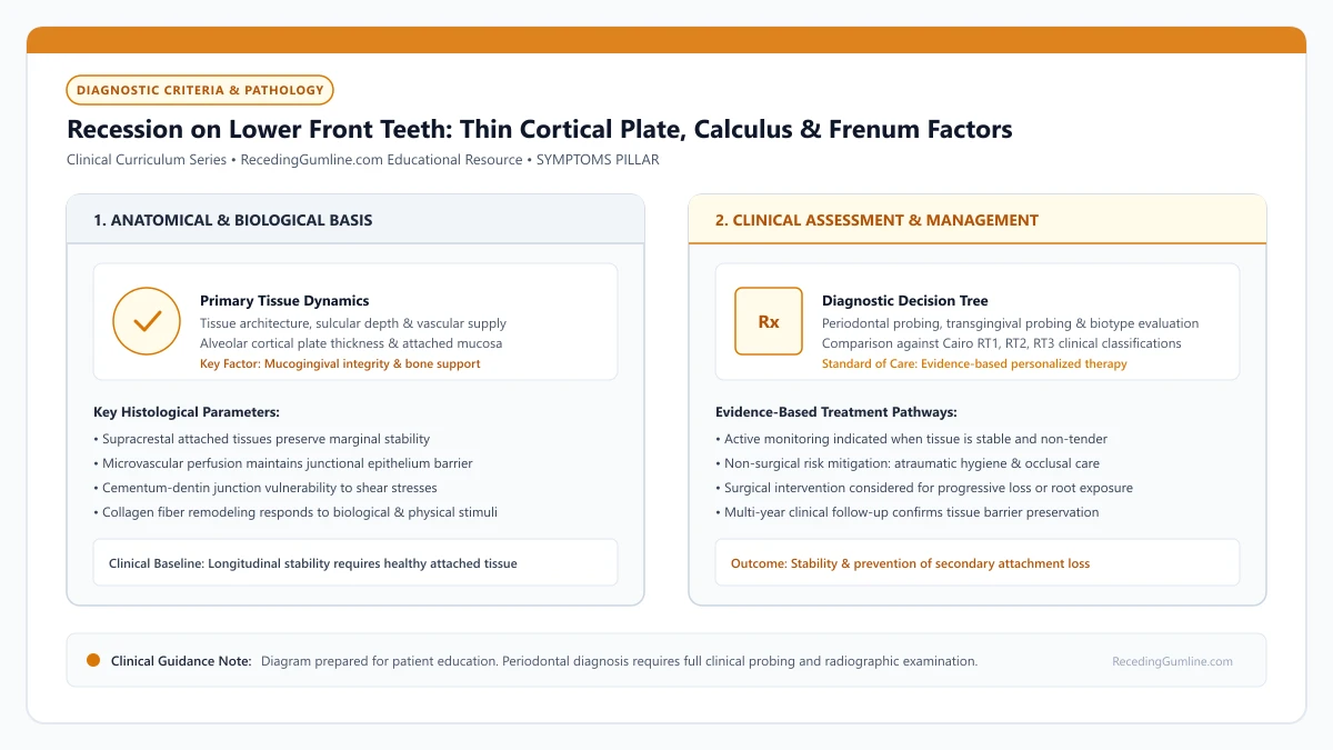
Educational illustration: Recession on Lower Front Teeth: Thin Cortical Plate, Calculus & Frenum Factors. Clinical management requires comprehensive periodontal evaluation rather than isolated self-assessment.
Source: RecedingGumline.com Clinical Editorial Team (Proprietary educational diagram for RecedingGumline.com)
Key Clinical Distinctions & Diagnostic Boundaries
- Lower incisor recession is functional rather than purely aesthetic; without treatment, teeth can lose bony anchorage and become mobile.
- Free gingival grafts are widely accepted on lower incisors because the pale tissue appearance is hidden behind the lower lip.
- Calculus buildup along the lingual and interproximal surfaces acts as a continuous microbial reservoir driving bone loss.
- Tight lower lip frenums require concurrent surgical release (frenectomy) when soft-tissue grafting is performed.
The Tripartite Vulnerability: Bone, Frenum & Saliva
Lower front teeth face a "perfect storm" of biological vulnerabilities. First, the facial cortical bone plate over mandibular incisors is razor-thin—often under 0.3 mm—and frequently contains developmental dehiscences caused by crowded eruption.
Second, the lower labial frenum and mentalis muscle insert directly into the movable mucosa near the tooth margin, pulling on the gums every time you talk or chew. Third, the salivary ducts (Wharton's and Rivinus' ducts) empty directly behind these teeth, saturating the area with calcium and phosphate that turns soft plaque into rock-hard calculus within 48 hours.
Mandibular anterior incisors possess the thinnest alveolar bone plates and the narrowest bands of keratinized gingiva in the entire human dentition. Due to gravity and salivary pooling, dense subgingival calculus forms rapidly on the lingual surfaces of lower incisors, driving localized inflammation.
Clinical Considerations:
- Cortical bone over lower incisors is the thinnest in the mouth (< 0.3 mm)
- Lower labial frenum and mentalis muscle fibers exert direct downward pull
- Sublingual salivary ducts promote rapid calculus deposition that harbors bacteria
Clinical Consequences: From Root Exposure to Tooth Mobility
Because mandibular incisor roots are exceptionally narrow and short compared to molars or canines, losing 4 or 5 millimeters of attachment represents a catastrophic loss of total periodontal support. As recession extends through the narrow band of attached gingiva into the alveolar mucosa, the tissue margin separates from the root.
Once bacterial plaque penetrates the apical third of the root, alveolar bone support is lost circumferentially. The tooth begins to exhibit Class I or Class II mobility (looseness), signaling imminent tooth loss if clinical intervention is not promptly rendered.
High lingual and labial frenum attachments frequently insert directly into the marginal gingiva of lower central incisors. Continuous muscular pull from the lower lip combined with thin bone dehiscences can cause rapid, deep recession within a matter of months.
Clinical Considerations:
- Narrow incisor roots have less total surface area; 4 mm of recession represents over 40% attachment loss
- Loss of attached tissue allows movable mucosa to pull away, creating deep pockets
- Progressive bone loss leads to tooth looseness (mobility) and eventual extraction
Surgical Stabilization: Creating an Impenetrable Tissue Band
The primary treatment objective on lower front teeth is not cosmetic beauty, but structural survival. The gold-standard treatment is the Free Gingival Graft (FGG). The surgeon takes a dense, keratinized tissue graft from the palate and stitches it at the base of the receded incisors.
This graft heals into a wide, tough, thick band of attached gingiva that buffers the tooth against muscle pull and toothbrush friction. Because the lower lip covers these teeth during smiling, the slightly paler cosmetic appearance of a free gingival graft is completely hidden, making it the ideal functional solution.
Periodontists often perform free gingival grafts or connective tissue grafts on lower incisors to create a robust collar of dense keratinized tissue. Thickening this tissue band halts progressive mucogingival migration and protects fragile mandibular teeth from premature loss.
Clinical Considerations:
- Free gingival graft creates a dense, thick collar of attached keratinized tissue
- Buffers the delicate tooth margins against downward mentalis muscle traction
- Pale tissue appearance is hidden behind the lower lip, making FGG the ideal choice
Anatomical Vulnerability: Paper-Thin Cortical Plates & Shallow Vestibules
Mandibular incisors represent the single most common site for progressive gingival recession in the human dentition. This extreme vulnerability is the direct consequence of unique regional anatomy.
The labial cortical bone plate covering lower incisor roots is naturally paper-thin and frequently features developmental fenestrations and dehiscences. Furthermore, the anterior mandibular vestibule is characteristically shallow, and the insertion of the mentalis muscle and inferior labial frenum is positioned immediately adjacent to the gingival margin.
Every time the patient speaks, chews, or swallows, dynamic muscle contraction pulls on the delicate mucosal margin. In the absence of a wide band of thick attached gingiva, this perpetual muscular pull causes rapid apical stripping of the gum tissue.
Clinical Considerations:
- Lower incisors are the most common site for progressive recession due to anatomical constraints.
- Labial cortical bone over mandibular incisors is paper-thin or congenitally absent.
- Shallow vestibular depth and mentalis muscle pull exert continuous mechanical tension on the margin.
Surgical Decision: Free Gingival Grafting vs. Tunnel CTG in the Mandible
Selecting the appropriate surgical modality for lower incisor recession depends on whether the primary clinical goal is functional tissue augmentation or cosmetic root coverage. Because lower incisors are rarely visible during normal smiling, functional stabilization is typically the primary objective.
When the zone of keratinized tissue is completely absent and high muscle tension is present, the Free Gingival Graft (FGG) is the treatment of choice. The FGG creates a dense, fibrous, immovable band of attached tissue that permanently resists muscle pull, effectively deepening the vestibule and halting progression.
However, if the patient exposes lower incisors during speech and demands aesthetic root coverage, a coronally advanced tunnel graft combined with a connective tissue graft may be considered, provided interdental bone is fully preserved (Cairo RT1).
Clinical Considerations:
- Functional tissue stabilization is the primary goal for lower incisors due to low aesthetic visibility.
- Free gingival grafts widen keratinized tissue, deepen the vestibule, and permanently resist muscle pull.
- Tunnel connective tissue grafting is reserved for cases requiring aesthetic root coverage.
Clinical Reality Check
Do not wait for lower front teeth to become loose before seeking care; once severe mobility occurs, bone loss is usually too advanced for soft-tissue grafting to save the tooth.
Questions to Ask Your Periodontist or Dentist
- How many millimeters of attached keratinized tissue remain on my lower front teeth?
- Are my lower incisors showing any measurable mobility or bone loss between the roots?
- Do you recommend a free gingival graft or a connective tissue graft to stabilize these teeth?
- Is my lower lip frenum pulling on the receded margin, and should it be released with a frenectomy?
Unsure What Your Gum Changes Mean?
Take our free, evidence-based Gum Recession Assessment — approximately 3 minutes. Identify potential risk factors, evaluate symptoms, and receive personalized discussion questions for your dentist or periodontist.
Non-diagnostic educational triage. Private, secure, completed in your browser.
Related Educational Topics
Clinical Evidence & Claim Traceability (3 Mapped Assertions)
Scientific Literature & Clinical Guidelines
3sources · Hide ▲
- Lang NP, Löe H (1972).
"The relationship between the width of keratinized gingiva and gingival health." Journal of Periodontology.
Clinical relevance: Landmark clinical investigation observing that gingival sites with less than 2 mm of keratinized gingiva (corresponding to less than 1 mm of attached gingiva) frequently exhibited clinical signs of persistent marginal inflammation despite plaque control, historically establishing the 2 mm keratinized tissue reference point.
- Kassab MM, Cohen RE (2003).
"The etiology and prevalence of gingival recession." The Journal of the American Dental Association.
Clinical relevance: Comprehensive epidemiological and etiological review establishing that gingival recession affects more than 50% of adults aged 18 to 64 and over 88% of individuals aged 65 and older, driven by interactions between predisposing anatomical factors and precipitating mechanical or inflammatory stimuli.
- Jepsen S, Caton JG, Albandar JM, Bissada NF, Bouchard P, Cortellini P, et al. (2018).
"Periodontal manifestations of systemic diseases and developmental and acquired conditions: Consensus report of workgroup 3 of the 2017 World Workshop on the Classification of Periodontal and Peri-Implant Diseases and Conditions." Journal of Periodontology.
Clinical relevance: Consensus report defining mucogingival conditions, gingival phenotype (replacing biotype), non-carious cervical lesions, and the multifactorial etiology of gingival recession; emphasizes that recession can occur without periodontitis and classifies recession by interdental clinical attachment loss.
Important Medical Notice
The contents of RecedingGumline.com, including text, graphics, self-assessment calculators, and other materials, are intended solely for educational and informational purposes. This content is not intended to replace professional dental examination, diagnosis, or treatment. Always seek the advice of a qualified dentist, periodontist, or other licensed oral healthcare provider with any questions you may have regarding a medical or dental condition. Never disregard professional medical advice or delay seeking it because of something you read on this website.