Frenectomy and Gum Recession: When Muscle Attachment Release Is Indicated
What is a frenectomy, when is it necessary for receding gums, and does it restore lost tissue?
A frenum is a fold of mucous membrane containing connective tissue and muscle fibers that connects the lips or cheeks to the alveolar mucosa and periosteum. When a frenum inserts too close to the marginal gingiva—a condition known as a high frenal attachment—normal lip movements generate mechanical tension that pulls the gumline away from the tooth, accelerating gum recession overview.
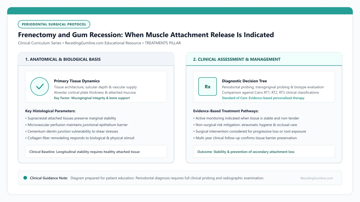
Educational illustration: Frenectomy and Gum Recession: When Muscle Attachment Release Is Indicated. Clinical management requires comprehensive periodontal evaluation rather than isolated self-assessment.
Source: RecedingGumline.com Clinical Editorial Team (Proprietary educational diagram for RecedingGumline.com)
Key Clinical Distinctions & Diagnostic Boundaries
- Frenectomy eliminates the mechanical pull causing recession, but cannot by itself restore soft tissue coverage over exposed root surfaces.
- A frenotomy involves simple incision and release, whereas a complete frenectomy excises the entire fibrous band and deep periosteal fibers.
- Frenectomies can be performed using traditional surgical scalpels, electrosurgery, or specialized soft-tissue dental lasers.
- On lower incisors, a frenectomy is frequently performed concurrently with a free gingival graft or connective tissue graft.
Biomechanics of Frenal Tension: The Blanching Test
In a healthy mouth, the labial frenum attaches comfortably within the alveolar mucosa several millimeters apical to the attached gingiva. However, when the frenum attaches directly into the marginal gingiva or interdental papilla, facial muscle actions—such as talking, smiling, or chewing—transmit physical tension directly to the tooth margin.
Clinicians evaluate this mechanical tension using the "blanching test." The clinician gently retracts the lip outward and upward. If the marginal tissue around the tooth exhibits ischemic blanching or separates from the root surface, pathological frenal pull is identified.
A high labial frenum attachment exerts continuous tensile traction on the marginal gingiva every time the patient talks, smiles, or chews. This persistent dynamic pulling separates the junctional epithelium from the root surface, creating an anatomical pathway for rapid recession.
Clinical Considerations:
- Healthy frenal attachments sit well below the zone of attached keratinized tissue
- High frenal insertions transmit direct tensile pull to the delicate marginal collar
- Blanching test demonstrates dynamic tissue ischemia and margin retraction
Surgical Techniques: Scalpel, Electrosurgery & Laser Excision
A frenectomy is a minor outpatient procedure performed under local anesthesia. The clinician can choose from three primary surgical modalities: traditional scalpel excision, electrosurgery, or soft-tissue dental laser (diode, Nd:YAG, or CO2).
Traditional scalpel excision allows precise removal of deep fibrous attachments down to the periosteum followed by fine sutures. Laser frenectomies offer the advantage of instantaneous coagulation, minimal bleeding, and often eliminate the need for sutures, resulting in faster post-operative recovery.
Soft-tissue diode or carbon dioxide lasers provide efficient frenum excision with minimal intraoperative bleeding and reduced post-operative edema compared to conventional cold scalpel techniques. Laser coagulation eliminates the need for extensive sutures, improving patient comfort.
Clinical Considerations:
- Scalpel excision removes deep fibrous and muscle attachments with suture closure
- Soft-tissue lasers vaporize tissue with instant hemostasis and minimal swelling
- The entire fibrous band must be released down to the bone to prevent re-attachment
Why a Frenectomy Alone Does Not Restore Receded Gums: Combined Protocols
Patients sometimes assume that having a frenectomy will cause receded gum tissue to regenerate spontaneously. A frenectomy merely eliminates the offending pulling force; it cannot regenerate lost bone or reconstruct attached tissue over an already exposed root.
For this reason, periodontists frequently perform a frenectomy simultaneously with a soft-tissue graft (such as a free gingival graft or coronally advanced flap). By releasing the muscle pull and placing a graft in the same surgical session, the clinician both removes the root cause and repairs the anatomical defect.
A frenectomy alone cannot restore lost gingival tissue or cover exposed cementum; it merely halts ongoing mechanical traction. In cases where significant recession has already occurred, the frenectomy is staged simultaneously or sequentially with a connective tissue graft.
Clinical Considerations:
- Frenectomy removes the pulling force but cannot replace lost attached tissue
- Combining frenectomy with a gum graft provides comprehensive structural repair
- Healing requires 2 to 3 weeks of soft food and gentle oral hygiene
Surgical Modalities: Conventional Scalpel vs. Diode & CO2 Lasers
Surgically releasing an aberrant labial frenum can be executed via conventional scalpel excision or soft-tissue laser ablation (diode, Nd:YAG, or CO2 lasers). The conventional scalpel technique involves diamond-shaped excision of the fibrous band down to the periosteum, followed by muscle fiber undermining and suturing.
While scalpel frenectomy is highly effective, soft-tissue lasers offer substantial clinical advantages. The laser energy simultaneously vaporizes the collagenous band and seals micro-capillaries and nerve endings, providing a bloodless operative field, zero requirement for sutures, and dramatically reduced postoperative swelling.
Regardless of the instrument used, the critical surgical objective is complete disinsertion of the muscular fibers (transverse fibers of the orbicularis oris) anchored into the periosteum above the marginal gingiva.
Clinical Considerations:
- Conventional scalpel excision requires surgical undermining and suture placement.
- Soft-tissue lasers vaporize fibrous bands while coagulating vessels, eliminating sutures and bleeding.
- Complete release of deep muscular insertions into the periosteum is essential to prevent recurrence.
Preventing Relapse: Muscle Retraining & Cicatrization Management
A significant complication following frenectomy is fibrous cicatrization and muscle re-attachment. As secondary intention healing occurs, scar tissue contracture can pull the mucosal margins back toward their original cervical position, re-establishing pathogenic tension on the gumline.
To prevent relapse, periodontists prescribe structured lip mobilization exercises beginning 48 hours postoperatively. Patients are instructed to pull the lip outward and upward several times daily for four weeks to stretch healing connective tissue fibers and encourage healing in an extended, apical orientation.
When frenectomy is combined with connective tissue grafting, the apical relocation of the frenal attachment must be secured with deep non-resorbable periosteal sutures to ensure the muscular attachment remains permanently positioned within the vestibular fornix.
Clinical Considerations:
- Scar contracture during healing can pull muscle fibers back to their original position.
- Postoperative lip mobilization exercises stretch healing tissues and prevent muscle re-attachment.
- Combined grafting protocols anchor released muscle fibers apically with deep periosteal sutures.
Clinical Reality Check
A frenectomy on its own does not recover an exposed root; it is a preventive intervention that stops progressive pulling, which must be paired with grafting if root coverage is desired.
Questions to Ask Your Periodontist or Dentist
- Did my blanching test indicate that my frenum is actively pulling on my receded gums?
- Do you recommend performing the frenectomy with a surgical scalpel or a soft-tissue laser?
- Should we combine the frenectomy with a gum graft during the same appointment?
- What exercises or precautions should I take after surgery to keep the frenum from re-attaching?
Unsure What Your Gum Changes Mean?
Take our free, evidence-based Gum Recession Assessment — approximately 3 minutes. Identify potential risk factors, evaluate symptoms, and receive personalized discussion questions for your dentist or periodontist.
Non-diagnostic educational triage. Private, secure, completed in your browser.
Related Educational Topics
Clinical Evidence & Claim Traceability (2 Mapped Assertions)
Scientific Literature & Clinical Guidelines
3sources · Hide ▲
- Jepsen S, Caton JG, Albandar JM, Bissada NF, Bouchard P, Cortellini P, et al. (2018).
"Periodontal manifestations of systemic diseases and developmental and acquired conditions: Consensus report of workgroup 3 of the 2017 World Workshop on the Classification of Periodontal and Peri-Implant Diseases and Conditions." Journal of Periodontology.
Clinical relevance: Consensus report defining mucogingival conditions, gingival phenotype (replacing biotype), non-carious cervical lesions, and the multifactorial etiology of gingival recession; emphasizes that recession can occur without periodontitis and classifies recession by interdental clinical attachment loss.
- Zucchelli G, Mounssif I (2015).
"Periodontal plastic surgery." Periodontology 2000.
Clinical relevance: Peer-reviewed review of periodontal plastic surgery modalities, detailing flap design, coronally advanced flaps, autogenous connective tissue grafting, tunneling techniques, and anatomical factors governing aesthetic and functional root coverage.
- Chambrone L, Salinas Ortega MA, Sukekava F, Rotundo R, Kalemaj Z, Buti J, Pini Prato GP (2018).
"Root coverage procedures for treating localised and multiple recession-type defects." Cochrane Database of Systematic Reviews.
Clinical relevance: Cochrane systematic review evaluating root-coverage procedures for localized and multiple recession-type defects. While subepithelial connective tissue grafts (SCTG) combined with coronally advanced flaps demonstrated higher rates of complete root coverage and keratinized tissue gain compared to flap advancement alone, evidence quality varied across outcomes and clinical decisions must balance donor site morbidity and patient-reported outcomes.
Important Medical Notice
The contents of RecedingGumline.com, including text, graphics, self-assessment calculators, and other materials, are intended solely for educational and informational purposes. This content is not intended to replace professional dental examination, diagnosis, or treatment. Always seek the advice of a qualified dentist, periodontist, or other licensed oral healthcare provider with any questions you may have regarding a medical or dental condition. Never disregard professional medical advice or delay seeking it because of something you read on this website.